Article Text

Abstract
The spectrum of diseases encountered in post-transplant liver pathology biopsies is broad. In this review, these have been divided as belonging to one of three categories: (1) new-onset/de novo post-transplant abnormalities (early and late), (2) rejection, and (3) recurrence of original disease. The clinical and pathological features of the entities making up each category, with the relevant differential diagnosis and overlaps between and within these groups, are discussed and illustrated. Recurrent or de novo neoplasms make up a fourth category not included in this review. Early new-onset conditions are mostly related to surgical complications, donor factors and ischaemia to the graft. These include reperfusion/preservation injury, lipopeliosis, small-for-size-syndrome, biliary sludge syndrome and hepatic artery thrombosis. The various forms of rejection (cellular, chronic, antibody-mediated, and late atypical rejection) are detailed. Most chronic liver diseases can and do recur in the graft. They may display features that overlap with de novo conditions (eg, primary sclerosing cholangitis versus chronic rejection). As with most cases of allograft biopsy interpretation, accurate diagnosis rests with careful correlation of histological features with clinical, imaging and laboratory findings, and often comparison with previous sequential and follow-up biopsies. Late-onset new diseases include biliary strictures, idiopathic chronic hepatitis and de novo autoimmune hepatitis, among others. This review provides a practical approach to the interpretation of these challenging biopsies. Selected difficult scenarios or conundrums are identified and discussed in the relevant sections.
Statistics from Altmetric.com
The spectrum of pathological findings and processes seen in the liver allograft is vast and transcends non-transplant-related and post-transplant-related pathology. Such pathology may be encountered in post-transplant liver biopsies and in the excised failed liver allografts. Traditionally post-transplant pathology has been divided anatomically or as to early or late occurrence in the post-transplant period. These approaches, while very useful, tend to generate long lists that may be difficult to remember. We have attempted to distil the questions posed to the pathologist interpreting such biopsies into thee broad categories: (1) is there rejection (2) is there recurrence of the underlying liver disease for which transplantation was needed, and (3) is there a new process occurring post transplant apart from rejection?
Needless to say, these are complicated patients and two or more clinicopathological processes may coexist. The pathologist plays an important role in defining these processes, especially since the patterns of liver enzyme abnormalities and other clinical parameters leading to a liver biopsy are not always clear-cut in differentiating between diverse conditions potentially affecting the allograft, and which not infrequently require diametrically opposite interventions.
Most diagnostically important pathological studies can be completed on routinely processed formalin-fixed, paraffin-embedded tissue sections. Routinely two H&E-stained slides are prepared from each biopsy, each of which contains three or four sections. Trichrome, iron, periodic acid–Schiff with diastase (PASD), and any other histochemical or immunohistochemical stains are ordered after reviewing the H&E findings. Immunofluorescence staining, to exclude antibody-mediated rejection, optimally requires fresh frozen tissue, but C4d staining on formalin-fixed, paraffin-embedded tissues can add important ancillary information in cases of suspected antibody-mediated rejection.1 In the post-transplant setting, immunohistochemistry is often performed in liver sections for localisation of viral antigens, identification of bile duct epithelium and post-transplant lymphoproliferative disease (PTLD) immunophenotyping.
Indications for liver allograft biopsy
Follow-up of transplanted solid organs involves monitoring for evidence of graft injury. Liver enzymes changes are quite sensitive to hepatocellular (transaminases) or biliary (alkaline phosphatase) injuries. In some cases, abnormalities of liver function (bilirubin, albumin and coagulation parameters) are present, either from failure to normalise post-transplant or as a result of severe or advanced-stage post-transplant injuries. In most cases, liver allograft biopsies are performed in response to changes in liver enzyme levels, abnormality in one or more liver function parameters, imaging abnormalities or functional abnormalities, to follow-up an earlier biopsy, or as part of a protocol that requires time-specific biopsies. The pathologist’s first role is to understand the clinical question(s) the biopsy is meant to answer.
Specific indications for liver allograft biopsy in an individual patient typically depend on the age of the graft (ie, time from transplant grafting) and they can be divided into early and late periods. Early graft dysfunction refers to changes occurring within the first 3 months of transplantation, while late changes refer to those occurring after 6 months.2 3 The period of 3–6 months represents an intermediate time, when early and late changes overlap. Summaries of the indications for allograft biopsy in early and late graft periods are given in boxes 1 and 2. The entities to consider in relation to each of these indications are listed, to be followed later by the histopathological characteristics of some of these the entities.
Box 1: Early indications for liver allograft biopsy and considerations
Worsening or failure of liver function or enzymes to normalise post-transplant (primary or secondary non-function):
Technical problems (anastomotic: duct or vascular; non-anastomotic vascular factors: eg, hepatic artery thrombosis)
Immunological (cellular rejection, ABO incompatibility/antibody-mediated rejection)
Donor factors (“marginal” grafts including fatty liver, long warm and/or cold ischaemic period; small-for-size syndrome in live donor grafts)
Extreme preservation/reperfusion injury
Rise in liver enzymes after initial fall (or unsatisfactory nadir):
Immunological factors (rejection)
Infection (new or reactivated)
Delayed manifestation of anastomotic problems
Adverse drug reaction
Recurrence of primary disease
Donor factors
Post-transplant lymphoproliferative disease
Less than expected normalisation of liver enzymes following a treated event:
Wrong initial clinical and/or pathological diagnosis
Correct initial diagnosis, but no response to treatment
Correct initial diagnosis, but missed or unmasked other pathology
Adverse reaction to medication
Patient non-compliance
Follow-up to a prior biopsy:
Compare response to prior intervention, progression and QA prior biopsy
Other factors dependent on indication for follow-up biopsy
Abnormalities of post-transplant imaging:
Poor flow (ischaemic parenchymal injury, vascular thrombi, bile duct necrosis/ischaemic cholangitis, outflow obstruction, sinusoidal obstruction)
Collections (haematoma, abscess, infarct, neoplasm)
Protocol biopsy (time defined):
Compare with prior biopsies if available
Document any pathology or absence of any
Fibrosis staging
Box 2: Late indications for liver allograft biopsy and considerations
New-onset abnormality in liver function/rise in liver enzymes from baseline:
Recurrent disease
Infection (new or reactivated)
Immunological (cellular rejection, ductopenic rejection)
De novo post-transplant neoplasm (post-transplant lymphoproliferative disease, other)
Recurrent neoplasm (usually hepatocellular carcinoma)
Adverse drug reaction
Newly acquired liver disease (eg. de novo hepatitis or any other form of liver disease seen in native livers)
Late anastomotical complications (eg, biliary stricture)
Vasculopathies (chronic rejection related, sinusoidal obstruction syndrome, cirrhosis)
Metastatic neoplasm
Liver involvement by another systemic disease
Less than expected liver enzymes normalisation following a treated event:
Wrong initial clinical and/or pathological diagnosis
Correct initial diagnosis, but no response to treatment
Correct initial diagnosis, but missed or unmasked other pathology
Adverse reaction to medication
Patient non-compliance
Follow-up to a prior biopsy
Imaging abnormalities:
Neoplasm (primary, recurrent, metastatic)
Non-neoplastic mass lesions
Protocol (time defined):
Compare with prior biopsies if available
Document any pathology or important negatives
Fibrosis staging
Early new onset diseases/injuries in the in liver allograft
Preservation and reperfusion injury
At the time of organ harvesting, preservation injury may occur. This refers to tissue damage causing graft dysfunction immediately after transplantation. Factors that contribute to preservation injury include donor and recipient hypotension and other causes of warm ischaemia, cold ischaemia during organ preservation, and reperfusion injury.
Warm ischaemia occurs when the organ is maintained at body temperature but is inadequately perfused with blood. It preferentially damages hepatocytes. However, if restricted to less than 120 min in duration, it is not usually problematic.4 5 Warm ischaemia occurs in livers harvested from cardiac death donors as well.
Cold ischaemia occurs during storage of the liver in preservation fluid and ice bath immersion. It preferentially damages sinusoidal endothelial cells.6 7 Cold ischaemia causes loss of mitochondrial respiration and, consequently, adenosine triphosphate depletion.6 8 9 10 Eventually there is lifting of the sinusoidal endothelial cells away from the underlying matrix with loss of sinusoidal microvascular integrity and function.
The pathophysiology of preservation/reperfusion injury has been reviewed elsewhere.1
Much of the injury that occurs with liver preservation is attributable to the reperfusion phase.6 8 9 10 A cascade of processes is triggered that leads to an imbalance of vasoconstrictors over vasodilators—an additional important factor that contributes to microcirculatory failure.6 8 9 10 Reperfusion injury thus manifests in the biliary tree and in the hepatocytic parenchyma. Bile duct cells are directly susceptible to preservation and reperfusion injury. The biliary sludge syndrome is a caused by the pathophysiological mechanisms relevant to preservation/reperfusion injury and wound healing in the biliary tree.11 12 Biopsy appearances of this will be discussed later in the section on biliary complications.
With regard to the hepatic parenchyma, biopsy samples obtained within hours of complete revascularisation are also referred to as “post-perfusion biopsies”. In severe preservation/reperfusion injury,7 there is zonal or confluent coagulative necrosis, sometimes with periportal or bridging necrosis, and severe neutrophilic exudation may be seen. The subcapsular parenchyma is especially susceptible to damage and drying. Histologically, this area may show a more severe pathological process than the deeper parenchyma and might not be representative.
Biopsies taken a few days after transplantation may show mild hepatocellular injury, such as microvesicular steatosis, rounding-up of hepatocyte cytoplasm with detachment from adjacent hepatocytes, and hepatocellular swelling.7 13 Repair responses in such cases are in the form of increased hepatocellular mitosis, thickening of the cell plates and nuclear enlargement. Mild zone 3 hepatocellular swelling and canalicular cholestasis may be present. Portal inflammation and ductular reaction at the portal/periportal interface are usually absent in mild injury.
In more severe injury, if hepatocellular necrosis was mainly in zone 3, centrilobular hepatocyte dropout is seen. The adjacent viable zone 2 hepatocytes proliferate to restore the liver parenchyma, and mitoses are seen. If periportal necrosis and bridging necrosis are present, the parenchymal collapse triggers ductular reaction7 13 that can link adjacent portal tracts and distort the architecture. More severe injury is also usually accompanied by centrilobular hepatocellular swelling, and canalicular and cholangiolar cholestasis.7 13
The pathological differential diagnosis includes sepsis, biliary obstruction, antibody-mediated rejection and cholestatic hepatitis. Correlation is needed in such cases with the clinical history (eg, donor age, donation after cardiac death liver, details of cold and warm ischaemic times, operative note and microbiological studies).
Distinguishing between preservation injury and obstruction/cholangitis (table 1) requires careful examination of the bile ducts located within the portal tract connective tissue and comparing them with the ductules located at the interface zone. In obstruction or cholangitis, there is concentric periductal lamellar oedema, accompanied by neutrophils within the lumen or infiltrating between biliary epithelial cells. These bile duct changes are not seen in preservation injury. There is acute pericholangiolitis in preservation injury. Both disorders can show marked zone 3 hepatocanalicular and/or cholangiolar cholestasis and intralobular neutrophil clusters.1
Differential diagnosis of biliary stricture
Lipopeliosis
Lipopeliosis is a lesion that occurs in the early post-transplant period. It occurs when the engrafted donor liver is fatty, and is seen in approximately 5% of transplants.14 15 The liver sinusoids have the appearance of being engorged with fat. The mechanism by which the lesion develops is that hepatocyte necrosis occurs in a steatotic graft after transplantation due to ischaemia or preservation injury. The fat globules are then released from the injured hepatocytes and are sequestered in the sinusoids and/or the space of Disse.16 The clinical outcome of lipopeliosis can vary greatly and probably depends on the severity of the underlying hepatocellular necrosis that caused the release of fat droplets from hepatocytes in the first place.15 Lipopeliosis may be associated with prolonged post-transplant cholestasis,15 which, as discussed earlier, is an important indication for liver biopsy in the early post-transplant period. The clinical and pathological course of the reported cases suggests that lipopeliosis by itself is reversible and not toxic to the liver but is indicative of a more severe form of preservation injury.14 15
In our experience, the lesion is easily detectable when florid, but can be very subtle when mild, or when the biopsy is done later on in the course of the lesion, which by then may have started to resolve. Thus the pathologist needs to remember to think of the lesion and search for it. Trichrome stains help to make the extruded fat droplets stand out in contrast against the darker-staining surrounding hepatocytes (fig 1). Factor-VIII-related antigen and type IV collagen immunoperoxidase stains help to delineate the contours of dilated sinusoids, or may show that fat droplets are present just outside the sinusoid, in the space of Disse, and are compressing the sinusoid. CD68 immunoperoxidase stain demonstrates the cytoplasm of macrophages surrounding the “empty spaces” that represent the extruded fat droplets,15 16 indicating that the fat is no longer within the hepatocytic cytoplasm.

(a) Low-power view showing empty spaces representing extruded fat droplets that appear to be engorging sinusoids (Masson trichrome stain ×100). (b) Higher magnification of liver parenchyma showing findings similar to those in (a). Note that the appearances of nuclei abutting the extruded fat droplets are not those of hepatocyte nuclei but rather of macrophage nuclei (H&E ×200). (c) CD68 immunostain showing immunoreactivity (brown colour) in the cytoplasm abutting and surrounding the extruded fat droplets, confirming their location within macrophages (×400). (d) Type IV collagen immunostain showing linear staining of sinusoidal wall (arrow), with adjacent fat droplet (*) located outside the sinusoid in the space of Disse (×400).
Small-for-size graft syndrome
The portal hyperperfusion (PHP) or small-for-size graft syndrome (SFSS) is a complication that occurs primarily in living donor or reduced-size liver allografts. This complication occurs when the transplanted donor segment is less than 30% of the standard or expected liver volume of the recipient, or less than 0.8% recipient body weight,17 18 19 20 that is when a transplanted liver is not large enough to accommodate the markedly increased portal vein blood flow. Patients with cirrhosis coming to liver transplantation have markedly increased portal blood flow.21 The arterial buffer response regulates a balanced portal vein and hepatic artery inflow.21 22 23 There is reciprocal regulation between portal vein and hepatic arterial inflow. Increased portal venous flow diminishes hepatic artery flow, whereas decreased portal flow increases hepatic artery flow. A constant release of adenosine, a vasodilator substance, among the hepatic arterioles and portal venules maintains balanced inflows. Increased portal flow decreases local adenosine concentrations resulting in hepatic artery branch constriction and a reduction in arterial flow. This is observed in PHP/SFSS liver allografts. Conversely, decreased portal flow results in decreased adenosine and hepatic artery vasodilatation.21 22 However PHP/SFSS also occurs following transplantation of whole cadaveric livers and partial allografts that are greater than 0.8% body weight, thus factors other than graft size may be at play. The findings of Demetris et al suggest that portal hyperperfusion, venous pathology and the arterial buffer response contribute to early and late clinical and histopathological manifestations of the SFSS.24
The features of SFSS may be divided into early and late. Early findings include: portal hyperperfusion resulting in portal vein and periportal sinusoidal endothelial denudation and focal haemorrhage into the portal tract connective tissue. This dissects into the periportal hepatic parenchyma when severe; and poor hepatic arterial flow and vasospasm. In severe cases, this can result in functional dearterialisation, ischaemic cholangitis and parenchymal infarcts. Late sequelae seen in excised grafts that survive the earlier insults are small portal vein branch thrombosis with occasional luminal obliteration or recanalisation, nodular regenerative hyperplasia and biliary strictures.24 Thus, SFSS results in changes that are present in the peripheral and central liver. Since core biopsies sample the peripheral liver parenchyma, it follows that not all features of SFSS will be captured in a core biopsy. In peripheral core needle biopsies, affected grafts most commonly show the following triad: centrilobular hepatocanalicular cholestasis, centrilobular hepatocyte microvesicular steatosis, and a ductular reaction at the interface zone.24
However venous findings are uncommon in peripheral core needle biopsies.24 Detection in the liver core biopsy of the above mentioned in the proper setting such as early post transplant merits a recommendation to the clinician to investigate this possibility. It should be noted that the zone 3 changes and ductular reaction are not specific for SFSS. The differential diagnosis would also include suboptimal arterial flow because of hepatic artery thrombosis or bile duct stricturing not related to the SFSS, and systemic causes such as sepsis with or without systemic hypotension.
Hepatic artery thrombosis
The liver allograft, contrary to the native liver, is devoid of a collateral arterial circulation, especially early post transplantation, thus susceptibility to ischaemic injury is increased. Although with improving surgical techniques the incidence of hepatic artery thrombosis post-transplant has dramatically improved, it remains the most frequent cause of vascular complications after liver transplantation.25 26 27
Due to their predominant or exclusive dependence on arterial supply, the structures most commonly affected include are the hilum and large bile ducts, which are not routinely sampled in the liver biopsy. Peripheral core needle biopsies thus may show variable changes, but are not always reliable for establishing a diagnosis of hepatic artery thrombosis.13 Findings can range from completely normal to marked centrilobular hepatocyte swelling (later centrilobular hepatocellular atrophy and sinusoidal widening), ductular reaction, with or without bile plugs, and acute cholangiolitis or frank coagulative necrosis. In some cases, spotty acidophilic necrosis of hepatocytes, so-called ischaemic hepatitis, can mimic acute viral hepatitis, while duct ischaemia often leads to biliary tract injury and stricturing (discussed in Biliary complications).1
Rejection
As in other solid organ transplants, liver allografts are prone to immunologically mediated rejection, but the roles played by the major histocompatibility complex (MHC) antigens are not as well defined,28 and many programmes do not perform donor–recipient human leucocyte antigen comparison or cross-matches prior to transplant. Types of rejection in liver allografts include acute cellular (cell-mediated) rejection, “atypical” features associated with late cellular rejection, chronic rejection, and antibody-mediated rejection. Antibody-mediated rejection can be hyperacute (very rare outside of ABO mismatch) or it may occur days to weeks post-transplant. The parameters for recognising this entity, as discussed later, are largely dependent on the demonstration of relevant circulating donor-specific antibodies.
Acute cellular rejection
Acute cell-mediated rejection (ACR) remains the commonest cause of early graft dysfunction, with incidence ranging from 24–80%, with a mean of 49.8%. The reported incidence often includes ACR diagnosed clinically, with or without confirmatory biopsies. The Banff 1997 document defining criteria for scoring ACR on a scale of 0–9 is widely used and the total score of all the rejection features present in a given biopsy is known as the Rejection Activity Index (RAI) (table 2). At the University Health Network Laboratory Medicine Program, Toronto, Ontario, Canada, only 8.11% of 887 liver allograft biopsies performed between July 2007 and June 2009 showed histopathological features of rejection, 94.4% of which were either mild (RAI 3–4) or moderate (RAI 5–6) rejection. Severe rejection (RAI ⩾7) was less commonly seen, representing only 5.6% of biopsies showing rejection (unpublished data). Factors determining the incidence of ACR include site of transplant, type of immunosuppression, perioperative factors (ischaemia, infections), type of post-transplant surveillance, donor characteristics (including age, cadaveric versus living, etc), and other factors.29 30 31 32 33 34
Summary of the Banff 1997 criteria for scoring acute cellular rejection in liver allografts
The definition of ACR is not based on time of occurrence from transplant, but rather on characteristic morphological changes. Although most ACR occur in the early post-transplant period, late-occurring ACR, up to several years post-transplant, is not uncommon. As discussed earlier, late ACRs are those occurring 6 months post-transplant or later, and have been estimated to occur in 6–10% of adult patients, and are more likely to show “atypical” histopathological features, be resistant to treatment, require rescue therapy, or progress to ductopenic rejection.3 31 35 36 37 The atypical features in ACR are discussed below.
Histopathological features of ACR
The 1997 Banff scoring system for liver allografts is the most widely employed scoring system for liver allograft injuries, including ACR,38 and will be employed throughout this discussion. ACR is an immunologically mediated injury directed at specific targets, (ie, bile duct epithelium and vascular endothelial cells).39 40 41 This understanding underscores the resulting morphology. Therefore inflammatory infiltrates of cellular rejection are to be sought and found around these targets, namely portal tracts and in perivenular areas of zone 3. Notably the lobular regions between the portal tract and zone 3 venules show no significant involvement by the immune effector cells; this is a potentially helpful factor in differentiating typical ACR from hepatitis, where lobular inflammation with evidence of hepatocellular injury and death/apoptosis would be expected with absent or not-so-prominent endothelial and bile duct injury (table 2). In ACR, since the portal vein endothelium and bile ducts are targets, the infiltrates, except in the more severe forms, tend to cluster around these targets with little to no spillover to the lobule through the interface hepatocytes. When the terminal hepatic venules are involved, the infiltrates are seen under the endothelial cells (endotheliitis, phlebitis), but in the more severe forms of rejection these infiltrates involve the perivenular parenchyma, with or without hepatocellular necrosis.
The three histological parameters underlying ACR are each scored on a scale of 0–3 to give a total RAI on a scale of 0–9, using the Banff 1997 criteria.38 These parameters are portal inflammation, bile duct injury, and portal and/or terminal hepatic venule endothelial injury; the scoring parameters are charted in table 2, as defined by the Banff 1997 criteria.38
Portal inflammation
Portal infiltrates in ACR are usually but not always mixed, and may include activated lymphocytes (including blast forms), eosinophils and neutrophils. These infiltrates range from mild to severe depending on the density, and can involve few to all sampled portal tracts. As seen in table 2, the density and extent of portal tract involvement both factor into apportioning a score for this aspect of ACR, examples of which are illustrated in fig 2.

(A) Typical low-power view of acute cellular rejection (ACR) showing portal but no lobular inflammation (H&E ×50). (B) Higher magnification of the portal tract within the box in (A) showing bile duct injury (blue arrows), portal phlebitis and mixed infiltration that includes eosinophils and activated lymphocytes. Subendothelial inflammation with lifting of portal vein endothelial cells (green arrow) exemplifies endothelial injury (H&E ×200). (C) Hepatic vein phlebitis is another feature of acute cellular rejection, as shown in this illustration; perivenular inflammation is also present (trichrome stain ×200). (D) Inflammatory infiltrates from two portal tracts in a case with severe acute cellular rejection illustrate the typical mixed nature of cells that include activated/blastoid lymphocytes as well as eosinophils and neutrophils, but largely limited to portal tracts with some interface activity but otherwise quiescent hepatic lobule; note in addition prominent portal phlebitis in the right panel (top left panel and right panel, H&E ×100; bottom left panel, trichrome stain ×100).
Bile duct injury
Bile duct injury in cellular rejection is characterised by the presence of inflammatory cells within duct epithelial cells, but associated with evidence of epithelial injury, such as high N:C ratio, variation in nuclear size, cytoplasmic vacuolisation and disruption of lumen. Outright necrosis of ducts can be present in the most severe injuries (fig 2).
Endothelial injury
Injury to the portal and/or terminal hepatic vein endothelium comprises inflammatory infiltrates beneath the endothelium, variably referred to as endotheliitis or phlebitis. The authors prefer the term phlebitis as this seems to more aptly describe the process, and is used in the remaining portion of this article. Phlebitis can be seen in non-rejection processes including recurrent chronic hepatitis C virus (HCV); however, the rejection-related phlebitis is usually associated with morphological evidence of endothelial injury; this ranges from mere lifting of the endothelium that consequently assumes a more plump shape, to “embolisation” into the vascular lumen and/or nuclear atypia. In the more severe forms, phlebitis is accompanied by perivenular extension (usually around terminal hepatic venules) of inflammation into the lobule producing necrosis of surrounding hepatocytes (fig 2B–D)
Other helpful findings
Histological features of ACR can also be seen at the hepatic hilum, although this portion of the liver is rarely included in diagnostic liver allograft biopsies. However, when present, inflammation of hilar nerve twigs and/or large hepatic arterial intimal inflammation with endothelial injury are helpful findings, although not typically scored in the Banff schema (fig 3).

When the hepatic hilum is included, inflammation of nerves (top left) and/or hepatic artery intima with endothelia injury (blue arrow) can be seen in acute cell-mediated rejection (H&E ×100).
Evaluation of liver allografts for cellular rejection involves the recognition of diagnostic histopathological features as well as exclusion of non-rejection differentials and co-existing independent pathological processes. Since ACR is an inflammatory process, the most difficult and important differential diagnoses are other inflammatory processes, especially those due to viral infections, de novo non-viral and non-infective hepatitis, and lymphoproliferative diseases. The infective agents that pose the most problems are recurrent (or less commonly de novo) viral hepatitis B or C. However other viral infections (Epstein–Barr virus (EBV), cytomegalovirus (CMV) and others) should always be considered in this context of immunosuppression. Drug-induced hepatitis and de novo autoimmune hepatitis are diagnoses of exclusion when the inflammatory process shows a prominent hepatitic profile, and should be carefully correlated with clinical and serological markers. The history of temporal association of liver injury markers with exposure to potentially hepatotoxic drugs, pattern, rate and peak of enzyme changes, and presence or absence of relevant autoantibodies, should be employed in making this correlation. Table 3 highlights helpful histopathological (and clinical) features to consider in making the distinction between ACR and HCV; this represents the most commonly encountered diagnostic dilemma.
Histopathological and clinical differences between acute cellular rejection recurrent viral hepatitis C
Atypical features in late cellular rejection
ACR occurs late in transplant, but not as often as in early graft period. When late ACR occurs, the pattern and nature of inflammatory infiltrates sometimes differ from early ACR, in having more hepatitic features (interface activity, central perivenulitis and lobular inflammation), tendency towards monotypic/less mixed portal infiltrates, and less prominent duct injury.42 43 Late ACRs are those occurring after 6 months, but these atypical features are more likely to be observed in the much older grafts. Poor compliance to medication, biliary complications, and forced reduction in immunosuppressive regimen due to infections (such as tuberculosis, CMV, recurrent HCV) or PTLD, are some of the documented reasons for late ACR, although in many patients, no identifiable reasons are apparent.2 37 44
Of the features associated with late ACR, central perivenulitis (CPV) (fig 4) warrants more discussion. CPV can be mild, moderate or severe, and occurs with or without perivenular hepatocellular necrosis.42 Allograft CPV can be isolated or associated with hepatitic lobular inflammation, and/or portal-based features of ACR, including duct injury.45 46 47 When it occurs in association with portal features of ACR, the diagnosis is usually easy to make, and the treatment approach often follows established anti-rejection protocols. Isolated CPV is a more difficult feature to characterise as it can present with normal or only minimally elevated liver enzymes.46 When CPV is present in the biopsy with parenchymal or portal changes other than those of ACR, the aetiology of CPV in such a setting becomes the subject of debate. For example, when there are biopsy changes to suggest recurrent HCV, or when the infiltrate is plasma-cell rich, such as may be seen in recurrent or de novo autoimmune hepatitis (AIH), the issue becomes whether coexistent CPV in the biopsy is part of each of these processes (ie, one diagnosis in the biopsy), or whether CPV continues to represent ACR that is coexistent with other disease. This issue is further discussed below.

Late-occurring cellular rejection tends to be more hepatitic than typical acute cell-mediated rejection in the early post-transplant period; the inflammation involves portal, central perivenular (CV) and mid-lobular areas (upper panel, H&E ×200). As shown in the lower panels, the infiltrates in portal (left lower panel, H&E ×400) and zone 3 areas (right lower panel, H&E ×400) include mild duct injury (blue arrow) and rich perivenular plasma cell population (H&E prominent).
Chronic rejection
The 2000 Banff document describes the criteria for grading, recognising and scoring chronic rejection (CR) in liver allografts.48 CR occurs in less than 3% of liver transplant patients at 5 years, and the incidence does not appear to increase with increasing years post-transplant; this supports the view that factors determining its occurrence are determined early in transplant.43 49 50 These factors include repeated ACR, CMV infection, high donor age, long cold ischaemic period, and inadequate/suboptimum immunosuppression.49 51 52 CR is an immunological injury directed at the vascular endothelium of the hepatic artery and peribiliary plexus, as well as the bile duct epithelium. The resulting duct injury is characterised by epithelial senescence and ultimately loss of small bile ducts (ductopenia), with changes in the intima of hepatic artery that include thickening and accumulation of foamy macrophages. Most CR is diagnosed several months post-transplant, with a mean of 25.1 months in one large series.53 However, accelerated chronic rejection can also occur within few weeks post-transplant, although such events are fortunately rare, and usually seen in highly sensitised or suboptimally immunosuppressed patients.
Clinical history could include prior (multiple) or ongoing cellular rejection episodes, problems attaining satisfactory serum levels of immunosuppression, and rising alkaline phosphatase; bilirubin elevation is usually late. Needle biopsy should first be evaluated for adequacy in all cases of allograft biopsies, but more importantly in evaluating suspected CR, where diagnosis hangs on review of an adequate number of portal tracts (a minimum of seven fully sampled portal tracts is required). Histological features of CR are listed in table 4, and include senescence affecting the majority of interlobular bile ducts with less than 50% ductopenia in early CR, or duct loss in more than 50% in late ACR. Senescence or atrophy of bile ducts is characterised by epithelial disruption with irregular spacing, cytoplasmic eosinophilia, high nucleocytoplasmic ratio, nuclear hyperchromasia and luminal narrowing. There is none to minimally expanded portal tracts, and ductular proliferation or copper retention is not seen; inflammation is minimal (typically lymphoplasmacytic when present) (fig 5). Other helpful findings include zone 3 perivenular fibrosis, cholestasis that is typically hepatocellular with or without accentuation in zone 3, sinusoidal foam cells, and foam cell arteriopathy (if hilar structures are included).49 Although ductular reaction is typically absent in CR, this can be seen, as in the response recovery phase following increase in or switch of immunosuppressive agent.49

(A) Chronic rejection is characterised by none to minimal lymphoplasmacytic inflammation and cholestasis in the more severe cases (left panel, H&E ×100; right panel, H&E ×200). (B) Higher magnification shows a portal tract with senescent (atrophic) bile ducts (arrows) in portal tracts with sparse inflammation. Note the absence of significant proliferation. Senescence is characterised by irregular bile duct epithelial size with non-uniform spacing, cytoplasmic eosinophilia and high nucleocytoplasmic ratio. Note the absence of significant ductular reaction. Senescence in a majority of sampled bile ducts or duct loss in more than 50% is required for the diagnosis of chronic rejection (H&E ×200). (C) Portal tracts are seen with absent ducts (inset: confirmed by negative cytokeratin 7 stain ×50), minimal inflammation and early periportal fibrosis (trichrome stain ×50). (D) Other features of chronic rejection are highlighted. Left panel, perivenular fibrosis around the central vein (CV) probably reflecting healed foci of severe hepatic phlebitis from prior acute cell-mediated rejection (trichrome stain ×100). Right panels, hepatic artery from the hilum of an explanted allograft showing intimal thickening with aggregation of foamy histiocytes (inset), and luminal narrowing, called chronic graft arteriopathy (upper panel, H&E ×25; lower panel, elastic trichrome stain ×25; inset, H&E ×100).
Histopathological features of early and late phases of chronic rejection
Early duct senescence and/or loss can be subtle on routine stains and thus should always be searched for in patients with unexplained elevation of cholestatic liver enzymes. The use of cytokeratin 7 immunostain (or other biliary epithelial markers) for additional evaluation should be employed (fig 5C). CR is an evolving process and several biopsies may be needed before a definite histological diagnosis is feasible.
Recurrent primary sclerosing cholangitis (PSC) is difficult to distinguish from CR. This subject is discussed below in the section on recurrent PSC, and helpful distinguishing features are listed in table 5. Ductopenia in the liver allograft calls to mind CR, but other causes of the so called vanishing duct syndrome should always be considered, explored, and reasonably excluded, especially those emanating from drugs such as antibiotics like Septra®54 and ACE-inhibitors, to which many transplant patients are invariably exposed (see later sections).
Comparison of the features of recurrent primary sclerosing cholangitis and chronic rejection
Antibody-mediated rejection
There is no doubt that antibody-mediated rejection (AMR) occurs in liver allografts, either rapidly in the immediate perioperative period (hyperacute), usually but not only due to ABO incompatibility, or later but in the first few days following transplant.55 56 57 58 The incidence of these events is difficult to determine as they are both uncommon and under-recognised.
Compared with other solid organs, the liver is relatively protected from hyperacute AMR because the Kupffer cells of the liver are able to mop up deleterious antibodies, in addition to the helpful presence of dual circulation providing immense vascular reserve. Also many other immediate post-transplant complications, including sepsis and hepatic artery thrombosis, superimposed on prolonged preservation ischaemia, usually provide alternative explanations for primary non-function in liver allografts. However, most agree that the protection of the liver from antibody-mediated injury is not absolute, and that AMR does occur. When it does occur, it is usually within the first week post-transplant.57
The more aggressive (hyperacute) variant occurs immediately post-perfusion, usually but not only in the context of ABO isoagglutinins. The clinical features include hypotension, coagulopathy, progressive hyperbilirubinaemia, renal failure and refractory thrombocytopenia (ie, rapidly evolving and worsening hepatic failure). Imaging studies can reveal portal vein thrombosis and parenchymal necrosis. Serological demonstration of donor–recipient incompatibility at the ABO, MHC or other levels is present, as well as relevant donor-specific antibodies. The pathogenesis stems from transplantation into a sensitised host with preformed antidonor antibodies. The titre, class and specificity of these preformed antibodies determine the degree of injury, and the higher titres are more likely to result in more severe injury.59 60 Because of the associated coagulation problems, as well as the rapidity of progression, these cases hardly come to the pathologist as diagnostic needle biopsies, but are more likely to be seen as explanted failed graft or at autopsy. Pathologically the liver is enlarged up to twice the pre-engraftment weight,57 and congested/mottled on the capsular and cut surfaces. Patchy foci of haemorrhage and necrosis can be grossly recognised. Microscopically the liver shows haemorrhagic and coagulative areas of necrosis and, in the less affected areas, features mimicking preservation/reperfusion injuries (zone 3 hepatocellular “loosening” and cholestasis) can be identified. Ischaemic necrosis of bile ducts of all calibres is also present, as well as loss of small bile ducts, despite the short duration post-grafting. Findings in the vessels (small, intermediate and sometimes large) include thromboses and vasculitis, with evidence of neutrophilic exudation and fibrin deposits in and around vascular walls. Tissue demonstration of antibody activity by the presence of C4d, C1q or immunoglobulins (almost always IgG, ±IgM also) in vascular and sinusoidal walls can be seen, but is hardly necessary.
Less aggressive development of AMR as a cause of graft dysfunction can present in the setting of slow normalisation of liver enzymes post-transplant and/or increasing liver enzymes in the first week of transplant. The diagnosis of AMR in this context should include a combination of clinical, serological (demonstration of donor–recipient mismatch and presence of donor-specific antibody). The histopathological features of AMR are not as well defined as in ACR. AMR shows features that overlap with preservation/reperfusion injury, but are commoner and more pronounced in patients with preformed circulating donor antibodies.60 These features include zone-3-accentuated cholestasis, portal expansion with oedema and ductular proliferation (exclude obstruction/stricturing), as well as C4d deposition in the walls of portal capillaries and veins and hepatic venule; sinusoidal C4d can be seen, but its specificity is yet to be determined.57 60 If perihilar tissue is included in the biopsy, arterial injury and thrombi are present, as evidenced by endothelial hypertrophy and myocyte necrosis, vacuolisation and thickening, while necrosis of large bile ducts and congestion of peribiliary plexus complete the picture.60 61
Disease recurrence
Long-term survival after liver transplantation is now expected due to improvements in surgical techniques, patient selection and immunosuppression regimens. Approximately 85–90% of liver transplant patients are alive after 1 year and 75% after 5 years.1 Disease recurrence represents the major cause of graft dysfunction and loss after 6 months.29 The frequency of disease recurrence after liver transplantation is highly influenced by the aetiology of the primary liver disease29 62 63 64; for example hepatitis B and C almost universally recur. Furthermore, a different set of human leucocyte antigen (HLA) molecules present in the allograft may alter the recognition and response of the body to viral particles. Other factors influencing the prevalence and/or severity of disease recurrence include the immunosuppressive environment, which clearly facilitates viral replication and can underlie aggressive behaviour when the disease recurs.1 On a more positive note, the advent of effective prophylactic and therapeutic measures in the last 15–20 years has dramatically improved the post-transplantation outcome of patients transplanted for hepatitis B virus (HBV) infection.65 66 67 68 69 70
Primary biliary cirrhosis (PBC), PSC and AIH may all recur after liver transplantation and diagnostic criteria for recurrence may differ from the ones used for similar native liver disease.71 72 73 74 75 Recurrence of alcoholic liver disease, non-alcoholic fatty liver disease and non-alcoholic steatohepatitis have also been reported.1 30 76 77 78 Liver transplantation for primary hepatic malignancies (hepatocellular carcinoma or cholangiocarcinoma) is based on stage of disease, and the likelihood of recurrence is dependent on several risk factors, such as microscopic vascular invasion, multiple tumours and tumour burden, for hepatocellular carcinoma.1 70 79 80 81 82 83 84 Cholangiocarcinoma has a poor prognosis after liver transplantation and its recurrence influenced largely by disease stage.79 81 Liver disease is cured by transplantation in cases of α1-antitrypsin disease, Wilson disease and cystic fibrosis, and most metabolic liver diseases, do not recur after transplantation. Post-transplant outcome for patients with haemochromatosis is yet uncertain, but there seems to be little impact up to 5 years after transplantation.1 85
Recurrent HBV infection
In the past, liver transplantation for HBV-related liver disease was often followed by aggressive recurrence and reduced survival.62 63 65 66 67 68 70 86 87 88 89 Therefore, in the 1980s, HBV infection was considered an absolute or relative contraindication for orthotopic liver transplantation (OLT). Currently, combination therapy by oral antiviral agents (eg, lamivudine, adefovir) and hepatitis B immunoglobulin in the pretransplant and post-transplant setting achieves nearly 100% of protection against adverse outcomes from graft reinfection.65 67 70 86 87 90
The course of post-transplant HBV infection is well known.88 Similar to that which occurs in non-transplanted patients with HBV infection, reinfection also occurs in three phases, with an incubation period (approximately 3 months post-transplant), followed by acute infection (up to 6 months) and chronic infection (more than 6 months).91 HBV infection sources include the patient’s circulation and extrahepatic replicating sites.88 Disease activity and progression tend to be more severe compared with non-transplanted livers, but may be attenuated by coexistent HCV or hepatitis D virus (HDV) infection due to decreased viral replication.1 80 Nevertheless, fulminant recurrent HBV and HDV have been reported in patients with HDV infection and active HBV replication.92 The most common clinical feature of acute hepatitis B in the allograft is mild elevation of liver enzymes. Nausea, vomiting, jaundice and hepatic failure signal severe recurrent disease. Within the first month after transplantation, an atypical pattern of recurrent HBV infection, known as fibrosing cholestatic hepatitis (FCH), may rapidly lead to graft failure. Patients with chronic HBV infection may be without symptoms or may complain of fatigue or other non-specific gastrointestinal symptoms.92
Pathogenesis of recurrence
As in non-immunosuppressed patients, liver damage in allograft recipients is also secondary to virally directed immunological injury. Recognition of viral antigens by memory T helper cells leads to expansion and activation of antigen-specific TH1-type CD4+ lymphocytes.1 93 This results in macrophage activation and pro-inflammatory cytokine production. Interferon (IFN) γ and tumour necrosis factor (TNF) α cause damage by recruiting and activating non-specific inflammatory cells, upregulating TNF-receptor expression, exerting a direct cytotoxic effect on the hepatitis B surface antigen (HBsAg)-expressing hepatocytes, and inducing local mediators of tissue injury such as nitric oxide.1 It has been suggested that despite immunosuppression, HLA-class-I-independent immune mechanisms have a significant pathogenic role in liver damage associated with HBV recurrence after liver transplantation.93 In the setting of fibrosing cholestatic hepatitis, which usually affects overimmunosuppressed patients and is associated with massive viral replication, liver injury may be attributable to a direct HBV-mediated cytopathic effect.1
Histological findings
The pathological features of HBV infection in liver allografts are similar to those seen in non-allograft livers. However, in liver transplant patients, antiviral therapy can decrease viral replication and therefore alter disease histology.1 81 Inadequate antiviral treatment or the development of resistant viral strains may result in progression from acute to chronic hepatitis and cirrhosis. Resolution of disease can sometimes occur after a bout of acute hepatitis.
Scattered nuclear and cytoplasmic staining for hepatitis B core antigen (HBcAg) can be demonstrated by immunohistochemistry 2–5 weeks after transplantation in biopsies reinfected by HBV. Subsequently, surface antigen is expressed, but ground glass cells are not easily found during the acute phase.92 This generally corresponds to the onset of necroinflammatory activity, Kupffer cell hypertrophy, lobular disarray and portal inflammation.62 69 92 94 Clinically, these changes are associated with graft dysfunction. Individual hepatocytes undergoing eosinophilic or ballooning degeneration (spotty necrosis) can be found sporadically in the lobules. Patients with low immunosuppression may develop bridging or submassive necrosis, which is a reflection of more robust immune response against the virus.
Recurrent chronic HBV infection has a more aggressive course with more rapid progression of fibrosis and more severe activity, and it is most likely to be due to enhanced viral replication and attenuated host response.92 Liver biopsies show lymphoplasmacytic portal inflammation and fibrosis, interface hepatitis, ductular reaction, lobular disarray, Kupffer cell hypertrophy and variable lobular necroinflammatory activity. There may be ground glass cells or sanded-appearing nuclei corresponding to HBV surface antigen or core antigen expression, respectively. These can be demonstrated by immunostaining for HBsAg and HBcAg. A complete absence of stainable HBcAg should raise the possibility of other causes of graft dysfunction, including coinfection with HCV or HDV.
FCH is an atypical pattern of liver injury associated with HBV infection, and it can also affect renal transplant recipients.92 Histologically, FCH is characterised by diffuse hepatocellular swelling, cholestasis, prominent ductular reaction and perisinusoidal fibrosis, but lack of significant inflammatory infiltrate (fig 6). The swollen hepatocytes often show cytoplasmic and nuclear immunoreactivity for HBcAg as indicative of viral replication.92 Some other atypical forms of recurrent HBV infection associated with heavy viral load have been described as fibrosing cytolytic hepatitis, fibroviral hepatitis B and steatoviral hepatitis B.69 92

Fibrosing cholestatic hepatitis B virus infection in the liver allograft. (A) Diffuse hepatocellular swelling, without accompanying inflammatory reaction (H&E ×100). (B) Lobular disarray with several eosinophilic apoptotic bodies (H&E ×200). (C) Perisinusoidal fibrosis (Masson trichrome stain ×200). (D) Staining of hepatitis B surface antigen (orcein ×200).
In HDV/HBV coinfection, hepatitis D antigen can be identified in the nuclei of infected hepatocytes by immunohistochemistry.92 Recurrent HDV infection can also be confirmed by HDV RNA detection by molecular studies or the presence of anti-HDV IgM.92 HDV hepatitis associated with non-replicative HBV infection can result in hepatitic lesions similar to fibrosing cholestatic, fibrosing cytolytic or steatoviral hepatitis, but without HBcAg expression. In contrast, in the presence of active HBV replication, combined HBV/HDV hepatitis in allografts is histologically similar to that in non-allograft livers.1 81
The differential diagnosis of recurrent HBV infection includes HCV and non-hepatotropic viruses, drug-induced liver injury and immune-mediated hepatitis. There may be some overlapping features between recurrent HBV infection and acute or chronic rejection. Preferential lobular involvement and serological data are helpful in distinguishing HBV infection from rejection.94
Recurrent HCV infection
HCV infection is the most common indication for OLT, and disease recurrence in the allograft is among the leading causes of graft loss and the need for re-transplantation.62 63 88 89 90 91 95 96 97 98 99 100 101 102 103 104 105 106 107 108 109 110 111 112 113 114 115 116 Viral recurrence is universal and graft injury occurs routinely. Reinfection occurs during allograft reperfusion, and pretransplant viral titres are reached in about 72 h.95 Histological recurrence with hepatitis due to HCV occurs in up to 90% of individuals by 5 years after transplant.95 96 116 However, progression of HCV infection is variable: some patients will present an indolent course, whereas others rapidly progress to cirrhosis and graft failure. Overall, progression of HCV infection is accelerated after liver transplantation as compared with patients without transplants.95 101 102
The clinical presentation of allograft recipients with recurrent HCV hepatitis is similar to that of non-allograft patients with primary infection. Liver enzymes increase in parallel with histological evidence of hepatitis, usually within 3–6 weeks after transplantation. Severe recurrent HCV can cause fibrosing cholestatic hepatitis. This is usually associated with overimmunosuppression and is clinically manifested by fatigue, jaundice and a marked increase of serum bilirubin, alkaline phosphatase (ALP) and γ glutamyl transpeptidase (GGT). The presence of markedly elevated HCV RNA levels is helpful to establish a correct diagnosis. Cirrhosis will develop in 5–20% of patients because of recurrent HCV hepatitis.62 97 98
Pathogenesis of recurrence
Several factors related to the virus (ie, genotype 1b, viral genomic heterogeneity), the host, the environment and the donor are implicated in the outcome.101 102 104 107 The immune status is likely to be the more important factor influencing disease severity: more intense immunosuppression leads to worse outcomes.95 99 Some other predictors of HCV infection after liver transplantation include donor age at time of transplant, donor steatosis, length of cold ischaemic time at transplant, host immunogenetic background (ie, HLA matching), and timing of recurrence and early histological findings.95 102 Furthermore, rapid tapering doses of steroids and steroid-free immunosuppression, with or without induction antibodies, have been thought to reduce the likelihood of severe recurrent HCV infection.95 102 117 118 Pretransplant viral eradication by antiviral therapy prevents disease progression and improves survival, whereas post-transplant treatment before or after histological recurrence has shown variable outcomes.95 102 107 The presence of coexistent CMV infection after transplant and a history of acute allograft rejection are also associated with increased severity of HCV recurrence.1 Obesity and alcohol influences are likely to be similar to those in non-transplanted patients.95
As mentioned earlier, the initial biochemical and histological hepatitis usually occurs between 1 and 3 months after transplant. The acute phase is marked by a peak of HCV replication and induction of hepatocyte apoptosis and proliferation, CD8/NKT cellular infiltrate in the graft, and specific anti-HCV CD4 response.119 Persistent HCV infection in the allograft most often evolves to chronic hepatitis (6–12 months). At this point, the increased viral load seems to overcome the inhibitory effects of immunosuppressive therapy on the immune system with subsequent events characterised by: (1) enhanced inflammatory response and upregulation of IFNγ-inducible genes, (2) induction of antiviral IFNα-inducible genes that are not associated with reduced viral replication, and (3) a HCV-driven enhanced proliferation, apoptosis and fibrosis response in the allograft.103 Infiltrating inflammatory cells often lack a specific HCV-directed antigen response.1
Less than 10% of patients may develop severe liver injury (ie, fibrosing cholestatic hepatitis C). In this particular scenario, a reduced immune response with undetectable HCV-specific CD4 response and stable quasispecies takes place as a result of overimmunosuppression.103 These allografts typically show a non-specific TH2 cytokine response, with high levels of interleukin (IL) 10 and/or IL4. Together, these events are believed to allow rapid HCV replication resulting in extremely high viral burdens (HCV RNA levels in serum are typically more than 30 million IU/ml), and cytopathic allograft injury.1 92 Early hepatic stellate activation has been shown to occur in patients at greater risk to develop progressive fibrosis and more aggressive recurrent HCV infection in the allograft.90 108 114
Histological findings
The pathological features of HCV in liver allografts are similar to those of primary infection in non-allograft livers (fig 7A, B). Histological recurrence may be evident within 3–6 weeks after transplant, or sometimes as early as 10–14 days. Liver biopsies performed during the acute phase of recurrent HCV infection may show lobular disarray, Kupffer cell hypertrophy, hepatocyte apoptosis, mild sinusoidal lymphocytosis and mild mononuclear portal inflammation. Periportal and mid-zonal large droplet steatosis is often seen.1 Mild bile duct injury may be present in the form of intraepithelial lymphocytes and scattered biliary epithelial reactive changes. Such mild duct injury needs to be interpreted with caution so as not to overcall ACR in this setting. As disease progresses into a chronic phase, usually beginning at 4–12 months after transplant, the portal inflammation increases, often with lymphoid aggregates, and interface hepatitis of variable severity, lobular disarray, and mild necroinflammatory activity. If present, inflammatory bile injury is, most commonly, mild and focal. Bile duct loss is not a feature of recurrent HCV infection. Perivenular (zone 3) inflammation can be present but it typically involves a minority of hepatic veins.42 Prominent interface activity can occur in aggressive conventional recurrent HCV infection.

Recurrent hepatitis C virus infection in the liver allograft. (A) Portal inflammation with interface activity and mild bile duct injury (H&E ×200). (B) Foci of necroinflammation in the lobule (H&E ×100). (C) Fibrosing cholestatic hepatitis C infection: marked hepatocellular ballooning with minimal inflammation, and ductular reaction (H&E ×100). (D) Portal expansion and perisinusoidal fibrosis (Masson trichrome stain ×50).
Fibrosing cholestatic HCV
This is an aggressive variant of recurrent HCV that occurs in occasional patients with rapid deterioration. This is characterised histologically by extensive dense portal fibrosis with immature pericellular/sinusoidal fibrous bands, extensive hepatocyte swelling and degeneration, ductular reaction, marked canalicular and cellular bilirubinostasis, and moderate mononuclear inflammation (fig 7C, D).96 It is very important to recognise FCH in order to ensure proper therapy.
It has been shown that a small subset of patients with recurrent HCV infection present clinical and morphological features that overlap with AIH (ie, post-liver transplant AIH-like hepatitis).97 In these particular cases, the allograft biopsies show a prominent portal, periportal and lobular plasma-cell-rich infiltrate, and perivenular (zone 3) necrosis. Some of these patients also have positive autoimmune serology with increased serum globulins, presence of anti-nuclear antibody and anti-smooth muscle antibody. Its recognition is clinically important because of increased fibrosis progression; however, this may be difficult to separate from de novo AIH and atypical acute rejection.
Differential diagnosis
Distinction between ACR and recurrent HCV infection is very important because treatment for ACR with corticosteroids and OKT3 is associated with increased risk of allograft cirrhosis and mortality. On the other hand, if left untreated, ACR may progress to chronic rejection, especially in IFN-treated patients.42 Not only do these two conditions often share similar clinical and histological features, but they may also coexist in the liver allograft. A careful review of the post-transplant clinical course, including liver enzymes results and HCV RNA levels when available, should be considered in parallel to interpretation of the biopsy findings. Mononuclear portal inflammation and lymphocytic cholangitis are common features of recurrent HCV infection and ACR. However, in ACR, inflammatory bile duct injury tends to involve a majority of bile ducts. Perivenular (zone 3) inflammation involving a majority of hepatic veins also favours rejection.36 91 116 Lobular necroinflammatory activity and interface hepatitis with ductular reaction tend to be more prominent in recurrent HCV infection (table 3). If ACR and recurrent HCV coexist, the predominant process should be identified. In doubtful cases where low-grade ACR cannot be reliably excluded, patients should be monitored closely with re-biopsy recommended if liver enzymes continue to rise.36 120 Chronic rejection with or without coexistent recurrent HCV infection is identified by biliary epithelial degenerative changes or small duct loss or perivenular (zone 3) inflammation and fibrosis involving a majority of bile ducts/terminal hepatic veins.36
Distinguishing recurrent HCV from other viral hepatitides, de novo AIH, drug-induced hepatitis, PBC and PSC is based primarily on a combination of clinical, biochemical, serological and histopathological findings. AIH usually shows more prominent plasma cell inflammation and less steatosis compared with recurrent HCV infection.
FCH due to HCV needs to be distinguished from large duct obstruction and hepatic artery thrombosis (ischaemic cholangitis). Bile duct obstruction is identified by portal oedema and ductular reaction with or without acute cholangitis.121 A far more challenging scenario is when recurrent HCV (ie, non-FCH) coexists with a biliary problem or another cause of cholestasis (eg, adverse drug reaction or sepsis). In these situations, the histological features may be very similar to FCH. A careful review of the clinical course to rule out an infectious process, or history of new medications, and imaging of the biliary tree may be helpful to exclude or confirm a second problem. Still, in some instances, especially when the actual viral load cannot be determined by reliable quantitative methods, FCH cannot be excluded with certainty, and the decision whether or not to treat HCV infection will depend on weighing the risks and benefits. Anti-HCV therapy may be given as the only feasible intervention and, on occasion, response to anti-HCV therapy or lack thereof provides the answer in hindsight.
Recurrent autoimmune hepatitis
Autoimmune hepatitis is a relatively uncommon indication of liver transplant. Outcomes are good with 1-year and 5-year patient survival rates of about 87% and 80–90%, respectively. Graft survival rates at 1 year and 5 years are 84% and 74–76%, respectively.88 122 123 124 125 126 The reported recurrence rate for AIH in most studies is in the range of 17–42% at 5 years.71 72 74 82 In general, recurrent AIH appears at variable time periods after transplantation, and progression seems to be slow.122 Recurrent AIH responds well to increases in immunosuppression or addition of corticosteroids.72 The pathogenesis of AIH is unknown. Whether or not an autoimmune response will perpetuate seems to be influenced by a genetic susceptibility to present self or cross-reacting antigens, a sensibility to aetiological triggers (ie, viruses or toxins), and the composition cytokine environment.74 123 Aberrant exposure of HLA-II antigens and enhanced presentation of normal constituents on hepatocytes with subsequent activation and proliferation of cytotoxic T lymphocytes may take place. Hepatocellular damage seems to be secondary to proinflammatory cytokines released by sensitised T cells.71 127
There seems to be no consistent risk factors for recurrence, but recurrent AIH has been shown to be more common in transplant recipients who were HLA-DR3 positive or HLA-DR4 positive in one study.74 Furthermore, suboptimal immunosuppression, the presence of type I autoimmune disease, and severe inflammation in the native liver before transplantation, may also be associated with a greater incidence of recurrent disease.122 124
Diagnostic criteria for recurrent AIH are similar to those used in the non-transplanted liver and include biochemical, serological and histological abnormalities, and steroid dependency (see box 3).128 However, these criteria are more difficult to apply in the allograft liver for a number of reasons including biochemical and histological overlap with ACR, the immunosuppressive environment, and the possibility of alloimmune disease directed against allograft antigens.124 Because of the lack of reliable disease markers, a liver biopsy is often the main or sole diagnostic tool for identifying recurrent AIH in the allograft.
Box 3: Criteria for the diagnosis of recurrent autoimmune hepatitis, primary sclerosing cholangitis and primary biliary cirrhosis
Recurrent autoimmune hepatitis
Liver transplant for autoimmune hepatitis
Autoantibodies in significant titre (>1:40)
Sustained rise in serum aminotransferase activity ( more than two times normal)
Elevated serum immunoglobulins
Diagnostic or compatible liver histology
Corticosteroid dependency
Exclusion of other causes of graft dysfunction (eg, HCV infection, rejection)
Recurrent primary sclerosing cholangitis
Liver transplant for primary sclerosing cholangitis
Multiple nonanastomotic biliary strictures
Exclusion of other causes (ie, rejection, infection, ischaemia)
Diagnostic or compatible liver histology
Recurrent primary biliary cirrhosis
Liver transplant for primary biliary cirrhosis
Persistence of antimitochondrial antibodies
Elevated immunoglobulins
Diagnostic or compatible liver histology
Exclusion of other causes of graft damage
Histological findings
The histological changes attributed to recurrent AIH are not specific and need to be distinguished from other causes of chronic hepatitis, ACR, chronic rejection, adverse drug reactions and recurrent PBC and PSC. Early changes include lobular hepatitis with hepatocyte “rossetting”.42 74 124 The chronic phase is usually marked by portal infiltrate composed of lymphocytes and plasma cells with prominent interface activity. A plasma-cell-rich infiltrate directs attention to the possibility of AIH, but it is not a requirement for diagnosis. Lobular necroinflammatory activity is variable, and confluent and bridging necrosis are not uncommon. Perivenular (zone 3) inflammation can be present in a majority of hepatic venules similar to rejection. If present, bile duct inflammatory damage involves a minority of ducts.42 124
Recurrent PBC
PBC is considered to be a disease of disordered immune regulation characterised by progressive loss of interlobular and septal bile ducts leading to cholestasis and cirrhosis. Antimitochondrial antibodies are present in 95% of patients. Liver transplantation is indicated for advanced PBC, with excellent overall patient and graft outcomes. The 5-year survival rate after deceased donor liver transplantation is approximately 80%. Recurrent PBC after transplantation is controversial, but has now become accepted. Recurrent PBC is seen in 17% of patients at a mean of 36 months, and 30% at 10 years. The reported median time recurrence is between 3.7 and 5 years.71 72 73 74 75 129 130 131 132 The role of ursodeoxycholic acid in the treatment and prevention of recurrent PBC is controversial.73 It is not clear whether donor and recipient age, cold and warm ischaemia time, and type of immunosuppression used, may influence disease recurrence. However, it seems to be more common after living-related liver transplantation and after corticosteroid withdrawal.130
The diagnostic criteria for recurrent PBC are summarised in box 3. A diagnosis of recurrent PBC is made in the setting of characteristic histology and absence of other causes of graft damage. Elevated serum immunoglobulins and persisting antimitochondrial antibodies are not sufficient for the diagnosis of disease recurrence. Histological changes may be present in the allograft, even in the absence of biochemical abnormalities.73
Histological findings
Changes of recurrent PBC are similar to those present in native livers. Liver biopsies with recurrent PBC may show one or more of the following features: variable portal inflammation with mononuclear (or mixed) infiltrate, lymphoid aggregates with germinal centres, lymphocytic cholangitis with biliary epithelial eosinophilia, and periductal epithelioid non-necrotising granulomatous reaction.121 132 The diagnostic lesions (ie, epithelioid granulomas and florid duct lesions) are often focal, and therefore may not be present in needle biopsies in the early stages (fig 8). As disease progresses, there is development of lymphoplasmacytic interface activity resembling AIH, and biliary interface activity with cholate stasis. Additional features include ductular reaction, portal and periportal fibrosis, small bile duct loss and periportal oedema (halo sign). The parenchyma may show spotty necrosis or even scattered foci of lytic necrosis, and deposition of copper and copper-associated proteins at the portal/parenchymal interface provides supportive evidence of recurrent PBC if other causes of biliary tract disease have been excluded.

Recurrent primary biliary cirrhosis. (A) Predominantly portal-based changes with marked inflammatory infiltrate and interface activity (H&E ×50). (B) Portal tract with a granulomatous bile duct lesion (H&E ×100).
Bile duct injury or loss due to recurrent PBC needs to be distinguished from ACR, chronic rejection, adverse drug reaction, CMV and HCV infection, recurrent PSC, ischaemic cholangitis, recurrent or de novo AIH, and graft versus host disease. Usually, the clinical scenario, serological investigations and imaging results are very important to make the diagnosis clear. A diagnosis of recurrent PBC can be definitive when granulomatous bile duct destruction and/or florid bile duct lesions are present in the proper clinical context. In the absence of these features, the presence of a prominent but focal lymphocytic cholangitis, accompanied by portal-based lymphoid aggregates with germinal centres and bile ductular reaction, are highly suggestive, although not diagnostic, of recurrent PBC.121 Sometimes the time frame of rise of ALP is a clue: a sudden rise in ALP is unlikely to be due to recurrent PBC.
Recurrent PSC
PSC is a progressive cholestatic disease of unknown aetiology that usually involves both the extrahepatic biliary tree and the intrahepatic biliary tree, and has a close association with inflammatory bowel disease. The hallmark clinical lesion of PSC is an abnormal cholangiogram. Endoscopic retrograde cholangiopancreatography and magnetic resonance cholangiopancreatography typically show irregular strictures, beading, diverticular outpouching, and pruning of bile ducts. Liver transplantation is indicated for patients with end-stage disease. The long-term outcome after transplantation is very good, with survival rates of 86% at 5 years, and 70% at 10 years.62 A higher incidence of acute and chronic and steroid-resistant rejection in PSC patients has been reported, especially in the presence of coexistent inflammatory bowel disease.49 62 Recurrence of PSC after transplantation ranges from 9% to 47%.62 63 70 71 74 133 134 135 136 137 138 139 Risk factors for disease recurrence include donor–recipient gender mismatch, male gender, and intact colon at the time of transplantation.49 140 The presence of hilar cholangiocarcinoma before transplantation significantly decreases survival after transplantation.135 As a matter of fact, the presence of cholangiocarcinoma is considered an absolute contraindication to transplantation at most centres due to the high risk of recurrence in the graft.136 There is yet no effective treatment to delay the presentation or progression of recurrent PSC in the allograft.136 Selective elevation of ALP and GGT due to PSC recurrence usually manifests 1 year after transplantation. It is very difficult to separate recurrent PSC from other causes of biliary strictures (eg, choledochojejunal anastomotic stricture, hepatic artery thrombosis, preservation injury, chronic ductopenic rejection, ABO blood group incompatibility, viral/bacterial biliary tract infection, SFSS in living donors, non-heart-beating donors).49 136 The diagnostic criteria for recurrent PSC are summarised in box 3. Non-anastomotic intrahepatic strictures that develop within 90 days after transplantation are not considered recurrent disease. The diagnosis of recurrent PSC requires cholangiographic and histological evaluation.
Histological findings
The histological features of recurrent PSC are identical to those seen in the native livers with PSC. Early changes in the peripheral liver include mild non-specific acute and chronic “pericholangitis” and mild ductular reaction.42 72 As disease progresses, there is periductal lamellar oedema with increased ductules and mixed portal inflammation with eosinophils and neutrophils, periportal oedema, ductular reaction and scattered small duct loss.121 Later stages are featured by biliary cirrhosis, cholestasis, marked copper deposition, and Mallory bodies in paraseptal hepatocytes. Periductal concentric fibrosis and duct loss involve small and medium-sized bile ducts (fig 9A–E). These so-called “fibro-obliterative duct lesions” can also be seen in patients with ischaemic cholangitis (hepatic artery thrombosis) and other post-transplant causes of secondary sclerosing cholangitis. The large intrahepatic and extrahepatic bile ducts may show ulceration, biliary sludge and marked periductal lymphoplasmacytic infiltrate.49

Recurrent primary sclerosing cholangitis in a failed liver allograft. (A) Portal tract with mild changes, including periductal oedema and biliary epithelial degenerative changes (H&E ×100). (B) Characteristic periductal lamellar oedema (Masson trichrome stain ×100). (C) Fibro-obliterative duct lesions (“fibrous knots”) (Masson trichrome stain ×100). (D) Marked portal expansion ductular reaction and fibrosis (Masson trichrome stain ×100). (E) Periportal hepatocyte copper accumulation (rhodamine stain ×200). (F) Features of early chronic rejection are present in the same liver allograft: marked biliary epithelial senescent changes (H&E ×100). This patient also had multiple episodes of acute cellular rejection during the early and late post-transplant period.
The distinction between recurrent PSC and chronic rejection may be challenging, as both cause a cholestatic pattern of liver enzyme elevation and duct loss (fig 9F). The clinical history, evaluation of serial biopsies and histopathological findings are useful to separate these two conditions (table 5).121
Recurrent alcoholic liver disease, non-alcoholic fatty liver disease and non-alcoholic steatohepatitis
Alcoholic liver disease represents a leading cause indication for liver transplantation with short-term survival rates comparable to those for patients who undergo liver transplantation for other conditions.76 77 78 The rate of alcohol relapse is considered low, and resumption of alcohol seems to begin within the first year after transplantation.141 142 There is no significant evidence supporting a detrimental effect on graft or patient survival associated with recidivism. Fatty liver and steatohepatitis are the main histological features of alcohol relapse.142 More severe recidivism can lead to frank alcoholic hepatitis with Mallory’s hyaline, foamy degeneration of hepatocytes and perivenular fibrosis.121
Accurate data on the percentage of liver transplants performed for non-alcoholic steatohepatitis (NASH)-related cirrhosis are not available, in part because many cases identified as cryptogenic cirrhosis may in fact represent “burnt out” NASH.143 Steatosis has been reported to occur within 6–12 months and cirrhosis within 2 years of transplantation in patients undergoing liver transplantation for NASH.143 144 Recurrent NASH seems to occur at later times than fatty liver alone, with increasing incidence over time during follow-up.
Recurrent metabolic diseases
In disorders such as type 1 tyrosinaemia, α1-antitrypsin deficiency, Wilson disease, neonatal haemochromatosis, and glycogen storage disease types 1, 3 and 4, the liver is replaced by a genetically normal one that is not susceptible to recurrent disease.85
The risk of recurrence is higher in patients with metabolic defects involving extrahepatic sites, and the effects on the liver are largely secondary in those that are at highest risk of recurrence (eg, Niemann–Pick disease, Gaucher’s disease, cystinosis and erythropoietic protoporphyria).85
Later new-onset diseases/injuries in the in liver allograft
Biliary complications
At the time of transplantation, reconstruction of the biliary tract occurs in the form of a duct-to-duct anastomosis or choledochojejunal anastomosis. Mucosal and/mural damage may occur in the process and lead to biliary tract complications, such as bile leaks, and anastomotic or intrahepatic strictures.145 146 The process of biliary wound healing occurs and may or may not be ineffectual. The general concepts of wound healing as they apply to the biliary tract, interleukin 6/gp130 signalling pathways, and ineffective wound healing, are discussed in detail elsewhere.12 This can affect the small extrahepatic biliary tree and/or the large extrahepatic biliary tree. Briefly, in the extrahepatic large bile ducts, biliary healing may lead to scarring and stricture formation. In the small extrahepatic bile ducts, impaired proliferation of the bile duct epithelium or exuberant responses can contribute to liver injury.12 Radiological tests such as MRI and/or allograft biopsies may be performed in the course of investigation of biliary complication post-transplant. Biliary complications or their sequela may also be seen in biopsies performed for other reasons (eg, protocol surveillance biopsies for HCV in a patient with biliary stricture). Biliary complications occur early and late in the post-transplant course. All are discussed here for convenience.
Biliary sludge syndrome
Cold ischaemic-preservation injury depletes energy stores in microvascular endothelial cells and bile duct epithelium. As a result, metalloproteinases are activated. Biliary epithelium and endothelium are detached from underlying matrix. In the microvasculature, detachment of endothelium predisposes to thrombosis after reperfusion.8 10 Reperfusion brings leucocytes that become activated by tissue damage, and they release effector molecules, causing more tissue damage and further promote thrombogenesis.8 10
Several factors, including increased sensitivity of bile duct cells to reperfusion injury, poor functional recovery after ATP depletion, invasion of polymorphonuclear leucocytes into bile ducts, and hydrophobic bile salts, appear to contribute to preservation-related injury of bile ducts.8 10 147 148 Damaged biliary epithelial cells are sloughed into the bile. Exposure of the underlying stroma to bile appears to serve as a nidus for crystallisation of biliary sludge.12 Injury of bile ducts is associated with hyperbilirubinaemia and underlies the long-lasting phase of reperfusion graft injury.8 10
Morphological changes produced by bile sludge syndrome are present in the extrahepatic large bile ducts and intrahepatic large bile ducts, as well as in the small intrahepatic ducts. Since biopsies usually sample the peripheral liver, it is the latter that are encountered in biopsies. There is prominent ductular reaction consisting of biliary cells and periductal myofibroblasts. This occurs because of increased pressure in the biliary tree distal to the point of luminal obliteration. The proliferating ductules and myofibroblasts form a wedge of tissue that arises from the portal tract and distorts the liver architecture.
Morphological changes of the large bile ducts are usually seen at the time of re-transplantation in the excised failed graft. There is biliary sludge, mucosal ulcers and inflamed granulation tissue and myofibroblast proliferation in the wall of extrahepatic bile ducts and large intrahepatic bile ducts.11 Exposure of the underlying stroma serves as a stimulus for inflammation and activation of myofibroblasts. This leads to wound contraction and fibrosis, and eventually to strictures in large-calibre ducts. Complete fibrous obliteration of the bile duct lumens by concentric rings of fibrous tissue occurs.
Bile duct strictures
To appreciate the pathophysiology of bile duct stricture formation, the blood supply to bile ducts is summarised as follows. Blood is supplied to intrahepatic bile ducts and extrahepatic bile ducts exclusively through hepatic arteries (HAs).149 Over 50% of the blood conveyed by HAs is primarily destined to the bile ducts.150 Intrahepatic arteries course in close proximity to the bile ducts. They drain into the peribiliary plexus, which is a rich microvascular network surrounding bile ducts.150 Blood supplying the bile ducts drains into the sinusoids via the portal system. Ischaemia-induced bile duct lesions have been collectively labelled as ischaemic cholangitis.151 The biliary epithelium is dependent on arterial blood flow, unlike the hepatic parenchyma with its dual arterial and portal venous blood supply. As such, the biliary epithelium is susceptible to injury when arterial blood flow is compromised. Ischaemic cholangitis manifests as segmental strictures with resultant mechanical impairment of bile flow and, occasionally, secondary infection of the biliary system. Biliary strictures in liver transplant recipients are related to a combination of large HA occlusion related to surgical reconstruction or sepsis, and/or damage to small-sized arteries and peribiliary plexus—due to preservation, reperfusion, rejection, ABO incompatibility or CMV infection.152 Biliary strictures may be anastomotic or non-anastomotic. Anastomotic biliary strictures are thought to result from technical surgical problems or local ischaemia; the incidence has been reported to be <10% for deceased donor liver transplantation, and to be higher, up to 30%, for living donor liver transplantation.153 154 155 156 157 158 159 Most anastomotic strictures appear within the first several months after transplantation, but they may also develop, less frequently, many years after transplantation. Intrahepatic biliary strictures are included in non-anastomotic strictures; they result primarily from hepatic artery thrombosis.152 Intrahepatic biliary strictures, without hepatic artery occlusion, are related to chronic ductopenic rejection, ABO incompatibility, ischaemia–reperfusion injury, or recurrence of primary disease, such as PSC or AIH.146 147 159 160 161 162 Non-anastomotic strictures tend to occur later after transplantation, are less amenable to treatment, are generally progressive, and adversely impact graft and patient survival. The biopsy features of duct obstruction in allografts are the same as those encountered in native livers. Most complications show predominantly neutrophilic portal inflammation, periductal oedema, and intraepithelial and intraluminal neutrophils within true portal bile ducts. Mild ductular proliferation, centrilobular hepatocanalicular cholestasis and small clusters of neutrophils throughout the lobules are also commonly seen.163 Chronic biliary tract strictures may be associated with chronic portal inflammation and biliary epithelial cell senescence. The differential diagnosis of early duct obstruction (within 6 months post OLT) is preservation injury with biliary sludge syndrome and ACR; the differentiating histological features are compared in table 1.
After 6 months post transplantation, biliary obstruction/stricture mimics a broader spectrum of processes that include acute and chronic rejection, viral hepatitis and recurrent autoimmune disorders. Chronic intermittent biliary obstruction or cholangitis, as in patients with the biliary sludge syndrome, can be associated with a mixed, or a predominantly mononuclear, portal infiltrate and biliary epithelial cell senescence changes. Portal fibrosis with mild duct proliferation, mild portal neutrophilic or eosinophilic inflammation, and mild centrilobular cholestasis are features that suggest obstructive cholangiopathy1
Infectious complications
Apart from recurrence original viral infections, usually viral hepatitis B and C (described above), other viral infections can affect liver graft, including EBV and CMV. Also newly acquired hepatitis B or hepatitis C infection should be borne in mind in patients transplanted for other reasons other than viral hepatitis. The scope of infections in liver allograft patients is broad, and also includes several non-viral agents that either constitute new or re-activated (mostly opportunistic) infections. The histological features in most cases are not different from those seen in the other organ systems, and therefore they are not discussed further in this review. However, the syndromes associated with EBV and CMV warrant further discussion in that they are not only well studied in liver patients, but they also represent important differential diagnoses to other more commonly encountered histopathological problems, and are therefore discussed in the following sections.
Post-transplant EBV infection
Post-transplant liver biopsy in the setting of EBV disease may be encountered in the context of clinical hepatitis, or in the setting of suspected or known PTLDs, for example: (a) post-transplant EBV infection including hepatitis, (b) PTLD in the liver, (c) spectrum of allograft conditions associated with extrahepatic PTLD.
The incidence of EBV infection post-transplant varies depending on whether the patient was exposed to EBV prior to transplantation. EBV infection post-transplant occurs in 63–80% of patients who are seronegative at the time of transplantation. Reactivation infections occur in 20–22% of patients exposed to the virus before transplantation.164 165 It appears that many patients are asymptomatic. In a paediatric series, only 15% of patients with primary post-transplant EBV hepatitis were symptomatic.165
A range of histological patterns can be seen in the allograft biopsies in EBV hepatitis.166 167 168 These range from the characteristic picture described in the immunocompetent host in infectious mononucleosis to a distinctive constellation of features.166 At one end of the spectrum is the typical EBV hepatitis pattern that usually shows mild portal and sinusoidal mononuclear infiltrates (fig 10). These infiltrates are composed of small and mildly atypical lymphocytes. The lymphocytes arrange themselves in a single file pattern within the sinusoids, and this pattern should suggest an EBV-related disorder. In situ hybridisation for EBV-encoded RNA (EBER) is confirmatory. Other lobular changes include focal hepatocellular swelling, acidophilic necrosis of hepatocytes and mild lobular disarray. Granulomas may occur.169 Another pattern is that of non-specific reactive hepatitis. The liver sinusoids show mild lymphocytosis. Portal mononuclear inflammatory infiltrates may be of variable severity. Portal tracts with sparse infiltrates coexist with dense portal lymphocytic infiltrates elsewhere in the same biopsy. At the other end of the spectrum is a characteristic constellation of features described by Randhawa et al that consists of mixed mononuclear portal and sinusoidal infiltrates containing atypical large non-cleaved mononuclear cells and immunoblasts, associated hepatitic lobular activity, and relatively mild duct damage disproportionate to the severity of the infiltrate.166 Any of these patterns should arouse suspicion for EBV infection, and this should be relayed to the treating physician so that serological confirmation can be obtained. The patterns at the ends of the histological spectrum described above are sufficiently characteristic to raise suspicion for EBV. The pattern of non-specific reactive hepatitis carries a wider differential diagnosis that should include EBV, especially if serology for usual hepatotropic viruses (HBV, HCV) is negative.

Epstein–Barr virus hepatitis. Low-power view of parenchyma showing sinusoidal lymphoid infiltrate in a single file pattern (H&E ×100). Insert: high-power view of portal tract showing moderately dense portal infiltrate including small and mildly atypical lymphocytes (H&E ×400).
The differential diagnosis of a mononuclear infiltrate in the post-transplant setting is cellular rejection. In favour of EBV is the monomorphous composition of the portal infiltrate in EBV hepatitis, the lack of eosinophils and neutrophils and presence of atypical large cleaved lymphoid cells, and associated hepatitic changes. While EBV-related bile duct damage and phlebitis may occur,170 the tempo of these is more brisk when the portal infiltrate is mixed in the setting of cellular rejection. It should be remembered that cellular rejection and EBV hepatitis may coexist. This is especially more prone to occur in biopsies taken as follow-up after treatment for EBV hepatitis that includes reduction of immunosuppression
When there is a pre-existing diagnosis of PTLD and there is clinical cause to perform a liver biopsy, the most important consideration is whether acute rejection is present. Possible findings in such biopsies include acute rejection, EBV hepatitis and recurrent disease. In one series, acute rejection was found in 54% of biopsies, and features indeterminate for rejection in 29%. EBV hepatitis was the primary diagnosis in 8% of biopsies, and secondary in an additional 25%. Non-specific reactive hepatitis was seen in approximately 16% of biopsies, and 20% showed recurrence of primary liver disease.171 Immunoperoxidase staining showed primarily T cells. EBER was detected in 58.3% of biopsies, with and without acute rejection.171 In the rejection cases, the presence of occasional EBV-infected cells was thought to reflect an increased circulating viral burden in these patients.171 Positive in situ hybridisation for EBER in liver biopsies is only seen in the context of a high viral load in patients with PTLD or those at high risk for developing this complication.168
Post-transplant CMV infection
In liver transplant recipients, the overall incidence of CMV disease has been described as being up to 29%.172 In transplant patients, the greatest risk of CMV hepatitis is transplantation of an organ from a CMV-serology-positive donor to a serology-negative patient (D+/R−). In liver transplant recipients, in the risk group of D+/R− the risk may be as high as 44–65% if no prophylaxis is given.173 174 CMV hepatitis is a significant complication of CMV infection after liver transplantation, with an incidence of 2–17%.172 175 176
CMV infection of the liver transplant is characterised by graft dysfunction. Thus the biopsy indication may be one of elevated serum transaminases. The diagnosis, however, should be based on liver biopsy.177 178 The histological features of CMV hepatitis are variable. In high-risk patients (ie, heavily immunosuppressed or naive recipients), CMV cytopathic effects may occur in any cell type of the liver. The characteristic CMV inclusions are large eosinophilic and intranuclear, surrounded by a clear halo (fig 11). Occasional small basophilic or amphophilic cytoplasmic inclusions may be present.179 CMV hepatitis is characterised by spotty lobular necrosis, and mild lobular disarray. There is mononuclear, or mixed, portal inflammation, focal bile duct damage and aggregates of macrophages (microgranulomas) scattered throughout the parenchyma. Hepatocytes containing CMV inclusions may be associated with small clusters of neutrophils (microabscesses).

(a) Low-power view of needle biopsy showing no obvious parenchymal inflammation (H&E ×50). (b) Higher magnification of the box in (a) showing a typical cytomegalovirus (CMV) intranuclear inclusion in an enlarged hepatocyte liver surrounded by few neutrophils. This was the only inclusion in the core biopsy and was seen on one level of one slide, emphasising the need for meticulous scrutiny of the biopsy in such cases (H&E ×630). (c) CMV immunostain showing immunoreactivity (brown colour) in the intranuclear CMV inclusions from another case, confirming their nature (×400).
Dilemmas arise when, as is commonly the case, none of the characteristic features are apparent, or when features of CMV overlap or coexist with those of cellular rejection.
Absence of characteristic positive signs
The histological alterations associated with intrahepatic CMV infection may cause differential diagnostic problems, as the characteristic CMV inclusions are rarely seen. Neutrophilic abscesses are not specific or sensitive, and again are not always present in the biopsy. Lamps et al found microabscesses in only 17% of post OLT biopsies done for graft dysfunction; of the 17% who had microabscesses, only 19% were proven to have CMV.180 Microabscesses were seen in other conditions, including infectious conditions (bacterial, viral and fungal), graft ischaemia and biliary obstruction, and they were of undetermined aetiology in 29% of cases.
Also, although CMV hepatitis tends to exhibit milder lobular disarray and hepatocyte swelling as compared with HCV or HBV hepatitis, CMV hepatitis can sometimes be difficult to distinguish from early recurrent HCV or HBV. CMV hepatitis may also posses overlapping features with EBV hepatitis: mild lymphoplasmacytic portal and lobular inflammation. If present, microabscesses and microgranulomas are helpful, as they are not generally associated with HBV or HCV infection, but immunoperoxidase staining for viral antigens (CMV, HSV and HBV) or in situ hybridisation for EBV should be performed, as either is usually diagnostic in these difficult cases.
Overlapping features with, or associated rejection
Other features of CMV hepatitis (eg, portal lymphocytic infiltration, some degree of endotheliitis and cholestasis) may lead to the misinterpretation of mild acute rejection. Follow-up biopsies after CMV treatment may be difficult to interpret as to the presence of residual CMV versus evolving acute or chronic rejection possibly related to lowering of immunosuppression in the face of the initial diagnosis of CMV hepatitis. In practice, the demonstration of CMV inclusions or its antigens by immunostaining takes precedence, resulting in reduction in immunosuppression and initiation of ganciclovir therapy. Correlation of time lines of enzyme improvement or worsening with immunosuppression lowering and/or initiation of ganciclovir treatment can be helpful in resolving the differential diagnosis.
There is reported association between CMV and chronic rejection.181 182 It appears that persistence of CMV DNA, and not acute CMV hepatitis, is associated with chronic rejection. Successful antiviral treatment of CMV infection does not exclude the persistence of the virus and the risk of chronic rejection.
De novo hepatitis
Idiopathic chronic hepatitis
Unexplained chronic hepatitis (idiopathic chronic hepatitis (ICH)) in the adult liver allograft recipient is not uncommon, although its reported incidence is quite variable between different centres.183 The incidence ranges from <10% and up to approximately 50% in different series.43 144 184 185 186 187 188 189 ICH is characterised by a predominantly portal mononuclear inflammatory infiltrate associated with interface hepatitis. Lobular inflammation is of variable degrees, and hepatocyte necrosis or apoptosis is frequently present. By definition, the immune-mediated pattern of inflammation designated as ICH also requires that bile duct injury or vascular lesions characteristic of acute or chronic rejection are minimal or absent.190 Thus, the features are largely similar to those seen in chronic hepatitis in the non-transplant setting.191
There are several potential reasons for the variable incidence of ICH among transplant centres. These include factors that could affect long-term outcome or late biopsy findings.183 To name some of these factors: the use of extended-criteria organs with influence from donor factors and the early postoperative course, severity of early acute rejection, low-grade rejection going unrecognised in centres where protocol biopsies are not routinely performed, and variation in terminology and histological threshold for diagnosis of ICH (“portal and lobular mononuclear inflammation” at one centre may be diagnosed as ICH, whereas a similar infiltrate may be termed as “non-specific inflammation” in another centre).
The differential diagnosis includes a number of immune-mediated entities that include cellular rejection, infections and de novo post OLT AIH. A chronic hepatitic form of rejection is probably the leading consideration.190 Centrilobular-based acute rejection is another possibility in those cases of ICH where central perivenulitis involving a majority of hepatic veins is present.42
Recurrent or newly acquired HCV or HBV should always be considered. These are discussed in detail in the section on recurrent disease. Other potential viral hepatitides include EBV (see section on post-transplant infections) and hepatitis E virus (HEV). Chronic HEV infection can develop in immunosuppressed patients, who may then serve as long-term carriers of the virus. It has been hypothesised that HEV may be a cause of chronic hepatitis in liver transplant recipients. Haagsma et al diagnosed chronic HEV infection in two liver transplant recipients.192 Fourteen cases of acute HEV infection developed in patients receiving solid organ transplants: three liver transplants, nine kidney transplants, and two kidney and pancreas transplants. All patients were positive for serum HEV RNA. Histological features of chronic hepatitis were present.193 Unidentified hepatotropic viruses have been suggested as a possible cause of chronic hepatitis,183 194 but HEV may be one of these.
New-onset AIH diagnosis is based on a combination of the histopathological pattern of injury, autoimmune serology, and increased serum gammaglobulins.190 194 195 This was mentioned above as a consideration in the diagnosis of late-occurring cellular rejection, and is further discussed below as a clinical entity.
The histological features of ICH on routine stains are non-specific and thus, often, the aetiology cannot be determined on histological grounds alone. Correlation of histological findings with clinical events and time lines of enzyme elevation, if any, is required (eg, recent reduction in immunosuppression, recent illness interfering with absorption of medications, introduction of new drugs that may cause drug-induced liver injury or affect levels of immunosuppressants). Autoimmune and viral (including hepatotropic and non-hepatotropic viruses, eg, EBV) serology and immunoglobulin levels, particularly IgG, are an integral part of evaluation to complement histology. A careful search for ground glass hepatocytes to suggest infection with HBV is needed, and immunoperoxidase stains for HBsAg can further confirm the nature of such cells. This can be helpful in situations where a risk factor for HBV infection is not readily known. Immunostains for other viruses, such as herpes simplex and CMV, may be performed. In situ hybridisation for EBV performed on the liver biopsy should also be considered.
In most series of long-term biopsy follow-up studies it appears that ICH has a benign rather than a significantly adverse clinical outcome,43 144 184 185 186 187 188 189 although there are a few studies that show evidence to the contrary. Among the latter, in their retrospective study, the Birmingham group reported mild inflammatory activity and mild to moderate fibrosis in the initial biopsies of 12 of 30 recipients with ICH. Approximately 41% of these 12 recipients subsequently developed marked graft dysfunction, new or progressive fibrosis was noted in approximately 50%, and three patients developed cirrhosis.194
Seyam et al found histologically proven graft cirrhosis in 48 (representing 3.7%) of patients in this series.196 Ten patients at a median of 7 years after transplantation had no apparent aetiology for cirrhosis; in all 10 patients, previous biopsies had shown features of chronic hepatitis of uncertain aetiology. The authors concluded that chronic hepatitis was the most frequent underlying pathological process in cases where the cause of cirrhosis remains uncertain, as they found in 20% of patients with graft cirrhosis. The prevalence of “cryptogenic” post-transplant cirrhosis was significantly higher in patients initially transplanted for fulminant seronegative hepatitis (6%) than in those transplanted for other diseases (0.3%).196 Therefore, until there is a better understanding of the true impact of ICH, the histological pattern of chronic hepatitis should be reported and should trigger pathological and histological investigation as much as possible to exclude an underlying cause.
De novo AIH
In native liver, AIH does not have a pathognomonic feature, and its laboratory, serological and histological manifestations are found in acute and chronic liver disease of diverse causes. Thus, the diagnosis of autoimmune hepatitis requires confident exclusion of other causative factors.
In 1993, the International Autoimmune Hepatitis Group proposed diagnostic criteria, which were revised in 1999.128 197 198 199 Due to the complexity of these criteria and their insufficient validation, the International Autoimmune Hepatitis Group devised a simplified scoring system for wider applicability in routine clinical practice.198 Histologically, mononuclear infiltrates that are plasma-cell predominant and interface activity are reasonably good markers of AIH in native livers.199 200 Thus, de novo AIH in the liver allograft is diagnosed by a plasma-cell-rich infiltrate showing significant necroinflammatory interface and perivenular activity (fig 12). However, in the post-transplant setting, a new diagnosis of AIH is complicated by the need to distinguish this entity from recurrent HCV and rejection.

(a) De novo post-transplant autoimmune hepatitis: low-power view of needle biopsy showing central perivenulitis with zone 3 hepatocyte drop out and portal inflammation. There is milder inflammation of the parenchyma outside zone 3. The edge of the zone 3 areas with dropped-out hepatocytes is marked by arrows (H&E ×50). HV, hepatic vein; PT, portal tract. (b) De novo post-transplant autoimmune hepatitis: higher magnification of zone 3 showing concentric inflammation and parenchymal necrosis around the hepatic vein (ie, central perivenulitis) (H&E ×400). (c) De novo post-transplant autoimmune hepatitis: higher magnification of the portal tract, showing plasma-cell-rich infiltrate (H&E ×400).
HCV infection appears to induce a genetic susceptibility to autoimmune processes, including in the liver.201 202 203 When differentiating HCV from de novo AIH post-transplant, the difficulty stems from the fact that HCV infection by itself in the non-transplant setting can be associated with multiple immune-mediated extrahepatic manifestations, and chronic HCV liver disease can be associated with AIH-like features in native liver.199 204
Czaja et al reported higher serum levels of gammaglobulin and immunoglobulin G, higher frequency of cirrhosis, a higher mean Knodell score, a higher frequency of HLA-DR3, and a high titre of smooth muscle antibodies associated with the AIH-like pattern of HCV-induced liver injury in the general population.199
There is evidence in the literature to suggest that patients transplanted for HCV liver disease, like non-transplant patients with HCV, can develop AIH-like features in the graft associated with recurrent HCV or, after successful HCV therapy, with HCV RNA clearance.
Khettry et al reported “AIH-like” HCV based on histological hepatitis with moderate to severe portal, periportal and lobular necroinflammation with prominent plasma cells in 10% (nine patients) of HCV recipients in their programme. There was a significantly higher incidence of CPV.97 Serological evidence of autoimmunity was present in six of nine (66%) of the AIH-like HCV patients, supporting the hypothesis that prominent plasma cells are a marker of autoimmunity in liver allograft biopsies with chronic hepatitis.97
Berardi et al reported a series of 9 of 44 HCV liver allograft recipients who developed what they termed “de novo autoimmune hepatitis” after at least 6 months of anti-HCV therapy for HCV recurrence.205 Although HCV RNA clearance was achieved in all but one case, these patients developed significant graft dysfunction and hepatitis. Extensive investigations excluded other causes including infectious ones, and led to a diagnosis of de novo AIH according to clinicopathological criteria defined by the International Autoimmune Hepatitis scoring system.128 Prednisone treatment and cessation of antiviral therapy resulted in varying outcomes: five remissions, and four graft failures with two deaths. That the liver allograft damage in this series was related to autoimmunity was reasonably convincing given HCV clearance.
In a study of highly selected patients, Fiel et al use the phrase “plasma cell hepatitis (de novo autoimmune hepatitis)” to describe what they eventually conclude to be a variant of acute rejection.206 The authors concluded that plasma cell hepatitis (PCH) represents a form of acute rejection because PCH in these patients frequently developed in association with suboptimal immunosuppression; their cohort had a high incidence of acute rejection prior to developing PCH (which was felt to indicate a tendency toward rejection), and had a better outcome when treated with increased immunosuppression.
The conundrum: CPV in recurrent HCV and de novo AIH—rejection or what is it?
CPV (zone 3 inflammation surrounding the hepatic vein) with or without associated zone 3 necrosis is usually immunologically mediated, and mostly represents rejection in the context of liver transplantation.207 208 209 210 211 Besides AIH, other causes of perivenular necrosis and inflammation are much less likely.212
In a series of 100 patients in a study published by the University of Pittsburgh Medical Center,46 40 patients had CPV, of which 12 were classified as ACR because of associated portal-based findings. Follow-up revealed that five of the remaining 28 patients subsequently developed portal-based ACR on separate biopsies, two patients developed de novo AIH, and two developed chronic ductopenic rejection. In general, and until new insight emerges, CPV accompanied by portal-based features of ACR with or without “lobular hepatitis” is best regarded and treated as late ACR. When CPV is isolated, serological evidence for autoimmunity should be sought and, if present, (for now) is best regarded as AIH in an allograft, and classified as de novo or recurrent as the case may be. The remaining cases probably represent a variety of conditions not easily lumped together, and as these patients are followed-up a better understanding of the contributing disease process(es) should become clear, but should nevertheless be reported as “idiopathic” hepatitis, discussed with the treating clinicians and, if possible, followed up closely with early re-biopsy, the goal being to detect fibrosis progression and/or development of ductopenia.
It remains uncertain whether plasma-cell-rich centrilobular lesions (fig 12b) in HCV liver allograft recipients represent rejection, as concluded by Fiel at al,206 or an altered immune variant of HCV, or de novo AIH. The arguments for and against have been recently discussed extensively by Demetris et al,203 but their conclusion was that perivenular inflammation and necrosis involving a majority of central veins, plasma-cell-rich or not, are immune-mediated injuries (cellular rejection and AIH), and responsive to increased immunosuppression, regardless of the HCV status of the patient, and irrespective of whether or not the patient was post-transplant.
In recurrent HCV, prominent lobular inflammation with zone 3 necrosis in HCV transplant patients should prompt the consideration as to whether these changes represent a severe or aggressive form of HCV recurrence, or HCV with superimposed cellular rejection with CPV, or de novo AIH. It would appear that lobular changes in de novo post-transplant AIH are more prominent than in the native liver AIH.213 214 The features that favour post-transplant AIH are nevertheless similar to those considered in the native liver and include: portal inflammation with numerous plasma cells, prominent interface hepatitis, and lobular inflammation (plasma cell rich) with zone 3 necrosis. Perivenular inflammation, and necrosis involving a majority of central veins, are not typical features of recurrent HCV in allografts; when they are found, an alternative explanation that includes an accompanying immune-mediated injury/cellular rejection should always be considered and further investigated clinically.
The decision to treat rejection or autoimmune phenomena in HCV-positive patients requires close clinicopathological correlation and an informed discussion with the clinician. In our experience at a large transplant centre, and similar to the opinion of Demetris et al,203 this decision depends on the severity of the perivenular damage and whether the short-term gain of less liver damage offsets a diminished ability to eventually clear the HCV infection. If left untreated, significant (moderate or severe) perivenular damage, whether plasma-cell-rich or not, promotes the occurrence of bridging fibrosis (portal-to-central and central-to-central).42 97 205 206
Drug-induced liver injury
Drug-induced liver injury can mimic many patterns of transplant-related and non-transplant-related liver pathology. A detailed description of drug-induced liver injury is beyond the scope of this review and is given elsewhere.215 Brief comments will however be made on some of the drugs commonly used in the post-transplant setting: immunosuppressants and sulfamethoxazole–trimethoprim (Septra).
Ciclosporin (CyA) and tacrolimus (KF506) hepatotoxicity were reported in liver allograft recipients in the early 1990s. However, newer insights indicate that many of the features thought to represent CyA and/or FK506 toxicities, such as perivenular necrosis/fibrosis, bile duct epithelial changes and sinusoidal foam cells, are in fact due to chronic rejection.48 216 217 218 In practical terms, these calcineurin inhibitors are more likely to present with renal toxicity leading to dose adjustments before hepatic injury becomes clinically apparent, and may explain why they are almost never reported in clinical practice. Sirolimus and azathioprine probably cause hepatotoxicity; the prevalence of these injuries is unknown partly because the drugs are infrequently used compared with the calcineurin inhibitors. However, in cases of liver allografts biopsied for elevated liver enzymes, including acute hepatic injury in patients on sirolimus (alkaline phosphatase and transaminases) or preferentially alkaline phosphatase elevation in the case of azathioprine, in the absence of other contending explanations and with non-specific histopathological findings (such as centrilobular cholestasis, focal feathery degeneration of hepatocytes, peliosis and sinusoidal dilatation), drug toxicity should be considered and discussed with the clinicians.219 220 221 222 Importantly the tendency by azathioprine to cause endothelial injury, sometimes resulting in veno-occlusive disease (VOD) should be borne in mind,223 224 as is discussed more in the section on VOD below.
Sulfamethoxazole–trimethoprim (Bactrim, Septra) is a frequently post-transplant primarily for infection prophylaxis, and is unfortunately a known cause of cholestatic liver injury, which may be prolonged to 1–2 years after discontinuation, and sometimes presenting with pruritus.225 Liver biopsy in Septra toxicity is primarily cholestatic, sometimes causing “vanishing ducts” but minimal hepatocellular necrosis or inflammation.54 226 Most times the cholestatic nature of injury is apparent, but sometimes cytokeratin 7 or other bile duct epithelial markers should be obtained once this possibility becomes relevant in the individual case.
Vascular abnormalities
Nodular regenerative hyperplasia
The original description of nodular regenerative hyperplasia (NRH) is one of innumerable small parenchymal liver nodules usually 1 mm in diameter, with minimal or no parenchymal fibrosis.227 228 The hepatocyte cell plates are often two cells wide surrounding a central portal tract. NRH is associated with obliteration of small portal veins and occasionally small hepatic veins. The pathogenesis is felt to be ischaemic atrophy of the parenchyma, with secondary nodular hyperplasia in areas with favourable blood flow.228 229
NRH has been reported to develop up to 20 years after stem cell230 and other solid organ transplantation. However, it is increasingly recognised in liver allografts and, as discussed earlier, especially in the context of SFSS.24 As in the non-transplant setting, NRH is presumed to be due to chronic low-grade vascular injury, which in the post-transplant setting may be partly due to abnormal activation of the immune system. A localised imbalance of portal vein and hepatic arterial inflow leads to periportal hepatocyte proliferation and NRH changes that may be regional or focal rather than diffuse. Regional hypoperfusion of small portal vein branches late after transplantation because of thrombosis and luminal obliteration can locally increase hepatic arterial flow.231 The diagnosis of NRH can be difficult in small biopsy; therefore a reticulin stain is a recommended tool when NRH is suspected; this stain helps to outline the typically subtle nodularity of liver parenchyma, with compressed reticulin fibres forming the boundaries of each nodule (fig 13).

Nodular regenerative hyperplasia: reticulin stain showing the nodular parenchymal architecture. There are nodular hyperplastic areas of parenchyma (*) centred on portal tracts and not bound by fibrosis alternating with atrophic areas (×50).
Veno-occlusive disease (sinusoidal obstruction syndrome)
VOD is a recognised complication of bone marrow transplantation232 and kidney transplantation in the context of azathioprine immunosuppression.233 234 The term VOD of the liver refers to a form of liver injury characterised clinically by the development of hepatomegaly, ascites and jaundice. The histological features are marked sinusoidal fibrosis, necrosis of zone 3 hepatocytes, and narrowing and eventual fibrosis of small hepatic venules. Recent studies suggest that the primary site of the toxic injury is sinusoidal endothelial cells, and this is followed by a series of biological processes that lead to circulatory compromise of centrilobular hepatocytes, fibrosis and obstruction of liver blood flow, primarily at the sinusoidal level, making “sinusoidal obstruction syndrome” a more appropriate term.235 236
VOD has been mostly reported anecdotally after liver transplantation, especially in patients with azathioprine-based immunosuppression.237 238 In the first large series of about 1000 consecutive liver transplant patient patients followed over a 9-year period, the incidence of VOD was only 2%, but with generally poor outcome.239 VOD occurred between 1 and 20 weeks post-transplant and showed an association with ACR. The venous endothelial inflammation was significantly enhanced compared with the bile duct inflammation in the study group, suggesting endothelialiitis-induced damage to the hepatic vein wall as a key pathogenetic pathway. In addition to azathioprine and prior cellular-rejection-related endothelialitis, patients who have had chemotherapy for PTLD are also at risk for developing VOD.239
The histological diagnosis of VOD can be difficult. It is characterised by small hepatic vein obliteration and centrilobular congestion with or without haemorrhagic necrosis, which may occur together or in separate foci. Obliterative venous lesion, although considered the main feature of VOD, can be difficult to recognise, especially if the biopsy is performed after the acute phase when the lesions have organised. Since the injury starts in the sinusoids and because terminal hepatic vein involvement is typically patchy rather than uniformly involved, patent small hepatic veins do not preclude this diagnosis. Therefore false negativity due to sampling or inadequately developed features is a potential problem in interpreting liver allograft biopsies. Also, haemorrhagic necrosis in zone 3, when present, could mask the injury of the corresponding hepatic vein. In suspicious cases, additional sections treated with elastic trichrome and reticulin stains are recommended as they are more often than not helpful (fig 14). Other causes of centrilobular/zone 3 necrosis include preservation injury, hepatic artery thrombosis and AMR.210 These other considerations are discussed in earlier sections of this review.

(a) First biopsy from patient treated with previous episodes of cellular rejection and azathioprine therapy: hepatic vein with lumen obstructed by fibrous tissue (*) from markedly thickened intima (Masson trichrome stain ×400). (b) Higher magnification of the biopsy in (a) showing severe sinusoidal congestion in zone 3 around the hepatic vein (Masson trichrome stain ×400). (c) Second biopsy from the same patient 4 years later: low-power view showing organisation of collapsed parenchyma by fibrosis that is severe (Masson trichrome stain ×25). (d) Second biopsy, as in (c), showing healed hepatic veins. There are subtle remnants of structural collagen from the distorted wall of a small hepatic vein (arrows) (Masson trichrome stain ×400).
The scenario of the focal change
In this section, we would like to offer personal observations and viewpoints, and propose an approach to some of the biopsy findings when vascular and biliary abnormalities are present focally in the liver graft. These are opinions based on local experience from our large-volume transplant centre.
Biliary strictures, non-specific inflammation, advanced fibrosis and intrahepatic obstruction of a branch of hepatic vein or portal vein with or without liver infarct can occur, and lead to one of the following scenarios:
There are no enzyme abnormalities and the features are discovered incidentally in a biopsy done for other reasons (eg, features of biliary obstruction and advanced biliary fibrosis in a protocol biopsy for HCV surveillance with normal ALP). The explanation may be an intrahepatic stricture of a bile duct branch, without diffuse biliary disease, but the biopsy originated from the area drained by the affected duct. In this case the biliary features in the biopsy are discrepant with the non-cholestatic, hepatitic biochemistry.
Hepatitic liver biochemistry with biopsy showing minimal or no changes and thus does not explain the abnormal enzymes. Focal necrotic hepatocellular lesions such as liver infarct may be the cause. Because these are focal and thus not sampled in a non-targeted biopsy, they may cause global elevation of transaminases. As discussed in the section on VOD, hepatic vein involvement in VOD is patchy and the characteristic lesions may not be sampled.
Biopsies originating at the edge of an asymptomatic, and as yet unrecognised infarct, may show portal and parenchymal inflammation including neutrophils, and suggest a diagnosis of cellular rejection. In these situations the inflammatory changes are uneven in the biopsy, and may be focally accentuated at some point along the length of the core or clearly accentuated towards one end of the core with a decreasing gradient in severity away from that end (fig 15). Correlation with clinical setting and additional radiological investigation are necessary to resolve the situation.
Significant but patchy biliary disease (eg, multiple intrahepatic strictures from ischaemic cholangitis, recurrent PSC) causes cholestatic enzyme pattern leading to a biopsy if imaging is non-conclusive. If the biopsy originates from an area not drained by an affected duct, the biopsy may show non-specific changes or normal architecture. In our experience, if such a biopsy follows a negative magnetic resonance cholangiography, and histology shows no infiltrative process or chronic rejection, an endoscopic retrograde cholangiopancreatography should be considered, as this usually shows the subtle stricture(s) missed by magnetic resonance cholangiography.
Changes adjacent to a mass lesion: a biopsy originating next to an intrahepatic mass lesion of any kind frequently produces inflammation local to the immediate lesion vicinity. A diagnosis of hepatitis in these instances should be made with caution, and only with appropriate clinical correlations. Also, fibrosis could be accentuated adjacent to a mass lesion, and not generalised to the whole liver. It should be noted that cirrhosis in the allograft rarely develops without prior documentation of progressive fibrosis, and rarely in association with CR. In making a diagnosis of cirrhosis in a needle biopsy out of context with prior (especially more recent) biopsies or clinical history, the pathologist should understand the scenario around the biopsy, especially potential origin next to a mass lesion.

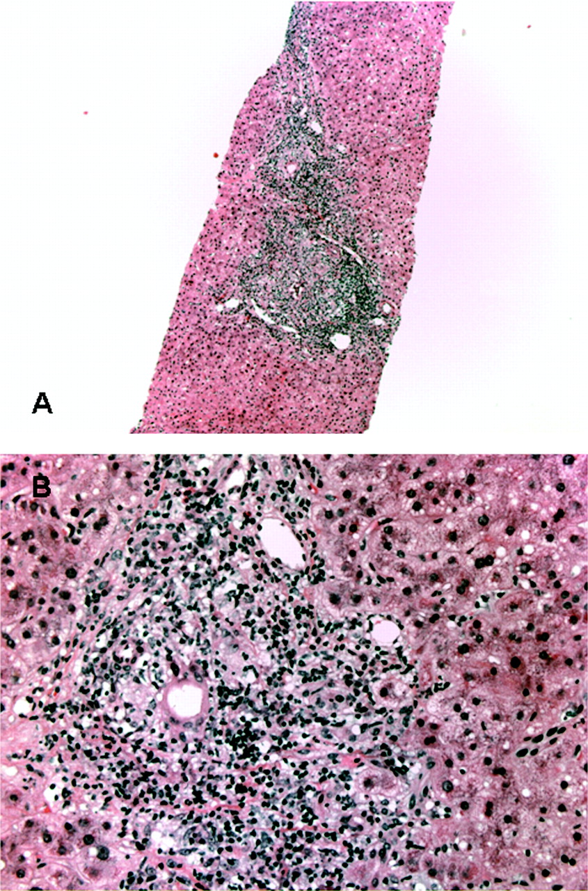
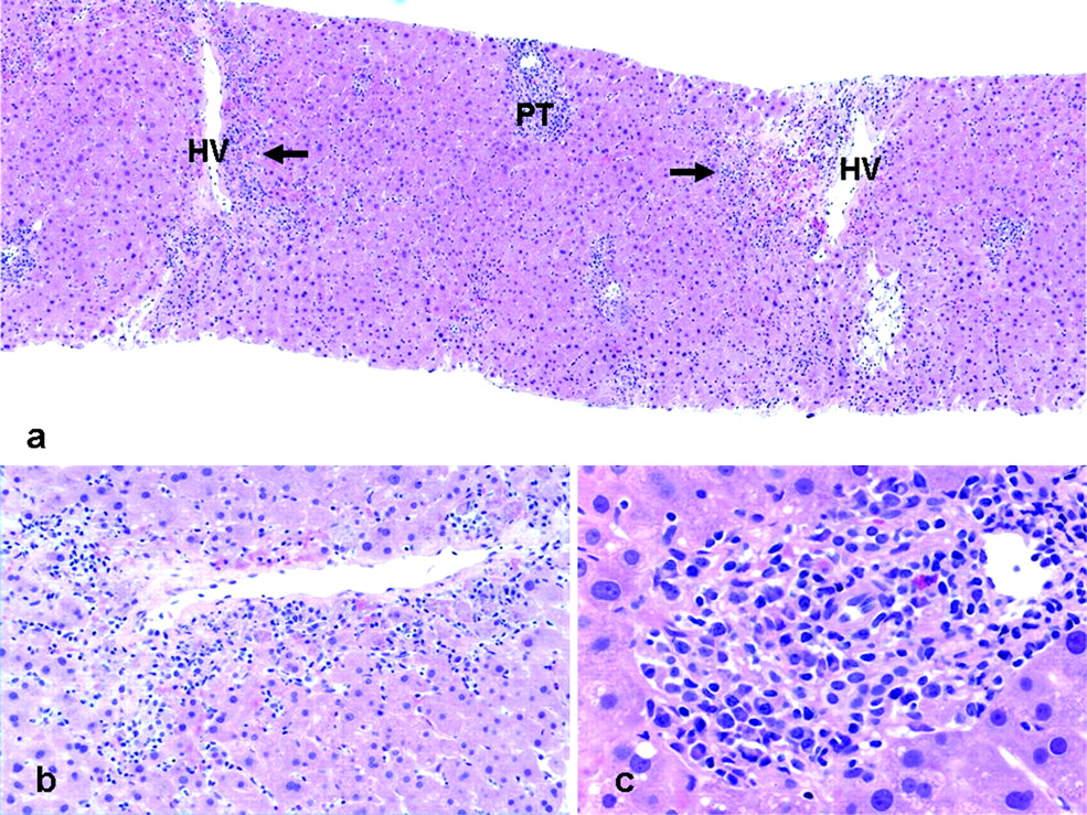
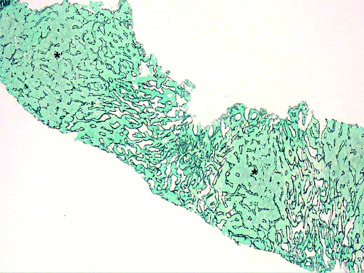
(a) Low-power view from a post-transplant liver biopsy of a patient with recurrent hepatitis C virus and recent elevated transaminases (H&E ×25). Note mild portal inflammation accentuating towards one end of the core (arrowhead). Insert: high-power view of the portal tract with accentuated inflammation. Note that the inflammation here is mixed and there is bile duct injury by lymphocytes (arrow); these are features that mimic cellular rejection (H&E ×400). (b) Repeat biopsy after the abscess was searched for by imaging: low-power view of organising abscess with portal tract at the edge (PT) (H&E ×50). (c) Higher-power view of portal tract at periphery of abscess showing bile duct injury as a bystander next to an inflammatory lesion biopsy from the same patient 4 years later: low-power view showing organisation of collapsed parenchyma by fibrosis that is severe (H&E ×200). (d) Higher-power view of abscess showing inflammatory cells including neutrophils (H&E ×200).
Take-home messages
Successful interpretation of liver allograft is dependent on adequacy of sampling, familiarity with clinical context, working knowledge of differential diagnoses, clinical aspects of transplant hepatology, understanding of the clinical implication of every diagnosis, and close interaction with the clinical team.
The scope of liver allograft pathology is broad but it can be divided into recurrence of original disease and de novo diseases; because pathogenesis in both categories is via immunological injury in many instances, there is overlap between and within these categories histologically. The predominant injury should be identified and communicated appropriately.
Acute cellular rejection occurs usually in the first 3–6 months post-transplant and is characterised by mixed portal inflammation, phlebitis and duct injury, each graded on a scale of 0–3 for a total index of 0–9.
Rejection sometimes shows atypical features that should be differentiated from other causes of inflammation other than rejection. Distinction of acute cell-mediated rejection from recurrent hepatitis C virus (HCV) infection is critical, as the treatment for one has deleterious effects on the other. Importantly, if the biopsy features are indeterminate or the biopsy shows features of both, this should be conveyed very clearly in the biopsy report.
Of the recurrent diseases, viral hepatitis can be difficult to differentiate from rejection; active lobular inflammation, less-mixed portal inflammation, and none or occasional duct inflammation typify viral hepatitis but they overlap with features of atypical late-onset cellular rejection and may be especially difficult to distinguish from recurrent HCV. If testing is negative for the presence of HCV RNA, it is helpful in obtaining a correct diagnosis.
Fibrosing cholestatic hepatitis is an aggressive form of recurrent HCV or hepatitis B virus, and is characterised by minimal inflammation, but marked hepatocellular injury, rapidly progressing fibrosis, and cholestasis resulting from heavy viral burden.
The jury is still out as to whether plasma-rich central perivenulitis represents de novo autoimmune hepatitis or rejection; however, the consensus is that the injury needs to be treated by immunosuppression, as outcome is not favourable in many series.
Bland cholestasis in the early post-transplant period has a broad differential diagnosis that includes sepsis, small-for-size graft syndrome, drug reaction and early duct obstruction. The biopsy is often obtained to confirm or exclude cellular rejection. For the remainder of differential diagnoses, clinical, microbiological and imaging correlation is required.
REFERENCES
Footnotes
Competing interests None.
Provenance and Peer review Commissioned; not externally peer reviewed.