Article Text

Abstract
On behalf of the NHS Breast Screening Programme Pathology Coordinating Group we present recommendations for terminology and diagnostic criteria for a number of key areas of practice in breast pathology where terminology can be confusing and where accurate communication will ensure appropriate clinical management. These recommendations cover columnar cell lesions and the spectrum of changes that can be seen in these epithelial proliferations, lobular neoplasia, micrometastases and isolated tumour cells in axillary lymph nodes, the use of basal/myoepithelial markers in diagnostic practice and oestrogen receptor testing in ductal carcinoma in situ.
- Breast
- CERB 2
- breast cancer
- steroid receptors
- immunohistochemistry
- apoptosis
- breast pathology
- histopathology
- diagnostic screening
- cytology
- diagnosis
- cancer
Statistics from Altmetric.com
- Breast
- CERB 2
- breast cancer
- steroid receptors
- immunohistochemistry
- apoptosis
- breast pathology
- histopathology
- diagnostic screening
- cytology
- diagnosis
- cancer
Introduction
The research subgroup of the NHS Breast Screening Programme (NHSBSP) Pathology Coordinating Group have compiled overviews and recommendations for topics that were either not considered or had minimal discussion in NHSBSP publication number 58 Pathology Reporting of Breast Disease. These will be included as appendices in the pending revision of the non-operative reporting guidelines. In conjunction with NEQAS the HER2 reporting guidelines have been updated and published: Walker RA, Bartlett JM, Dowsett M, et al. HER2 testing in the UK: further update to recommendations. J Clin Pathol 2008;61:818–24.
These topics are emerging issues in diagnostic breast pathology and are presented below as a series of short monographs.
Columnar cell lesions: NHSBSP Coordinating Group recommendations for terminology and diagnostic criteria
Background
In recent years there has been growing interest and awareness of epithelium within breast biopsies having a columnar morphology. One of the common accompaniments of this change is the presence of secretions within lumina, which are often distinctly eosinophilic and associated with microcalcifications.1 These microcalcifications are detectable at mammography and consequently more of these lesions are being detected with the introduction and expansion of breast screening programmes.
The diverse terminology employed for this epithelium, its increased detection at screening and the uncertainties regarding its biology continue to cause concern among both diagnostic breast pathologists and their clinical colleagues in the breast care team. In this document, we present a proposal for a simplified terminology for lesions with columnar epithelium and histological criteria for recognising these, and recommendations for classification using the ‘B’ grading system in core biopsies. These proposals are not set in stone and may change as more knowledge is acquired regarding the pathobiology of these lesions and their relationship to malignancy.
Terminology
It should be noted that a columnar/flat morphology is often seen in the epithelium of benign breast lesions outside the columnar change/flat epithelial atypia (FEA) spectrum and in cytological preparations from a number of conditions, for example, intraductal papillomas.2 Tall columnar cells are also a feature of a number of more unusual carcinoma variants, for example, a tumour recently described as resembling the tall cell variant of papillary thyroid carcinoma.3
Here we refer to a group of lesions deduced by several authors to lie within the constellation of fibrocystic change.4 ,5 Within this group, a more focused spectrum of lesions has been delineated—namely, columnar cell change (CCC), CCC with hyperplasia, CCC with atypia and columnar cell hyperplasia with atypia.6 The morphology of those with atypia corresponds to FEA, as detailed in the latest WHO classification.7
CCC5 ,8–10 has been otherwise variously described and/or illustrated over the years and lesions with attributes of columnar lesion have been referred to under a great diversity of names.1 ,4 ,7 ,11–23 Some of these terms are detailed in figure 1. However, in assessing these it is worth noting, as Ho et al comment, that ‘one must be extremely careful when comparing outcome of FEA and its equivalents in the literature, without first having determined if authors are referring to the same lesion’.23 This statement equally applies to all forms of the overlapping columnar cell lesions (CCLs).
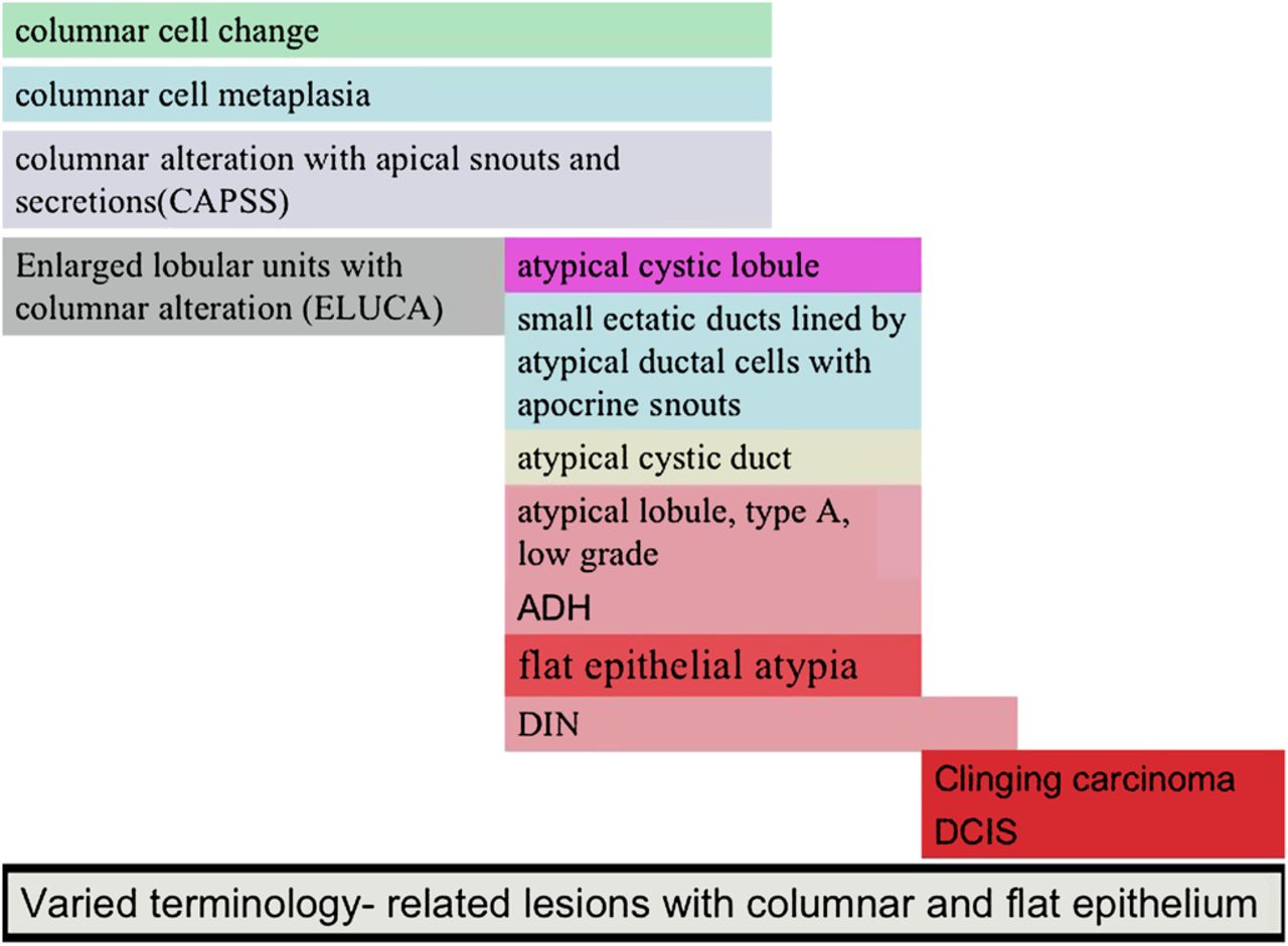
Varied terminology-related lesions with columnar and flat epithelium. ADH, atypical ductal hyperplasia; DCIS, ductal carcinoma in situ; DIN, ductal intraepithelial neoplasia.
The proposed ductal intraepithelial neoplasia (DIN) spectrum described by Bratthauer et al ranges from usual type ductal hyperplasia through to high-grade ductal carcinoma in situ (DCIS),24 although molecular studies do not support this as a continuum and this system of classification is not widely used. The territory mapped out relevant to the present discussion is the DIN 1-flat type which equates to FEA.24
More recently, terminological practice has been generally simplified (figure 2), reflecting current guidelines.

Most simple classification of columnar lesions (after Schnitt and Vincent-Salomon6). DCIS, ductal carcinoma in situ.
Microscopic appearances of CCLs (Table 1)
CCCs without atypia
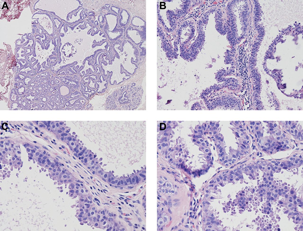
The classic CCL is comprised of lobular acini lined by epithelial cells that are tall and snouted in a manner similar to that observed in tubular carcinoma. Commonly this is associated with luminal secretions and/or microcalcifications (figure 3A,B). If there is a piling up of several layers the term ‘columnar cell hyperplasia’ is used, assuming that the stratification is real (figure 3D) rather than artefactual as a consequence of cross-cutting (figure 3C). There is morphological diversity within these groups, for example, the hyperchromasia of the nuclei can vary, as well as nuclear shape and the ‘tallness’ of the cells; thus in some cases some lesions are more cuboidal than columnar.
Comparison of pathological features of the spectrum of entities from columnar cell change through to ADH/DCIS

Columnar cell change without atypia (A-D) Columnar cell change affects lobular units which show variably dilated acini often with microcystic change and calcification (figure 3A, B). The lining epithelial morphology varies from low cuboidal to the classic columnar type with apical snouts (3C) In more florid cases the epithelium is multi-layered and classified as columnar cell hyperplasia without atypia (3D).
Flat epithelial atypia
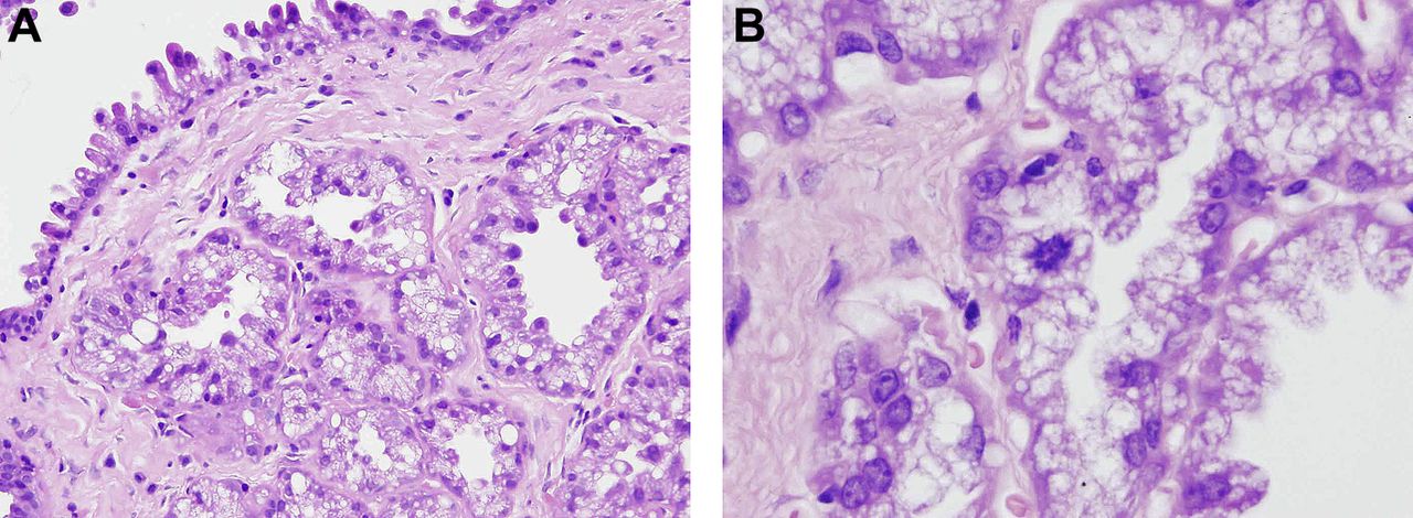
If cytological atypia is present in CCC or columnar cell hyperplasia, the lesions should be classified as FEA (figure 2).8 ,33 There is, however, morphological diversity in this group of lesion; in some cases the atypia manifests as relatively uniform rounded evenly spaced nuclei with a similar cytomorphology to that displayed by the cells of low and some intermediate grade DCIS (figure 4C). In this form of FEA, polarisation of the cells may be lost. Nuclei are often more centrally placed. In others, distinct nuclear pleomorphism is evident, falling short of high nuclear grade but, equally, without the relative nuclear uniformity described above (figure 4A,B). Finally, some FEA display a distinctly endometrioid appearance with tightly packed tall hyperchromatic fusiform nuclei (figure 4D).

Flat epithelial atypia (4 A-D) Flat epithelial atypia usually shows more exajurated involvement of the breast parenchyma (4A). The cells show a loss of nuclear polarity but may retain a columnar morphology (4B). The nuclei are often more rounded and less ovoid than in columnar cell change (4C0. The degree of atypia can become more marked but is not high grade (4D)
Exclusions
Atypical ductal hyperplasia
FEA by definition does not have a complex architecture; if micropapillary or cribriform structures are seen, the diagnosis of atypical ductal hyperplasia (ADH) or low-grade DCIS should be considered.34 The process may involve more than one terminal duct lobular unit. Sporadic atypical architecture may be widely spaced. Rather than result in overdiagnosis of DCIS, it is proposed that the current size criterion guidelines for differentiating DCIS and ADH (>2 mm or >2 duct spaces)25 be relaxed and the term FEA be applied under these circumstances. Whether most FEA is biologically equivalent to most ADH is, as yet, uncertain but likely. In practice, FEA is often seen merging with the more elaborate architecture of ADH (figure 5) and DCIS, demonstrating the close relationship of FEA to these lesions. Lesions which fulfil the criteria for low-grade DCIS by showing ≥2 ducts fully involved by an architecturally and cytologically atypical process without intervening gaps of non-atypical epithelium should still be regarded as such and not as FEA.

Atypical ductal hyperplasia. Flat epithelial hyperplasia, particularly when florid can show both cytonuclear and architectural feature of low grade DCIS but fail to have sufficcent extend for classification as DCIS. The diagnosis of atypical ductal hyperplasia arising in a background of FEA can be made in these circumstances.
Ductal carcinoma in situ
Where flat epithelial proliferative epithelium within a terminal ductal lobular unit (TDLU) possesses high cytonuclear grade nuclei, the lesion should be categorised as high-grade flat DCIS, not FEA (figure 6). Care should be taken in the diagnosis of flat high-grade DCIS in core biopsies and it may be prudent to categorise this as B4 suspicious in such limited samples.

High-grade ductal carcinoma in situ may show mono- or oligo-layering within the inner surface of an involved duct. High-grade nuclear pleomorphism such as this is not seen in flat epithelial atypia.
Differential diagnosis
It should be borne in mind that there are many phenomena with low or ‘flat epithelium’ that do not fall under the category of the CCLs—for example, the tall monolayered epithelium of in situ papillary neoplasia.
Apocrine change
Apocrine epithelium shares with columnar change the presence of apical snouts and sometimes associated secretions. However, the cytoplasm of apocrine cells is typically copious and granular and, unlike most columnar cells, nucleoli are readily seen. Apocrine cells are usually androgen receptor positive but oestrogen receptor (ER) and progesterone receptor (PR) negative; the converse is true for columnar epithelium.
Lactational change
Another lesion that may be mistaken for FEA is focal lactational change, which may appear both monolayered and mildly cytologically atypical with enlarged hyperchromatic nuclei. Typically, the epithelial cells are more cuboidal than tall and the foamy appearance to the cytoplasm produced by finely divided lipid should alert the pathologist to this diagnosis (figure 7).

(A) Lactational changes can be mistaken for flat epithelial atypia and (B) in some rare variants show distinct cytological atypia. The epithelium tends to be cuboidal and the presence of foamy clear cytoplasm should alert one to this diagnosis.
Markers in CCLs
Producing a clear idea of the marker profile in columnar lesions is bedeviled by the variety of terminology used in different publications and the uncertainty about their equivalence. What follows is a distillation of what is known, under the broad umbrella of the names indicated above.
Unlike the normal breast and usual ductal hyperplasia (UDH) where ER and PR staining is heterogeneous and limited to about 10–15% of cells,25 in CCC and related lesions there is extensive and uniform positivity of the nuclei of all lesional cells.4 ,5 ,15 ,20 Most cells stain positively for keratin 1915 and osteoprotegerin26 and a high proportion stain for cyclin D1.15 Kusama et al demonstrated a Ki67 labelling index of 0.3% in columnar lesions versus 13.7% in adjacent DCIS.18 Unlike UDH, the lesional cells do not express basal markers such as CK5/6.27 As indicated above, immunohistochemistry (IHC) may be useful in separating apocrine from columnar epithelium, the former being androgen receptor positive and ER and PR negative. The utility of GCF15, typically positive in apocrine epithelium, in making this distinction has yet to be documented in the literature. It is worth noting that McLaren et al found that patients with ER negative CCLs were at increased risk of malignancy compared with those whose lesions were ER positive,23 but as yet there are no confirmatory studies.
In conclusion, at present the main use for IHC is in the differential diagnosis of columnar from some apocrine lesions.
Significance of CCLs
CCLs of the breast encompass a morphological spectrum which may coexist within a wide range of lesions that may include in situ and invasive carcinoma.9 However, the overall biological significance of these lesions is unknown.28 There is supporting evidence for a precursor role including an association with tubular carcinoma9 ,12; the finding that these lesions are found more commonly in cancerous than non-cancerous breasts17; are often continuous with cancerous lesions18; commonly coexist with lobular in situ neoplasia22; and share similar cytological characteristics and immunohistochemical profile with coexistent malignancies.15
Despite the above, the certainty and speed of progression of these lesions to malignancy is unknown. Eusebi et al11 discovered 25 cases (0.26%) of what was called low-grade clinging carcinoma of flat type, but which appears morphologically equivalent to FEA. During an average follow-up interval of 19 years, none of the 25 women with low-grade clinging carcinoma of flat type developed invasive carcinoma and only one had a recurrence, which was described as consisting of low-grade clinging carcinoma of flat type.29
At a more fundamental level, there is evidence that, in general, there is a progressive accumulation of allelic damage in FEA through to DCIS and invasive carcinoma, implying a molecular continuum from at least some lesions with a columnar morphology (which is not the case for UDH and carcinoma).28 ,30 Despite the very limited follow-up data to date and the need for more evidence, this implies that the biological progression of an individual columnar lesion may be slow. However, it seems very likely that FEA is one of the earliest forms of neoplastic change in the breast.
CCLs in breast core biopsies
Cores bearing CCLs are typically sampled for the histological assessment of mammographic microcalcifications. As for other such specimens, these should be examined at multiple levels (at least three). If CCC or hyperplasia only is found without atypia, the lesions should be regarded as within the constellation of fibrocystic change and categorised as B2, benign.
CCLs with atypia should be regarded as FEA and classified as B3, of uncertain malignant potential. Lesions with more complex architecture should also be regarded as an atypical epithelial proliferation and also regarded as B3, of uncertain malignant potential.
As for all such screen-detected lesions, multidisciplinary discussion should be undertaken to correlate radiological, clinical and histopathological findings. Data on risk of finding adjacent associated malignancy are extremely limited.
CCLs in surgical excision specimens
Thorough sampling and histological examination of surgical specimens bearing FEA should be performed to search for more established neoplasia. In association with some DCIS and/or invasive carcinoma, FEA may be present and may be extensive and/or may extend to the margins of the specimen. This is particularly problematic for lesions which are well recognised as being associated with FEA such as tubular carcinoma9 and, in such cases, it may be uncertain as to whether the CCL represents the precursor process. There is a very limited evidence base for guidance but, at present, it is recommended that whole tumour size (DCIS plus invasive carcinoma) and margin status should include only those areas regarded as established DCIS or invasive tumour using conventional criteria.
Reproducibility of current classification of CCLs
The reproducibility of diagnosis of CCL varies in the published literature. One study looking at the assessment of images by a mixed population of assessors showed poor agreement.31 Another demonstrated ‘excellent agreement’ for a specified task within the CCL group after a PowerPoint directed training session undertaken by a group of pathologists with an interest in breast pathology.32 Agreement was slightly better for determining the absence of FEA (92.8%, 95% CI 84.1% to 97.4%) than for determining its presence (90.4%, 95% CI 79.9% to 96.7%). The latter authors concluded that the diagnosis of FEA and its distinction from CCLs without atypia was highly reproducible if the presently available diagnostic criteria were adhered to. This simplified classification in the latter study forms the basis of the recommendations presented here.
Issues of poor reproducibility can confound diagnosis and lead to inconsistent patient care. This is more likely when complex and diverse terminology with shades of possible interpretations exist. This is no more apparent than in the case of lesions possessing columnar epithelial morphology and is the strongest argument for the simplified classification of these lesions presented in this document.
Key points and recommendations
The terms columnar cell change and columnar cell hyperplasia are regarded as benign lesions falling within the constellation of fibrocystic changes and in core biopsy should be classified as B2, benign.
Columnar cell lesions with cytological atypia should be regarded as FEA and, in a core biopsy, categorised as B3, of uncertain malignant potential.
Sporadic atypical architecture may be widely spaced. Rather than result in overdiagnosis of DCIS, it is proposed for the current guidelines that the size criterion for differentiating DCIS and ADH (>2 mm or >2 duct spaces)25 be relaxed and the term FEA be applied under these circumstances. In needle core biopsy samples these should be regarded as B3, of uncertain malignant potential.
Columnar cell lesions with high cytonuclear grade should be regarded as flat high-grade DCIS.
In situ lobular neoplasia: NHSBSP Coordinating Group overview and recommendations
Lobular carcinoma in situ (LCIS) was first described in 1941 by Foote and Stewart33 and was considered to be akin to DCIS with regard to clinical behaviour, with the recommended management being mastectomy. Subsequently, lobular neoplasia, the term introduced by Haagensen et al,34 was felt to differ from DCIS, being a risk indicator of subsequent development of breast cancer34–36 rather than a true precursor, so radical surgical treatment fell out of favour. The advent of mammographic screening, further follow-up studies and molecular analysis of lobular neoplasia has resulted in the need to re-address its significance and management.
Terminology
Atypical lobular hyperplasia (ALH) and LCIS have been considered distinct entities. They are both characterised by a proliferation within the TDLUs of poorly cohesive, monotonous cuboidal or polygonal cells with clear or light cytoplasm. Intracytoplasmic lumina may be present. Mitotic figures are infrequent. Pagetoid spread of cells between the surface epithelial cells and the basement membrane may be present. The differentiation between the two forms relates to the extent and degree of distension of acini, but this is subject to intra- and inter-observer variability. Because of this the term ‘lobular neoplasia’ or ‘in situ lobular neoplasia’ was introduced, which does not differentiate between ALH and LCIS.
A further system proposed is ‘lobular intraepithelial neoplasia’37 with three subdivisions that relate to morphology and predicted clinical outcome. This grading system requires validation and has not been endorsed by the WHO at the present time.38
‘Pleomorphic lobular carcinoma in situ’ is a more recently described entity39–42 in which the cells show more marked pleomorphism with larger nuclei. Central necrosis and calcification may be present. Distinction from high-grade DCIS may, on occasions, be difficult. Comedo-type necrosis can be present in LCIS that has the typical cytological and architectural features42 and, although atypical, this should not be diagnosed as pleomorphic LCIS.
Other variants that have been described include endocrine, apocrine, histiocytoid and signet ring lobular neoplasia,43 mainly based on cytonuclear features.
Frequency
It is difficult to assess the frequency of lobular neoplasia since there are no specific clinical, mammographic and macroscopic features. It was considered to be present more frequently in the 40–50 age group34 but the incidence in postmenopausal women is increasing, probably due to detection by mammographic screening. The incidence in otherwise benign biopsies is reported as between 0.5% and 3.8%.34 ,44 It can be both multifocal and bilateral.
Immunohistochemical analysis
The characteristic alteration of lobular neoplasia is the lack of expression of the cell adhesion molecule E-cadherin, which is consistently present on the membranes of normal luminal epithelial cells and is also lost in invasive lobular carcinoma (ILC).45–47 Alterations are seen in both classical and pleomorphic lobular neoplasia and immunohistochemical analysis of E-cadherin can aid the diagnosis of solid in situ proliferations of indeterminate nature.48–50 Lack of E-cadherin supports a diagnosis of lobular neoplasia but, if a mixed pattern of staining is present, then it should be reported as combined DCIS and lobular neoplasia.50 Negative markers can, however, cause problems in interpretation. p120 catenin is a member of the cadherin-catenin complex that has a cytoplasmic staining pattern in lobular neoplasia and has been found to be useful in separating lobular neoplasia from low-grade DCIS.51
High molecular weight cytokeratins identified by the antibody 34βE12 have been reported to be consistently present in lobular neoplasia and could be used to differentiate it from DCIS,52 but a subsequent report considered staining to be an artefact.53 This antibody should be used with caution.
Lobular neoplasia consistently shows positivity for ER and PR, including the pleomorphic variant.41 ,47 HER2 amplification and overexpression have been found in pleomorphic LCIS.41 ,42
Molecular studies
The CDH1 gene that encodes for E-cadherin is inactivated in LCIS and ILC through both genetic (deletions and inactivating mutations) and epigenetic (gene promoter methylation) mechanisms.54–56 Concurrent mutations have been identified in LCIS and ILC.46 Mutations have also been found in ALH.56
Comparative genomic hybridisation analysis has shown that similar chromosomal changes are present in ALH and LCIS.57 Array comparative genomic hybridisation and mitochondrial DNA analyses have confirmed similar genetic alterations between matched lobular neoplasia and ILC.58 ,59
Overall, the molecular data provide strong evidence that lobular neoplasia is a non-obligate precursor of ILC.
Differential diagnosis
The main problems relate to poor fixation in a surgical specimen, differentiating low-grade solid DCIS from lobular neoplasia and pleomorphic LCIS.
In larger surgical specimens poor fixation can lead to artefactual discohesion within lobular units, resulting in an overdiagnosis of LCIS. This is more of a problem in mastectomies that have not been sliced sufficiently in the fresh state.
Differentiating low-grade solid DCIS from lobular neoplasia can be difficult in both surgical specimens and core biopsies and the use of E-cadherin staining can be particularly helpful.48–50 Coexistent low-grade DCIS and lobular neoplasia is not rare and a mixed pattern of E-cadherin staining will be seen in these circumstances. Cancerisation of lobules by DCIS can also cause problems and again E-cadherin staining is of value.
Pleomorphic LCIS39–42 can be difficult to differentiate from high-grade DCIS due to the degree of pleomorphism, the presence of necrosis and calcification, and has most certainly been classified as DCIS previously. Pleomorphic LCIS should be considered if there is poor cellular cohesion along with these other features. Again E-cadherin staining can be invaluable.
Since it is not possible reliably to assess the degree or extent of lobular changes in core biopsies, providing that DCIS has been excluded, the lesion should be reported as B3, of uncertain malignant potential, and recorded as lobular neoplasia, with no attempt to distinguish ALH and LCIS.
Significance
The significance of the diagnosis of lobular neoplasia in a needle core biopsy relates to the risk of there being associated invasive carcinoma (or DCIS) present at the time of the biopsy (see Management). When identified within a surgical biopsy/excision specimen, the questions of concern are different. These are: (1) What is the risk of subsequent development of invasive carcinoma? (2) Is lobular neoplasia a precursor lesion of invasive disease? (3) Does lobular neoplasia affect the risk of recurrence following breast conserving therapy?
It has been recognised for a long time that lobular neoplasia is an indicator of increased risk of developing invasive cancer,34–36 ,44 with the RR differing between ALH (3–5×)60–62 and LCIS (8–11×).34 ,36 ,44 ,63 ,64 The increased risk has been found in some reports to be evenly distributed between both breasts,34 ,64 ,65 although Page et al62 found invasive carcinoma to be three times more likely to arise in the breast with ALH than the contralateral. Collins et al66 found the risk of developing an invasive cancer to be greater for premenopausal women with ALH than ADH, whereas SEER data64 show an increased incidence after the menopause.
The evidence that lobular neoplasia could be a non-obligate precursor lesion comes from molecular and epidemiology studies. As described above, the genetic profiles of ALH and LCIS are similar to each other and to those of ILC.54–59 That there is a greater risk of invasive cancer in the ipsilateral breast62 ,66 ,67 and that the invasive cancer is more likely to be lobular than ductal35 ,67 also indicate that lobular neoplasia could be a non-obligate precursor. Although there are no long-term follow-up studies of pleomorphic LCIS, the small-size series that have been published have shown a higher incidence of associated invasive disease, usually ILC, which is suggestive of a precursor lesion.42
There is general agreement from published studies that the presence and extent of lobular neoplasia, including presence at the margins, does not increase the risk of recurrence following breast conserving surgery,68–71 although the NSABP study69 excluded cases of pleomorphic LCIS (described as DLCIS).
Management
Needle core biopsy
Issues relating to lobular neoplasia in needle core biopsies have recently been reviewed.72 ,73 The EUSOMA Working Group74 considered lobular neoplasia to be most frequently a coincidental finding in a core biopsy and therefore advised that multidisciplinary discussion was essential to determine management, as is advocated by others.75 Diagnostic surgical excision of lobular neoplasia has been advocated.76 A recent review of the literature revealed an upgrade of 20% for LCIS and 13% for ALH to carcinoma when excised.77 Concerns have been raised about underestimation of cancer,77–81 even with stereotactic vacuum-assisted biopsy,79 and this has led to the recommendation of diagnostic surgical excision for all such lesions from some groups.73 It must be appreciated that much of the data is retrospective and that not all studies have considered radiological-pathological discordance as a factor resulting in upgrade, as found by some.75 ,82 What is required are large prospective studies with all factors included.
There is general agreement, although limited robust data, that pleomorphic LCIS should be subjected to therapeutic excision; in essence, treated as DCIS and therefore categorised as B5a on needle core biopsy. In one series of 12 cases diagnosed on needle core biopsy, ILC was found in 3 on subsequent excision.83 There is a need for larger studies to confirm that this does represent a more aggressive disease.
Surgical excisions
The presence and extent of lobular neoplasia in therapeutic excisions should be reported as well as its presence at margins. At present further surgery is not recommended if the invasive cancer is excised.68–71
If lobular neoplasia only is seen in a diagnostic excision biopsy and the clinical/radiologically relevant area has been excised, then no further surgery is required.
If pleomorphic LCIS is found at excision margins then it should be treated as would DCIS and further surgery undertaken to obtain complete excision with appropriate margin of clearance.
Key points and recommendations
Due to problems in assessing the degree or extent of changes in needle core biopsies, the terms ‘lobular neoplasia’ or ‘in situ lobular neoplasia’ should be used rather than ‘atypical lobular hyperplasia’ and ‘lobular carcinoma in situ’ (LCIS). In surgical specimens, atypical lobular hyperplasia and LCIS should be distinguished and recorded.
Lack of expression of the cell adhesion molecule E-cadherin can aid differentiation from low-grade solid DCIS.
Pleomorphic LCIS exhibits more marked pleomorphism, necrosis and calcification. It can be differentiated from DCIS by lack of E-cadherin but should be managed as DCIS when diagnosed on needle core biopsy (B5a) and if at surgical excision margins.
Lobular neoplasia is an indicator of increased risk of developing invasive carcinoma, which is greater for LCIS.
There is evidence that lobular neoplasia is a non-obligate precursor lesion, based on similarities of genetic profiles with ILC and greater risk of invasion in the ipsilateral breast.
Finding of lobular neoplasia in needle core biopsy merits multidisciplinary discussion and, for the majority, diagnostic surgical excision.
Presence of lobular neoplasia at surgical excision margins should be recorded but does not require re-excision.
Micrometastases and isolated tumour cells (ITCs): NHSBSP Coordinating Group overview and recommendations
Lymph node status is recognised as being a major prognostic factor for patients with breast cancer and forms part of staging systems such as the TNM (tumour, lymph node, metastases) classification system. That lymph node metastases could be ‘occult’ was recognised over 40 years ago,84 but their significance is still debated. The introduction of sentinel lymph node biopsy has raised further issues regarding the importance, or not, of these ‘occult’ metastases. These relate to their definition; how they are detected and measured; and their significance in predicting involvement of non-sentinel lymph nodes and prognosis.
Definitions
The term ‘occult’ metastasis has been defined as one missed by initial histological examination and identified on subsequent assessment, and also as a metastasis identified through additional evaluation of paraffin-embedded lymph node blocks.85 It has not been defined by size. Publications referring to occult metastases are therefore not necessarily comparable with regard to the size of these metastases. Some earlier studies did use 2.0 mm as the cut-off between micrometastasis and macrometastasis.86–88 Based on the evidence produced from these, the 5th edition of the AJCC Cancer Staging Manual introduced the definition of micrometastasis as being ≤2 mm (pN1a).89 The sixth editions of the AJCC/UICC staging manuals90–92 further defined micrometastases to be >0.2 mm but not >2.0 mm and introduced the term isolated tumour cells (ITC) that are ≤0.2 mm:
| Micrometastases |
|
| Isolated tumour cells (ITC) |
|
Connolly,93 in discussing changes and problematic areas in the AJCC Cancer Staging Manual, identified that there could be differences in interpretation of ITCs in different situations which would result in different classification of node status. The European Working Group for Breast Screening Pathology (EWG) undertook an evaluation of low volume lymph node involvement in order to test reproducibility of these categories for distinguishing ITCs and micrometastases.94 The first evaluation of images from 50 lymph nodes showed only fair reproducibility, better for micrometastases than for ITCs. Following discussions, refinements to the definitions were proposed (box 1). The use of these refinements improved reproducibility between participants. When these criteria were used by another less specialised group to categorise the same cases, consistency was achieved with the experts95 but the level of consistency was still moderate. A subsequent study to improve interobserver reproducibility was undertaken led by Turner,96 in which six experienced breast pathologists examined digital images of small volume nodal metastases before and after studying a training programme (criteria in box 2). The EWG then applied the two interpretations of the TNM definitions and found that the Turner system had better reproducibility but it may underestimate the rate of non-sentinel node involvement.97
European Working Group study classification rules (from Cserni et al97)
Capsular (including intravascular) lesions considered nodal, and any lesion with nodal and simultaneous extranodal involvement considered nodal, but lesions outside the node (afferent lymphatics, perinodal fat) not recorded as nodal involvement.
Tumour cells (multiple) localised in parenchyma considered micrometastasis even if <0.2 mm with no proliferation or stromal reaction.
Tumour cells localised in vessels or sinuses: if ≤0.2 mm isolated tumour cells, if >0.2–2 mm micrometastasis.
If multiple foci, only largest should be considered.
Single tumour cells or clusters arranged in a continuous manner or separated by 2–5 cells should be considered and measured as one focus and characterised by largest dimension.
Cells or clusters arranged in a discontinuous manner and dispersed homogenously in a definable part of the lymph node should be considered and measured as one focus.
Cells, clusters or foci (as defined above) arranged in a discontinuous manner and dispersed unevenly should be considered as one if the distance between the clusters or foci is smaller than the smallest cluster or focus.
Cells, clusters or foci (as defined above) arranged in a discontinuous manner and dispersed unevenly should be considered as distinct and multiple if the distance between the unevenly distributed tumour cells, clusters or foci is greater than the smaller cluster or focus and should be characterised by the largest cluster or focus.
In cases of doubt, the lower category (isolated tumour cell) should be given.
Turner study classification rules (from Cserni et al97)
Isolated tumour cells and micrometastases are distinguished by the size of the largest tumour cell cluster, regardless of microanatomical location within or adjacent to the lymph node and regardless of the number of clusters or single cells.
A cluster is a confluent focus of tumour cells touching other tumour cells.
Clusters or cells separated by a single benign cell or a spatial gap are measured as separate clusters, except when fibroblastic reaction has caused the separation.
Any lesion composed of non-cohesive cells or clusters not >0.2 mm represents isolated tumour cells.
Any lesion with largest cohesive cellular lesion >0.2 mm but not >2.0 mm represents micrometastasis.
Mitotic activity is not considered.
For borderline or indeterminate findings at the two ends of the spectrum, the lower stage category should be selected.
The evidence suggests that there are problems with using these definitions, particularly when discriminating between ITCs and micrometastases which will affect interpretation of their significance. The TNM definitions of low volume nodal metastases need to be better formulated with visual information.
Detection
The three areas that have been considered have been the extent to which lymph nodes should be examined histologically, the use of IHC and, to a lesser extent, the use of reverse transcriptase PCR to identify micrometastases and ITCs.
Histological examination of lymph nodes
How extensively to analyse a lymph node has become a question raised frequently with the increasing use of the sentinel lymph node biopsy technique.
There are differences in relation to the initial slicing of the node; the American Society of Clinical Oncology (ASCO) guidelines for sentinel lymph node biopsy in early breast cancer98 recommend slicing through the long axis whereas NHSBSS Pathology Reporting of Breast Disease99 recommends slicing the node perpendicular to the long axis. Both advocate thin slicing and embedding all pieces. When sentinel nodes are sliced at 2 mm intervals and totally embedded, the probability of identifying all metastases >2.0 mm is high.85 Cserni100 used a geometrical model of sentinel lymph node histopathology and found that sections 1 mm apart should identify metastases >2 mm. To identify micrometastases, step sectioning with levels of 250 μm or 200 μm would be required.
A review of the literature101 on the value of using step sectioning and IHC (see below) found upstaging to be reported after the former to range from 4.8%102 to 30.7%.103 However, it was difficult to assess in various studies whether this referred to macrometastasis or micrometastasis.
Immunohistochemistry (IHC)
Cytokeratin antibodies (eg, Cam5.2, MNF 116, Pan cytokeratin; AE1/AE3) have been used in many studies to aid the detection of metastases (macrometastases and micrometastases, ITCs), often in combination with step sectioning and on multiple levels in cases with negative H&E findings. Reviews of the literature101 ,104 indicate IHC results in upstaging in 2.6–19% of cases. One study105 found IHC to detect metastases in 4%, 2% being micrometastases (detectable on H&E section on review) and 2% being ITCs, and concluded it was of limited value. Concerns about interpretation of IHC were raised by Roberts et al106 who found that, as tumour cell numbers decreased, the ability of pathologists to recognise them also decreased. Other concerns have been raised about IHC-only detected tumour cells in lymph nodes.107 In a review of 4066 cases of sentinel nodes positive only on IHC, there was no association with vascular invasion, tumour size, tumour type and location but there was an association with method of biopsy, the frequency increasing in relation to the degree of manipulation.
Molecular analysis
The predominant method has been reverse transcriptase PCR for a range of markers (cytokeratin 19, 20, CEA, MUC-1, maspin, mammoglobin) using either frozen sections or half the sentinel node. One of the largest series with 123 patients and 146 nodes by Manzotti et al108 had a high sensitivity but lower specificity for any marker, with a higher specificity for two of CK19, maspin or mammoglobin. Molecular analysis has recently been developed commercially for the intraoperative assessment of metastases in sentinel lymph nodes109–111 but has not been evaluated extensively.
Significance
There are two topics of importance: (1) whether micrometastases and ITCs predict the involvement of non-sentinel axillary lymph nodes and (2) the prognostic significance of micrometastases and ITCs.
Predicting non-sentinel lymph node metastasis
The aim of the sentinel node procedure is to reduce the frequency of axillary node surgery in node negative women and so reduce morbidity. Identification of sentinel node involvement is considered an indication for further axillary surgery or radiotherapy, but the question is whether such treatment is required if only a micrometastasis is present in the sentinel node. Cserni et al112 undertook a literature review and meta-analysis of 25 studies published up to 2003 that considered non-sentinel node metastases associated with sentinel node micrometastasis. They concluded that the risk of non-sentinel node metastasis was around 10–15%, lower if sentinel node involvement was detected by IHC. Viale et al113 found differences in frequency of non-sentinel node metastases in relation to the size of the micrometastasis, being less if <1 mm. Other factors that have been found to relate to non-sentinel node metastases in the presence of sentinel node micrometastasis include tumour size,114–116 grade115 and vascular invasion.116 Since the meta-analysis undertaken by Cserni et al,112 further publications have reported differing frequencies in the incidence of non-sentinel node metastases. In tumours 15 mm or less, no further involvement was found for cases with ITCs, with 12% of cases with micrometastases having non-sentinel node involvement.117 Tumour size and type were found to be factors predicting non-involvement in the study by Houvenaeghel et al.118 Similar frequencies of involvement for cases with micrometastases have been reported in two papers from Holland (19%, 20%),119 ,120 although there were differences for ITCs (7%, 13%). In a study confined to ITC positive sentinel lymph nodes, a very low incidence of non-sentinel node metastases was found and no axillary recurrence if axillary surgery had not been performed.121
There is sufficient evidence to support the avoidance of axillary node dissection in cases with ITCs, but there is no agreement with regard to the further management of the axilla if micrometastases are identified.
Prognostic significance of micrometastases and isolated tumour cells (ITCs)
Many of the studies undertaken (reviewed by Cserni et al101) have considered occult metastases with only a small number commenting specifically on micrometastasis. Different combinations of H&E ± step sectioning ± IHC have been used. There is no strong evidence for a survival disadvantage. Some studies suggest that there is, but within these there are variations in relation to disease-free and overall survival and different patient groups. A study with a long follow-up that considered occult metastases found no difference in survival for patients with and without these,122 and emphasised the need for multivariate analysis in such studies. Using data taken from a cancer registry for a 25-year period, Kuijt et al123 found on multivariate analysis that patients with micrometastases had a significantly worse survival than those without, but there was no pathological review of lymph nodes. The same applies in two studies based on data from SEER which found the prognosis for micrometastatic disease intermediate between N0 and N1124 ,125 and concluded that a prospective study is required. A re-evaluation of node negative cases with 20-year follow-up using IHC identified 23% of cases with metastatic disease, predominantly ITCs.126 The prognosis of those with micrometastatic disease was poorer than node negative cases, which was more apparent after 10 years. Another centre examining node negative cases after 8 years of follow-up found 13% of cases with micrometastatic disease but there was no association with outcome.127
Key points and recommendations
Despite the extensive literature, there is still a lot of debate about the importance and significance of micrometastases and isolated tumour cells (ITCs), particularly with increasing use of sentinel node biopsy. This impacts on factors such as the routine pathological handling of sentinel nodes.
If a micrometastasis is identified in a single H&E section, levels should be cut to check whether it remains a micrometastasis or becomes a macrometastasis (pN1mi to pN1).
Sentinel lymph nodes should be sliced at 1–2 mm with all pieces embedded to ensure a full face H&E section. This is the minimum requirement. Step sectioning at 200–250 μm can be undertaken but is not mandatory. It would be appropriate for centres undertaking detailed clinical studies of sentinel lymph nodes.
There is no good evidence for the routine use of immunohistochemistry in evaluating sentinel lymph nodes; it could be of value for centres undertaking detailed clinical studies of sentinel lymph nodes.
Definitions—AJCC/UICC amended definitions to be followed; discrimination between micrometastases and ITCs can be aided by using criteria from the European Working Group and Turner studies.
Since there are variations in the incidence of non-sentinel node metastases in the presence of sentinel node micrometastasis, axillary surgery/radiotherapy is considered appropriate but is not indicated for ITCs.
There is no strong evidence for an effect of micrometastases on survival.
Further data are required in several areas to assess the significance and importance of micrometastases and isolated tumour cells.
Use of basal/myoepithelial markers in breast pathology: NHSBSP Coordinating Group overview and recommendations
The increasing use of needle core biopsy for non-operative diagnosis and the nature of lesions identified mammographically can result in diagnostic difficulties. Interpretation may be helped by the use of IHC to detect basal/myoepithelial markers. A recently circulated questionnaire showed that there were variations in the type and frequency of markers used diagnostically in the UK and Ireland. This paper summarises current knowledge of the various markers, discusses the diagnostic areas where they are of value and makes recommendations for a suitable diagnostic panel.
Basal/myoepithelial markers
The myoepithelial cell has traditionally been distinguished from luminal epithelial cells by the presence of smooth muscle fibres. However, many more proteins have now been identified that are expressed in myoepithelial cells and these fall into three groups (smooth muscle-related, cytokeratins and others).
Smooth muscle-related
Smooth muscle actin (SMA): antibodies detect actin microfilaments; good sensitivity but specificity poor since will detect myofibroblasts.128
Smooth muscle myosin heavy chain (SMMHC): structural component of smooth muscle myosin. SM2 isoform is expressed in breast myoepithelial cells. Sensitivity and specificity are high with SMMHC being present in periductal and periacinar myoepithelial cells and not stromal myofibroblasts,129 although sensitivity reported to be slightly lower than SMA.128
Calponin: 34 kD polypeptide, modulates actomyosin ATPase activity. Excellent sensitivity but present in a subset of myofibroblasts.128 ,129
H-caldesmon: SMA binding protein. Only detectable in myoepithelial cells of ducts but not found in myofibroblasts.129 ,130
Cytokeratins
Myoepithelial cells express cytokeratins (CK) characteristic of basal layer of stratified epithelium: CK5, CK10, CK14 and CK17.131 ,132
CK5/6 in normal breast can be detected in both myoepithelial and luminal epithelial cells.133
The antibody 34β12 recognises CKs 1, 5, 10 and 14 but is not specific for myoepithelial cells.134
Other markers
S100: has low specificity since detectable in epithelial cells.135
CD10 (Common Acute Lymphoblastic Leukaemia Antigen (CALLA)): endopeptide expressed in myoepithelial cells.136 Antibodies now available that work on fixed tissue; high sensitivity in normal breast.137
P cadherin: cell adhesion molecule with high sensitivity for myoepithelial cells in normal breast, no reactivity with myofibroblasts.138
P63-p53 homologue: differs from other markers in being nuclear139 with high sensitivity for myoepithelial cells.140
14-3-3σ: protein associated with apoptosis and cell cycle control, present in myoepithelial cells in normal breast.141
Maspin: serine protease inhibitor present in myoepithelial cells142 with both cytoplasmic and nuclear reactivity.
Diagnostic areas
Radial scar versus tubular carcinoma
Highly sensitive and specific markers are required due to myoepithelial cells in central parts of radial scars being attenuated, plus the presence of myofibroblasts. The requirements favour SMMHC and p63 as markers of choice. SMA and, to a lesser extent, calponin will be present in myofibroblasts.
Papillary lesions
IHC can reduce the variability in reporting of core biopsies of papillary lesions, particularly in relation to B3 and B4.143 Intraduct papillomas have a complete layer of myoepithelial cells, whether detected by SMMHC, calponin, p63 and CK5/6144 or SMA, p63, CD10 and CK14.145 However, findings for intracystic papillary carcinomas differ, with Collins et al144 failing to detect any myoepithelial cells and Tse et al145 finding them to be scattered and discontinuous. p63 had the highest sensitivity, CK14 was also present in florid hyperplasia, SMA showed stromal staining and CD10 epithelial and stromal staining.
Hyperplasia versus atypia versus DCIS
High molecular weight basal cytokeratins are of use particularly in interpretation of core biopsies enabling a B2 rather than a B3 diagnosis. Myoepithelial cells form a useful internal positive control. Hyperplasia of usual type and hyperplasia in papillomas show reactivity for CK5/6133 ,146 ,147 and CK14146 whereas atypical hyperplasia and DCIS epithelium do not.
Non-invasive versus invasive carcinoma
Myoepithelial cell markers can be of value in assessing whether or not invasion is present, but myofibroblast reactivity, discontinuity of staining of myoepithelial cells and staining of vascular smooth muscle cells can cause problems in interpretation. Discontinuous staining can occur with p63,140 ,148 but it has greater sensitivity and specificity. SMMHC has been found to be more sensitive than CD10 in assessing invasion149 but is in vascular smooth muscle cells. Owing to the advantages and disadvantages, it is better to use two myoepithelial markers that complement one another—for example, p63 and SMMHC or calponin.
Myoepithelial tumours
SMA, CK5/6, CK14, p63 and other markers are all suitable for analysing possible (adeno) myoepithelial tumours, both benign and malignant.150
Other lesions
Myoepithelial markers can aid differentiation of adenoid cystic carcinoma from collagenous spherulosis and cribriform carcinoma.
Basal-like carcinomas
There is increasing interest in basal-like breast carcinomas, partly due to the finding that BRCA1-related breast cancers can have these features151 and to their poorer response to anthracycline chemotherapy.152 There is no consensus as to the definition of basal-like, which probably reflects the view that they are a heterogeneous group of cancers. Nielsen et al153 consider them to express CK5/6, HER-1 (EGFR) and/or c-kit and lack ER and HER2. Others have used CK14154 or P-cadherin155 as a basal marker to identify these tumours. Rakha et al156 have proposed that basal-like carcinomas can be defined based on the expression of basal cytokeratins (CK5/6 and CK14) irrespective of other markers.
Recommendations for a diagnostic panel
It is important not to rely on just one marker.
Smooth muscle myosin heavy chain (SMMHC) or calponin are sensitive and specific cytoplasmic myoepithelial markers. Smooth muscle actin can be used but care has to be taken in interpretation in the presence of myofibroblasts.
P63 is a sensitive and specific nuclear myoepithelial marker but staining can be discontinuous, so use of a cytoplasmic myoepithelial marker such as SMMHC or calponin as well will aid interpretation.
When trying to identify the presence/absence of myoepithelial cells, use p63 and SMMHC/calponin.
CK5/6 is of value in several diagnostic situations such as analysis of hyperplasias or basal-like breast cancers.
Oestrogen receptor assessment in ductal carcinoma in situ (DCIS)
The response of invasive breast cancer to endocrine therapy is well recognised as being related to the hormone receptor status of the tumour.157 It is therefore logical to investigate the role of hormone receptors in determining the role of endocrine therapy in the management of DCIS. However, to date, there remain issues with regard to the latter and with the assessment of hormone receptors.
Evidence from clinical trials
The value of tamoxifen in the management of DCIS has been reported in two randomised studies:
The NSABP B-24 trial of the use of tamoxifen and radiotherapy in DCIS showed a benefit from endocrine therapy, primarily due to a reduction of ipsilateral recurrent invasive carcinoma.157 ,158 The 7-year risk of local recurrence in the treated breast after lumpectomy plus radiation was reduced from 11.1% without tamoxifen to 7.7% with tamoxifen (p=0.02). The risk of all breast cancer events (ipsilateral plus contralateral) was reduced from 16.9% to 10.0% (p=0.0003). Allred and colleagues retrospectively reviewed 628 cases from this trial and demonstrated that the benefit was confined to those patients whose tumours were ER positive; this was defined as 10% or more cells staining.159 However, it is not clear whether there was a difference in ipsilateral and/or contralateral recurrence with regard to ER status of the primary disease process as the data are available in abstract form only. It is also not clear how the figure of 10% was reached and, in particular, whether modelling was carried out to determine a cut-off value or range that best correlated with therapeutic benefit. The review included a mixture of both locally reported ER and central assays and concern was raised about possible false negative results from laboratories using diverse non-standardised assays. With the exception of the abstract referred to above, the study has not been published.
The UK/ANZ (UKCCCR) Trial160 comparing radiotherapy and tamoxifen in the treatment of DCIS showed after 53 months of follow-up a non-significant reduction in all breast events with the addition of tamoxifen but a RR reduction of 22%. It should be noted that the follow-up period in the UK/ANZ trial at the time of reporting was shorter than NSABP B-24 (4.4 vs 6.9 years) and receptor status of the DCIS was not measured prospectively in this trial. In addition, a high proportion of cases were of high cytonuclear grade; a relatively high proportion are therefore likely to be ER negative which will have influenced the effect of hormone treatment in this series (S E Pinder, personal communication).
A further study, the NSABP-P1 trial of tamoxifen for the prevention of breast cancer, studied 13 000 at-risk women.161 This cohort did not include patients with DCIS but illustrates the potential benefit of endocrine therapy in the prevention of breast cancer events. Invasive and non-invasive carcinomas were reduced by 50% at 69 months follow-up. There was a threefold increase in thromboembolic events, a marginal increase in cataracts and a non-significant increase in strokes.
Current guidance and options
A cut-off point for positive versus negative for ER in DCIS was set at <5% cells staining as an entry criterion for two UK clinical trials of endocrine therapy in DCIS (IBIS II and DCIS II, the latter discontinued). The UK breast screening guidelines162 recommend the use of <5% cells staining for DCIS and the use of the Allred score for invasive cancers. A cut-off of Allred score 3 has been used in a large study of pure DCIS examining the distribution of various different phenotypes162 and is the same as that applied to invasive breast carcinoma.157 ,162 It would be illogical to adopt a different scoring system from those already in use for invasive disease unless sound evidence to the contrary emerged. Furthermore, there is strong evidence that the ER status of invasive breast cancer parallels that of associated DCIS in individual cases.163 Individual practitioners benefit from familiarity with a particular scoring technique and clinical colleagues require consistency in the approach to reporting. The three scoring methods currently in use worldwide are: (1) a simple percentage of cells staining without reference to intensity; (2) the histochemical ‘H’ score which is the sum of the percentage of cells with three levels of staining, giving a range of 0–300164; and (3) Allred scoring (replacing previous ‘Quick Score’) which is the sum of an intensity score of 1–3 and the percentage of cells staining banded in a non-linear fashion using a score of 1–5 giving a final score between 2 and 8.165
Allred scoring and the histochemical score provide similar information about receptor status as both include an assessment of intensity as well as percentage of cell nuclei staining. Simple percentage scoring gives no information about staining intensity although low proportion scores tend to go hand in hand with low intensity scoring with ER scoring, as opposed to PR immunoreactivity.
There is no evidence base to support a preference for a particular scoring technique. However, there is evidence for assessment of proportion and intensity staining in invasive breast cancer and, as such evaluation is routine practice, it would be logical to mirror this practice in the reporting of DCIS. Evidence from the Sloane Project,166 a UK audit of screen-detected DCIS and atypical hyperplasias of the breast, points to considerable confusion in this area among pathologists in their reporting practice of ER status in DCIS. A wide variation is seen between and within laboratories of the scoring method used and also of the cut-off point for positive versus negative results. In this audit, 40% of reporting laboratories used one method and the same cut-off criterion throughout, 19% used one method but different cut-off criteria for different cases, 15% of laboratories used two methods for different cases with equivalent cut-off points for each method while 25% of laboratories used two methods and non-equivalent cut-off points from case to case.
Because there is such uncertainty about what cut-off is therapeutically relevant (if any) in the pharmacological management of DCIS and possibly as a more general prognostic marker for this disease process, it would be sensible to record the percentage of cells staining and the average intensity in the duct profiles available and correlate this information with clinical outcomes.
Patterns of ER expression in DCIS
From the data gathered by the Sloane Project, intermediate and low-grade DCIS are almost invariably ER positive whereas high-grade DCIS is positive in 69% of cases (table 2).
Grade of DCIS and ER status: Sloane Project data based on 1684 cases
Baqai and Shousha reviewed 60 cases of DCIS, 56 pure and four with associated microinvasion. DCIS was assessed in terms of grade, architecture and, among other things, ER positivity was defined as disease with >10% of cells showing dark brown nuclear staining (Allred score >4, Histoscore >20). The grade distribution seen was 52% high, 38% intermediate and 10% low, which is very similar to the distribution among the Sloane Project cases.168 There was a strong association between ER negativity and high nuclear grade. The number of cases in this study was small and the cut-off point was set relatively high. It is a pity that the opportunity to model different cut-off points against grade was not taken.
Ottesen et al169 showed in a series of 133 cases of DCIS that one-third displayed 5% or less staining for ER, the overwhelming majority of which were of large nuclear size. In this series, perhaps unusually, most cases were either strongly positive or negative.
Basal phenotype DCIS, seen in approximately 6% of high-grade lesions, is commonly negative for ER, PR and Her2.162 ,170 ,171
Future directions
It is likely that the IBIS II trial will give invaluable information about the validity of setting a positive/negative cut-off point at 5% or 10% for ER. Obviously there will be limited information about the relevance of staining intensity unless Allred scores are used and these are not obligatory. The Sloane Project, given its long-term goals of prolonged follow-up of DCIS patients, will also provide information about a range of cut-off points because of the variability that has already been demonstrated in the first cohort of patients reviewed.167 Because data are not available to give guidance on the clinical relevance of different proportion and intensity scores, it is recommended as a minimum to record both when reporting the hormone receptor status of DCIS. This will enable Histoscores and/or Allred scores to be derived without loss of the original scoring information.
There remains considerable scope for modelling ER cut-off points in DCIS against clinical outcomes in large cohorts of patients but, until such time as that is possible, it seems reasonable to apply the limited evidence available at the present time and use the currently accepted cut-off points to guide the endocrine management of patients with DCIS as for reporting ER status in invasive breast cancer.162
Key points and recommendations
>80% of DCIS is ER positive and there is a body of evidence indicating a therapeutic benefit for endocrine treatment in selected ER positive patients.
Previously a cut-off point of 5% of cells staining has been recommended to define positive and negative. It is recommended that both proportion of cells staining and intensity should be recorded.
Take-home messages
Columnar cell lesions may show a spectrum of abnormalities from simple metaplasia to ductal carcinoma in situ.
Lobular neoplasia or in situ lobular neoplasia are the preferred terms for core needle biopsy reports whereas the terms atypical lobular hyperplasia or lobular carcinoma in situ should be specified in excisions.
Metastatic tumour clusters in lymph nodes <0.2 mm are designated isolated tumour cells. Discontinuous clusters should be measured as one focus if in a definable part of a lymph node. Unevenly dispersed clustered should be characterised by the largest cluster.
When using basal/myoepithelial markers do not rely on a single marker. There is often mismatch between individual marker's staining profiles.
ER scoring for DCIS should follow practice in invasive disease recording both proportion and intensity of cells staining using, for example, a Histoscore or Allred type system.
Acknowledgments
The authors acknowledge the members of the UK National Coordinating Committee for Breast Pathology.
References
Footnotes
Competing interests None.
Provenance and peer review Commissioned; internally peer reviewed.