Article Text

Abstract
Aims The clinical significance of TOP2A as a prognostic marker has not been clarified. The aims of this study were to investigate the frequency of TOP2A copy number change; to correlate TOP2A with HER2 status, hormone receptor (HR) status and molecular subtype, and further to explore differences in breast cancer-specific survival according to TOP2A and HER2.
Methods In this study, TOP2A, HER2 and chromosome 17 copy number were assessed in 670 cases of breast cancer using in situ hybridisation techniques. Gene to chromosome ratios ≥2 were classified as amplification. TOP2A deletion (gene to chromosome ratio ≤0.8) or monosomy (only one signal for both gene and chromosome in more than 75% of nuclei) were classified as gene loss.
Results A strong association between TOP2A change and HR and HER2 status was found. During the first 5 years after diagnosis, the risk of death from breast cancer was significantly higher for cases with HER2 amplification irrespective of TOP2A status.
Conclusions TOP2A copy number change was strongly associated with HR and HER2 status and as a prognostic marker TOP2A is probably of limited value.
This is an Open Access article distributed in accordance with the Creative Commons Attribution Non Commercial (CC BY-NC 3.0) license, which permits others to distribute, remix, adapt, build upon this work non-commercially, and license their derivative works on different terms, provided the original work is properly cited and the use is non-commercial. See: http://creativecommons.org/licenses/by-nc/3.0/
Statistics from Altmetric.com
Introduction
The HER2 gene has a well-established biological and clinical role in breast cancer, and the HER2 amplicon on chromosome 17 harbours a number of genes involved in breast cancer pathophysiology. Copy number change among these genes is frequently observed though their significance remains to be clarified.1
TOP2A is one of the genes close to HER2 and its protein product, topoisomerase II α, is the molecular target of anthracycline treatment. TOP2A amplification status has been thought to be linked to response to treatment. However, data are conflicting and, as yet, unresolved.2 HER2 and TOP2A are associated with high histopathological grade3 and high proliferation,4 but the clinical significance of TOP2A and its relationship to HER2 have not been clarified.
The aims of this study were to investigate the frequency of TOP2A copy number change in a well-characterised cohort of women with breast cancer5 and to correlate TOP2A with HER2 status, hormone receptor (HR) status and molecular subtype. A further objective was to explore differences in breast cancer-specific survival (BCSS) according to TOP2A and HER2.
Materials and methods
Study population
A screening programme for early diagnosis of breast cancer was conducted by the Norwegian Cancer Registry between 1956 and 1959. The patients developed breast cancer in a time period with limited access to adjuvant treatment. None were treated with anthracyclines or trastuzumab. According to the guidelines at the time of diagnosis, 30.7% patients may have qualified for treatment with tamoxifen. The population has been described in detail previously.5–7 A total of 1393 women in the underlying population developed breast cancer in the follow-up period from 1961 to the end of 2008. Of these, 945 had tissue samples available at the Department of Pathology and Medical Genetics, St. Olav’s Hospital, Trondheim, Norway, and 670 were suitable for assessment of TOP2A and HER2 copy number. Survival data were generated after linkage between the Cause of Death Registry of Norway and the Norwegian Cancer Registry.
Specimen characteristics
All cases in this study have previously been classified according to histopathological type and grade and reclassified in molecular subtypes according to figure 15 using oestrogen receptor (ER), progesterone receptor (PR), Ki67, cytokeratin 5 and epithelial growth factor receptor (EGFR) 1 as surrogate markers for gene expression. HER2 status was assessed using chromogenic in situ hybridisation (CISH).
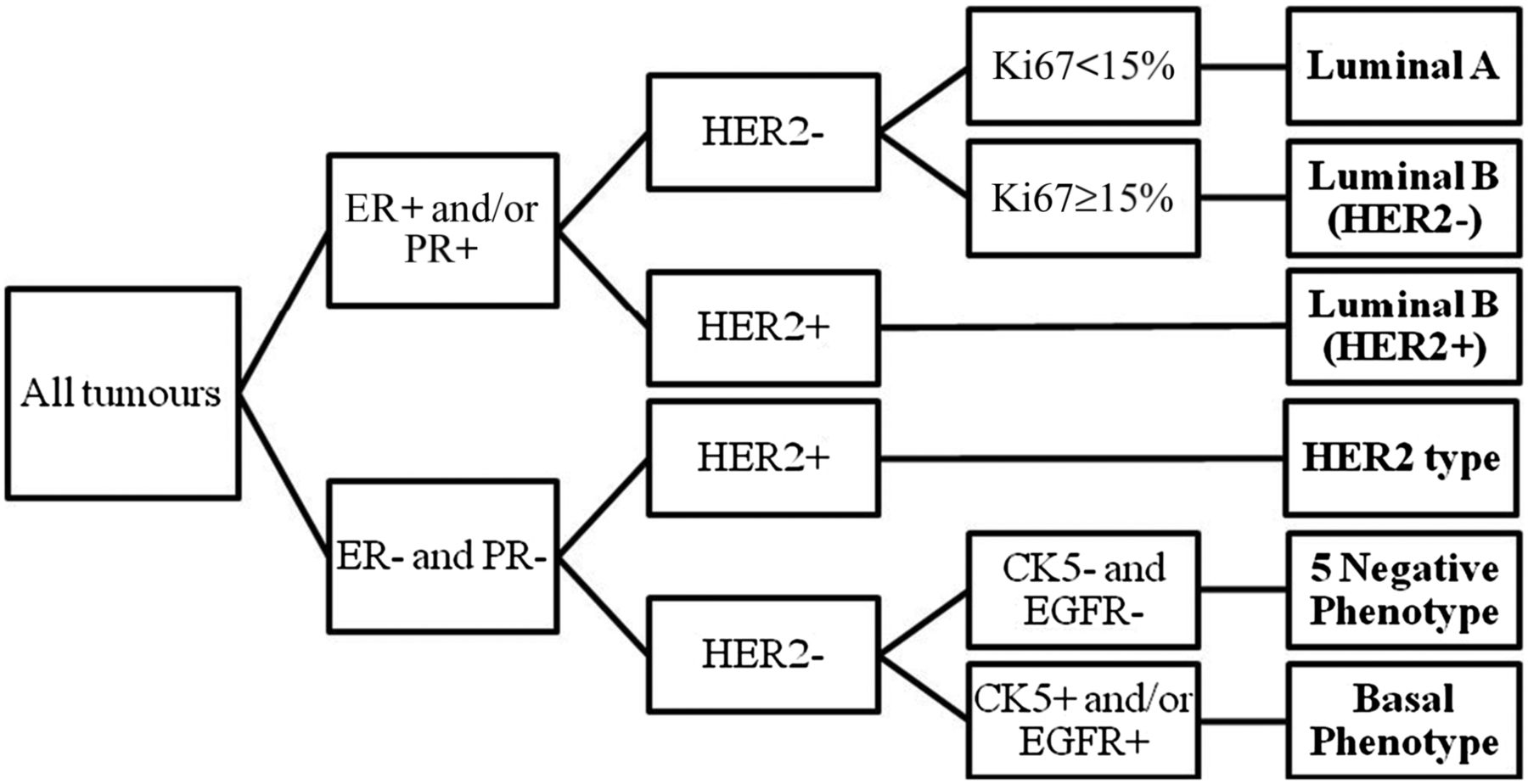
Classification algorithm for molecular subtyping.
Assay methods
For the present study, fluorescence in situ hybridisation (FISH) was employed for detection of TOP2A and chromosome 17 according to the manufacturer`s guidelines. Pretreatment was done using Histology FISH Accessory Kit, code K5799 (Dako). The probe mix (VYSIS TOP2A/CEP 17 FISH Probe Kit, code 03N89-020 Abbott Molecular Inc) was applied and denatured at 73°C for 5 min before hybridisation at 37°C overnight. For HER2 and chromosome 17, the HER2 CISH pharmDx Kit, code 109 (Dako), was used and immunostaining for ER (ER SP1 Cell Marqque 33 mg/mL 1:100) and PR (PR 16 Novocastra 360 mg/mL 1:400) was done in a DakoCytomation Autostainer Plus (Dako) using Dako REAL EnVision Detection System with Peroxidase/DAB+, Rabbit/Mouse, code K5007, as previously described.5
Scoring and reporting
TOP2A gene copy number was evaluated under a fluorescence microscope (Nikon Eclipse 90i) and HER2 gene under a bright field microscope (Nikon Eclipse 80i) by three of the authors (AMB, BY and MJE). A minimum of 20 non-overlapping tumour cell nuclei with signals for both chromosome and gene were counted in each case. Gene to chromosome ratios ≥2 were classified as amplification.8–11 TOP2A was considered to be deleted when the gene to chromosome ratio was ≤0.8.9 ,12 Cases with only one signal for both gene and chromosome in more than 75% of all nuclei were recorded as monosomy. In the analyses, deletion and monosomy were grouped together. ER and PR were classified as positive when ≥1% of the tumour cells showed positive nuclear staining.
Statistical analyses
Follow-up was from breast cancer diagnosis to death from breast cancer, death from any other cause or to December 31, 2010, whichever occurred first. BCSS was estimated using the Kaplan–Meier method, and Cox proportional hazards models were used to estimate risk of death from breast cancer. HRs were calculated with 95% CIs using Stata V.12.1 IC for Windows (Stata Corp).
Results
Description of breast cancer cases
Of the 670 cases, 251 (37.5%) died of breast cancer, 314 (46.9%) died of other causes, and at the end of the observation period, 105 (15.6%) were still alive. Mean age at diagnosis was 73.1 years (SD 9.8; range 41–96 years), and median follow-up was 6.6 years (IQR 9.42 years). Histopathological grade, tumour size and molecular subtypes are given in table 1.
Descriptive statistics of the 670 breast cancer cases
Amplification and deletion
Table 2 shows amplification of TOP2A was found in 41 cases (6.1%) and monosomy or deletion in 25 (3.7%). HER2 was amplified in 110 cases (16.4%) and co-amplified with TOP2A in 32 cases (4.8%). Of the 25 cases with TOP2A loss, 6 were amplified for HER2. The majority with TOP2A amplification (78.1%) were co-amplified with HER2, whereas 34.5% of the HER2 amplified tumours were either TOP2A amplified or showed TOP2A loss. The proportion of HR+ tumours was higher among cases with TOP2A amplification (75.6%) and TOP2A loss (68.0%) compared with HER2 amplification (55.5%).
Number of positive and negative cases for each marker
Amplification and loss according to molecular subtypes
With the exception of 5NP, TOP2A copy number aberrations were found in all subtypes and were associated with both HR and HER2 status. A majority of 56.1% of TOP2A amplified cases were Luminal B (HER2+). Loss of TOP2A was found among the HR+ and HER2 negative subtypes (Luminal A and Luminal B (HER2−)) (64.0%) or HER2 subtype (20.0%). One of four TOP2A deleted case was Luminal B (HER2+).
BCSS, TOP2A and HER2
The Kaplan–Meier plots in figures 2 and 3 show BCSS according to TOP2A and HER2, respectively, and in figure 4 the BCSS according to the status of both genes. Loss of TOP2A in the absence of HER2 amplification did not affect BCSS. The Kaplan–Meier plots show poorest survival in HER2-amplified cases and TOP2A aberrations did not affect this.
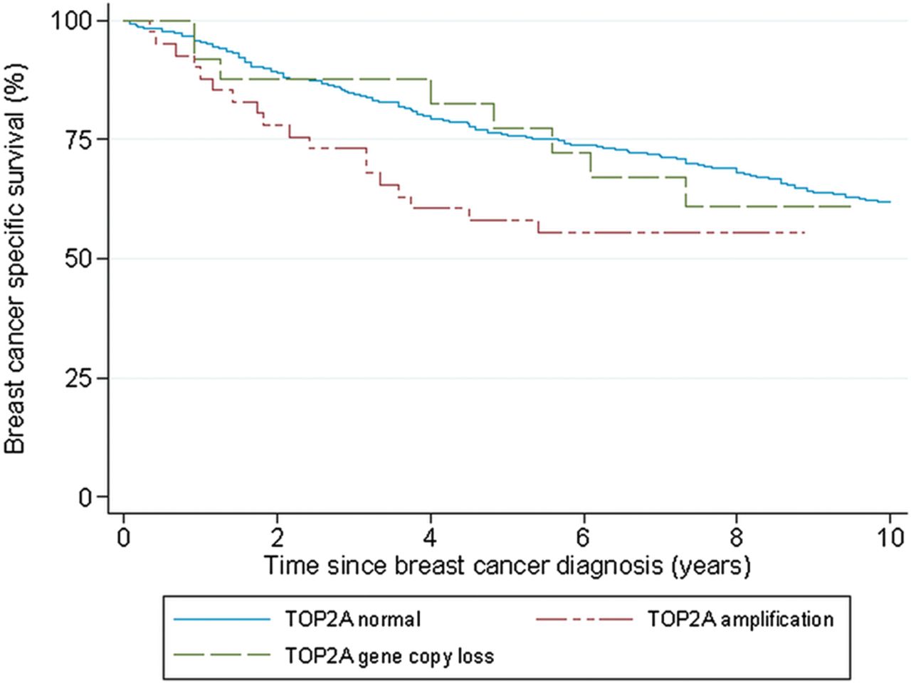
Kaplan–Meier plot. Breast cancer-specific survival (BCSS) according to TOP2A. p Value from log-rank test of differences in BCSS first 5 years after diagnosis was 0.02. After 5 years, the p value was 0.4.

Kaplan–Meier plot. Breast cancer-specific survival (BCSS) according to HER2. p Value from log-rank test of differences in BCSS first 5 years after diagnosis was <0.0001. After 5 years, the p value was 0.9.

Kaplan–Meier plot. Breast cancer-specific survival (BCSS) according to TOP2A and HER2. p Value from log-rank test of differences in BCSS first 5 years after diagnosis was <0.0001. After 5 years, the p value was 1.0.
Risk of death from breast cancer, TOP2A, HER2 and HR status
During the first 5 years, risk of death from breast cancer appears to be significantly higher in cases with amplification of TOP2A and HER2 when analysed separately. When compared with no amplification for TOP2A and HER2, respectively, the HR for TOP2A amplification was 2.03 (95% CI 1.22 to 3.360) and for HER2 was 2.77 (95% CI 1.97 to 3.89). Adjusting for age and stage did not change the results. For those who survived the first 5 years after diagnosis, there were no statistically significant differences in survival according to gene amplification status.
However, as shown in table 3 and figure 4, TOP2A did not exert an independent effect on prognosis. Adjusting for HR status in the Cox proportional hazards model did not change the results (data not shown). During the first 5 years after diagnosis, the risk of death from breast cancer was significantly higher for HR+ cases with HER2 amplification irrespective of TOP2A status. Among the HR− cases, the numbers in each category were low and the results must be interpreted with caution.
Risk of death from breast cancer according to TOP2A and HER2 amplification
Discussion
TOP2A gene copy number change in breast cancer is an infrequent finding and its significance has been difficult to establish. In this study of 670 cases of breast cancer with long-term follow-up, the number of cases with TOP2A amplification or loss was far lower than the number of HER2-positive cases. However, there was a large proportion of co-amplification. In contrast to others who have found that amplification of one or both genes entails a poorer prognosis compared with cases with no amplification,11 ,13 ,14 this study demonstrates that associations between BCSS and TOP2A copy number change are not independent of HER2 and HR status.
The most important finding in this study is the strong association between TOP2A copy number change and HR and HER2 status. These markers are well established as prognostic and predictive factors, and are to a high degree decisive for treatment after surgery. To the best of our knowledge, few studies have been designed to examine the prognostic value of TOP2A, though it has been shown that TOP2A amplification affects BCSS and risk of death from breast cancer15 and that TOP2A may be a prognostic marker in ER+ breast cancer.14 ,16 However, when the analyses include HR and HER2 status, the present study shows that TOP2A has no independent prognostic impact. TOP2A may still have some modulating effects on prognostication, but this is probably of limited benefit in clinical practice.
Twenty of twenty-five cases with TOP2A loss were PR−, and of these, 12 were ER+. PR negativeness is a predictor of poor prognosis and appears to be associated with TOP2A loss. However, in this study, survival tended to be better in PR− cases with loss of TOP2A compared with cases with normal or amplified TOP2A (data not shown).
The proportion of amplification and co-amplification of TOP2A and HER2 in breast cancer varies between studies. HER2 amplification is reported to be around 15%.2 For TOP2A, amplification varies from 5% to 19%.3 ,17 ,18 In HER2-positive breast cancer, amplification of TOP2A varies from 25% to 42%.1 ,19 Both amplification and deletion of TOP2A in the absence of HER2 amplification have been demonstrated.3 ,20 In the present study, 29.1% of the HER2-amplified cases were co-amplified with TOP2A. The proportion of TOP2A positive tumours in this study was lower than in other studies.2 However, the frequency of HER2 amplification is comparable with others, and this weighs against methodological problems. Furthermore, a short DNA probe for TOP2A was used to avoid overlap with HER2.21 This may in part account for the low number of TOP2A-amplified cases in this study compared with previous studies and may reflect the true frequency of this finding.
Assessment of loss should be carried out with caution in histopathological sections because nuclear truncation may lead to a falsely low estimation of copy number. The cut-off for amplification is usually set at a gene/chromosome ratio of ≥2.0, and for deletion the cut-off level ranges from 0.5 to 1.0.21 It is possible that monosomy may have an impact similar to loss of individual genes, but this is uncertain. In this study, only four cases showed deletion and monosomy and deletion were grouped together.
HER2-positive breast cancer has been shown to be more aggressive than HER2–negative breast cancer. Co-amplification with other genes, such as STARD3 and GRB7, may contribute to and possibly strengthen this aggressive behaviour.2 The proportion of amplification and co-amplification of TOP2A and HER2 in breast cancer is low, and even in a series of 670 patients, the numbers are too low to draw reliable conclusions. As a prognostic marker, TOP2A is probably of limited value. TOP2A aberrations are strongly associated with HR and HER2 status, and the importance of these markers in prognostication is still unchallenged.
Take home messages
-
TOP2A gene copy number change is an infrequent finding in breast cancer.
-
There is a strong association between TOP2A copy number change and hormone receptor and HER2 status.
-
As a prognostic marker, TOP2A is probably of limited value, and hormone receptor and HER2 status remain unchallenged.
Acknowledgments
The authors thank the Department of Pathology and Medical Genetics, St. Olav's Hospital, for making the archives available for the study and the Cancer Registry of Norway for providing the patient data.
References
Footnotes
-
Contributors MJE contributed to interpretation of in situ markers, carried out statistical analyses, interpretation of the results and drafted the manuscript. BY participated in planning and performing the laboratory work, contributed to interpretation of in situ markers and to discussion and review of the manuscript. LJV contributed to discussion of the study and review of the manuscript. SO participated in acquisition of data and tissue blocks, discussion of the statistical analyses and reviewed the manuscript. AMB contributed to conception and design of the study, interpretation of the in situ markers, interpretation and analyses of the data, and draft and review of the manuscript. All authors read and approved the final manuscript.
-
Funding The study has received financial support from the Liaison Committee between the Central Norway Regional Health Authority and the Norwegian University of Science and Technology and the Cancer Fund, St. Olav's Hospital, Trondheim University Hospital, Norway.
-
Competing interests None.
-
Ethics approval Approval of the study and dispensation from the requirement of patient consent was granted by the Regional Committee for Medical and Health Sciences Research Ethics (REK, Midt-Norge, ref. nr: 836/2009).
-
Provenance and peer review Not commissioned; externally peer reviewed.