Article Text

Statistics from Altmetric.com
Introduction
Oral tongue squamous cell carcinoma (OTSCC) is the most common oral cavity cancer.1 We have recently identified two cancer stem cell (CSC) subpopulations within moderately differentiated OTSCC (MDOTSCC): a p63+/NANOG+/SOX2+/SALL4+/pSTAT3+/OCT4− subpopulation within the tumour nests (TNs) and a p63−/NANOG−/SOX2−/SALL4−/pSTAT3−/OCT4+ subpopulation within the stroma.1
The renin–angiotensin system (RAS) is associated with blood pressure regulation,2 but recent publications suggest its role in cancer growth.2 Its components, ACE, angiotensin II receptor 1 (ATIIR1) and ATIIR2 have been demonstrated within different cancers,3 implicating a role for the RAS in carcinogenesis.3 This report demonstrated expression of components of the RAS by the CSC subpopulations within MDOTSCC.
Immunohistochemical staining
Four-micrometre-thick formalin-fixed paraffin-embedded sections from 10 previously characterised MDOTSCC tissue samples1 underwent 3,3-diaminobenzidine (DAB) immunohistochemical (IHC) staining for the primary antibodies SALL4 (1:100; cat#6E3, Cell Marque, Santa Cruz, California, USA), SOX2 (1:500; cat#PA1-094; Thermo Fisher Scientific, Santa Cruz, California, USA), OCT4 (1:30; cat#MRQ-10, Cell Marque), (pro)renin receptor (PRR; 1:2000; cat#ab40790, Abcam, Cambridge, Massachusetts, USA), ACE (1:100; cat#MCA2054, AbD Serotec, Raleigh, North Carolina, USA), ATIIR1 (1:30; cat#ab9391, Abcam) and ATIIR2 (1:2000; cat#NBP1-77368, Novus Biologicals, Littleton, Colorado, USA), as previously described.1
To determine co-expression, two selected MDOTSCC samples from the original cohort were subjected to immunofluorescent (IF) IHC staining using a combination of Vectafluor Excel anti-rabbit 594 (ready-to-use; cat#VEDK-1594, Vector Laboratories, Burlingame, California, USA) and Alexa Fluor anti-mouse 488 (1:500; cat#A21202, Life Technologies, California, USA) for combinations of PRR, and Vectafluor Excel anti-mouse (ready-to-use; cat#VEDK2488, Vector Laboratories) and Alexa Fluor anti-rabbit 594 (1:500; cat#A21207, Life Technologies) for combinations of ACE and ATIIR1.
Positive human control tissues for the primary antibodies were placenta for PRR; liver for ACE and ATIIR1; kidney for ATIIR2; and seminoma for SOX2, OCT4 and SALL4. Omission of the primary antibodies was used as negative controls. Images were captured and processed as recently described.1
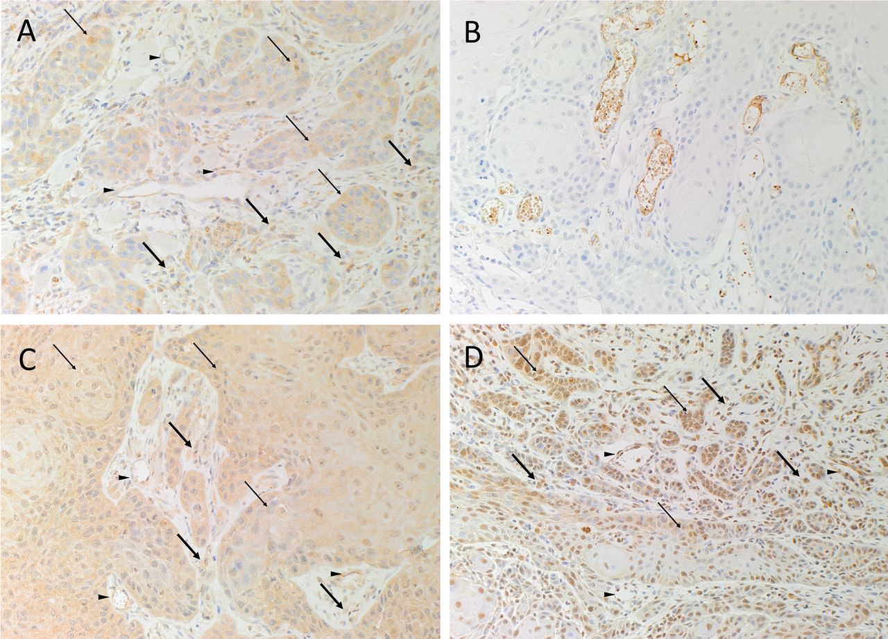
DAB IHC staining revealed cytoplasmic expression of PRR by cells within the TNs (figure 1A, brown, thin arrows), the stroma (figure 1A, brown, thick arrows) and the endothelium of the microvessels within the stroma (figure 1A, brown, arrowheads). ACE was expressed by the endothelium of the microvessels (figure 1B, brown). ATIIR1 (figure 1C, brown, thin arrows) and ATIIR2 (figure 1D, brown, thin arrows) were expressed by cells within the TNs, the stroma (figure 1C, D, brown, thick arrows) and the endothelium of the microvessels (figure 1C, D, brown, arrowheads).

Representative diaminobenzidine immunohistochemical-stained sections of moderately differentiated oral tongue squamous cell carcinoma samples showing cytoplasmic expression of PRR by cells within the tumour nests (TNs; A, brown, thin arrows), the stroma (A, brown, thick arrows) and the endothelium of the microvessels within the stroma (A, brown, arrowheads), between the TNs. ACE was expressed by the endothelium of the microvessels within the stroma (B, brown) and cells on the periphery of the TNs (B, brown). ATIIR1 (C, brown) and ATIIR2 (D, brown) were expressed by cells within the TNs. Both ATIIR1 and ATIIR2 were also expressed on the endothelium of the microvessels within the stroma (D, brown, arrowheads). Original magnifications: 400×.
IF IHC staining demonstrated expression of PRR (figure 2A, B, red) by the SALL4+ (figure 2A, green) CSCs within the TNs and the OCT4+ (figure 2B, green) cells within the stroma. ACE (figure 2C, green) was expressed on the endothelium of the microvessels but not the SOX2+ (figure 2C, red) cells within the TNs. ATIIR2 (figure 2D, E, red) was expressed by cells that expressed SALL4 (figure 2D, green) and OCT4 (figure 2E, green) within the stroma. ATIIR1 (figure 2F, green) was localised to the SOX2+ (figure 2F, red) CSC subpopulation within the stroma and the endothelium of the microvessels. PRR (figure 2G, red) and ATIIR1 (figure 2G, green) were expressed by cells within the TNs, the stroma and the endothelium of the microvessels.

Representative immunofluorescent (IF) immunohistochemical (IHC)-stained sections of moderately differentiated oral tongue squamous cell carcinoma showing PRR (A and B, red) were expressed by the cancer stem cells (CSC) within the tumour nests (TNs) that expressed SALL4 (A, green) as well as those that expressed OCT4 (A, green). ACE (C, green) was expressed on the endothelium of the microvessels within the stroma, but not the cells within the TNs that stained positively for SOX2 (C, red). ATIIR2 (D and E, red) was expressed by cells within the TNs that expressed SALL4 (D, green) but not those that expressed OCT4 (E, green). ATIIR1 (F, green) was localised to the CSC expressing SOX2 (F, red) within the TNs and cells within the stroma, between the TNs (F, green, arrowheads). PRR (G, red) and ATIIR1 (G, green) were colocalised to cells within the TNs and those within the stroma and the endothelium within the stroma. Scale bars: 20 μm.
Nanostring gene analysis
Six snap-frozen MDOTSCC samples from the original cohort were used to isolate total RNA for NanoString nCounter Gene Expression Assay (NanoString Technologies, Seattle, Washington, USA). Extraction, quantitation and analysis were performed as previously described.1 Probes for the genes encoding for PRR (ATP6AP2, NM_005765.2), ACE (NM_000789.2), ATIIR1 (AGTR1, NM_000685.3) and ATIIR2 (AGTR2, NM_000686.3) and the housekeeping gene clathrin heavy chain (CLTC; NM_004859.2) were designed and synthesised by NanoString Technologies. NanoString transcriptional profiling confirmed the presence of mRNA for PRR and ACE in all six samples (figure 3). ATIIR1 was detected in only one sample and ATIIR2 was below the detectable level.

Log10 expression of mRNA expression of the renin–angiotensin system-related genes in six moderately differentiated oral tongue squamous cell carcinoma samples showed the presence of ACE and PRR. ATIIR1 was detected in one sample. ATIIR2 was not detected. Target mRNA counts were normalised against nCounter internal positive and negative controls. Results are presented as a ratio over the CLTC housekeeper in relative units. The average across the six samples was calculated and logged.
Western blotting
Total protein extracts from three MDOTSCC samples of the original cohort were resolved by 4%–15% one-dimensional polyacrylamide gel electrophoresis (1D-PAGE; 30 µg total protein per sample) and transferred to polyvinylidene difluoride membranes (Bio-Rad) (n=2). The membranes were probed with the following primary antibodies: ACE (1:200; cat#sc-12184, Santa Cruz, Rockford, Illinois, USA), ATIIR1 (1:200; cat#sc-1173, Santa Cruz), ATIIR2 (1:200; cat#NBP1-77368, Novus Biologicals), PRR (1:500; cat#HPA003156, Sigma-Aldrich) and β-actin (1:5000; cat#ab8226, Abcam) and then incubated with the appropriate secondary antibody, goat anti-rabbit, horseradish peroxidase (HRP) conjugate (1:20000; cat#A16110, Thermo Fisher Scientific); donkey anti-goat, HRP conjugate (1:5000; cat#ab97120, Abcam); or rabbit anti-mouse, Alexa 647 conjugate (1:2000; cat#A21239, Thermo Fisher Scientific). HRP-conjugated secondary antibody detection was achieved using Clarity Western enhanced chemiluminescence substrate and a ChemiDoc MP imaging system (Bio-Rad).
Western blotting (WB) analysis of total protein extracts produced a single band in all three extracts that corresponded to the expected size of the PRR (figure 4A). ATIIR1 was detected in all three OTSCC extracts and based on appearance and mass shift the ∼55 kDa band detected in OTSCC_2 sample may represent a glycosylated species4 (figure 4B). ATIIR2 was detected in two of the three extracts (figure 4C). ACE was also detectable in all three samples (figure 4D). β-Actin confirmed approximately equal total protein loading for the three samples (figure 4E).

Representative western blot images of 1D-PAGE separated moderately differentiated oral tongue squamous cell carcinoma (OTSCC) total protein extracts probed for PRR (A), ATIIR1 (B), ATIIR2 (C) and ACE (D), and detected with HRP-conjugated goat anti-rabbit (A–C) or donkey anti-goat (D) secondary antibody. β-Actin (E) was used as the loading control, and detected using Alexa 647 rabbit anti-mouse secondary antibody.
Discussion
The localisation of PRR, ATIIR1 and ATIIR2 to both CSC subpopulations within MDOTSCC is novel: one within the TNs that express SALL4 and another within the stroma that express OCT4.1 It is intriguing that ACE is localised to the endothelium of the microvessels within the stroma, and it is exciting to speculate that these microvessels may represent vascular mimicry,5 although this requires further study.
Supplementary figure
The positive controls for PRR, ACE, ATIIR1 and ATIIR2 demonstrated expected staining in human placenta (A, brown), liver (B, brown), liver (C, brown) and kidney (D, brown) and the omission of the primary antibody in a moderately differentiated OTSCC sample provided an appropriate negative control (E, brown). Original magnifications: 400X.
The identification of ATIIR2 by IHC staining and a protein band at the corresponding molecular size in two of the three samples by WB in the absence of transcription message may be explained by the loss of transcriptional synthesis with ‘hangover’ protein still present, or that the primers used in our experiments did not cover all splice variants for ATIIR2. This remains the topic of further research.
The findings of this study suggest CSCs as a potential therapeutic target for MDOTSCC through modulation of the RAS, using existing drugs6–8 such as aliskiren, which targets renin, β-blockers that inhibit the production of prorenin, ACE inhibitors that prevent conversion of ATI to ATII and ATII receptor blockers.
Acknowledgments
The authors thank Ms Liz Jones and Dr Andrea Mikulasova of the Gillies McIndoe Research Institute for their assistance with IHC staining; and processing of tissues for NanoString analysis, respectively.
Footnotes
Handling editor Cheok Soon Lee
Contributors TI and STT formulated the study hypothesis. TI, PFD and STT designed the study. TI, HDB and STT interpreted the IHC data. JCD conducted the WB experiments and interpreted the data. AMC analysed and interpreted the NanoString data. TI, STT and PFD drafted the manuscript. All authors commented on and approved the manuscript.
Funding This work was funded, in part, by the Genesis Oncology Trust. TI was supported, in part, by the Foundation for Surgery ANZ Journal of Surgery Scholarship, Royal Australasian College of Surgeons.
Competing interests TI, PFD and STT are inventors of the PCT Patent Application (No. PCT/NZ2015/050108) Cancer Diagnosis and Therapy.
Ethics approval Central Regional Health and Disability Ethics Committee (reference no: 12/CEN/74).
Provenance and peer review Not commissioned; externally peer reviewed.