Article Text

Abstract
Objective To investigate the expressions of interleukin (IL)-21 and phosphorylated extracellular signal regulated kinase 1/2 (pERK1/2) in Kimura disease (KD) and to correlate the findings with clinical and prognostic variables.
Methods Immunohistochemical analysis of IL-21 and pERK1/2 was performed in 18 cases of KD and five gender- and age-matched control samples. Clinical data were extracted and patients followed up for a mean period of 32.1 months.
Results After a mean follow-up period of 32.1 months (range 1–102 months), recurrence was diagnosed as the end point for seven patients—that is, a 44% (7/16) cumulative recurrence rate. In comparison with gender- and age-matched controls, patients showed strong in situ expressions of IL-21 and pERK1/2, respectively (p<0.05). Patients with strong IL-21 staining intensity and overexpression of pERK1/2 had a lower recurrence rate than those with moderate staining intensity (p=0.049, p=0.019, respectively). However, differences were not statistically significant by gender, age, eosinophils, location, multiplicity, laterality, size, duration and primary outbreak. pERK1/2 was the independent prognostic factor (p=0.020), while age, gender, eosinophils, multiplicity, laterality, size, duration, primary outbreak and expression of IL-21 were not.
Conclusions This study suggests that the IL-21/pERK1/2 pathway is activated in KD, and pERK1/2 might be considered as a potential prognostic indicator in KD.
- INTERLEUKINS
- IMMUNOPATHOLOGY
- HEAD AND NECK
This is an Open Access article distributed in accordance with the Creative Commons Attribution Non Commercial (CC BY-NC 4.0) license, which permits others to distribute, remix, adapt, build upon this work non-commercially, and license their derivative works on different terms, provided the original work is properly cited and the use is non-commercial. See: http://creativecommons.org/licenses/by-nc/4.0/
Statistics from Altmetric.com
Introduction
Kimura disease (KD) is a rare, chronic inflammatory disease, clinically characterised by a triad of painless subcutaneous lump, infiltrative eosinophil in tissue and blood and elevated serum immunoglobulin E (IgE). This pruritic inert nodule attacks predominantly in the head and neck region, although in some patients it advanced to nephritic syndrome. It was reported that up to 50% of patients relapsed after the three mainstays of treatment.1 To reduce the chance of recrudescence, radiotherapy was recommended after surgical resection.2
Although a tendency to develop cancer and fatal outcome have not been reported in KD, it is longlasting and refractory.3 Patients treated with the duplicate protocol had different outcomes, with some not experiencing recurrence and others suffering.4 Elucidation and clarification of the pathogenesis might result in more efficient molecular precision medicine. The precise pathogenesis of KD might be multifaceted and the immunopathogenesis of KD is obscure, although much research suggests Th2 polarisation.5 Immune dysregulation was one of its characteristics.
Interleukin (IL)-21 is produced by activated T cells and exerts an influence on inflammation and autoimmunity.6 ERK1/2 is one of six distinct classes of the mitogen-activated protein kinase family and has myriad functions both in health and disease.7–9 To the best of our knowledge, few reports have examined whether IL-21 and phosphorylated extracellular signal regulated kinase 1/2 (pERK1/2) are associated with the immunopathogenesis of KD. To investigate the roles of IL-21 and pERK1/2 in the pathogenesis of KD, the expression of IL-21 and pERK1/2 was investigated immunochemically in patients with KD who had undergone postoperative radiotherapy, and their potential prognostic value in KD was assessed.
Material and methods
Approval from the institutional review board was obtained at the First Affiliated Hospital of Xinjiang Medical University before starting the study (20140521–01). Informed consent was obtained from each patient.
Clinical records were retrieved and excisional biopsy specimens were available in the pathology department. Assessed clinicopathological variables included age, gender, eosinophils, location, multiplicity, laterality, size, duration, primary outbreak and outcome.4 ,10 All patients underwent complete resection under general anaesthesia and postoperative radiation; the samples were fixed in formalin within 30 min. Radiotherapy regimens were from 36 to 40 Gray in fractions of 2 Gray.
Between 2006 and 2014, 18 patients with KD (14 male and 4 female subjects, aged 11–61 years, mean age 31.2) treated in hospital were included in this study. Patients who were exposed to any radiotherapy or oral steroids before admission were excluded. The control group for the immunohistochemistry study comprised five subjects (four male and one female, aged 17–60 years, mean age 35.4) with an adjacent normal area surrounding malignant parotid gland tumours.
The pathological diagnosis was established from the histopathological findings.11–13 All pathological sections presented similar histopathological features. One pathologist established the diagnosis, which was confirmed by another experienced pathologist on the basis of the following characteristic features: lymphoid tissue follicular proliferation, various degrees of proliferated vasculature and fibrosis as well as the scatter of eosinophils, lymphocytes, and mast cells’ infiltration.
Patients were followed up from discharge by mail or telephone according to the routine clinical practice. If any recurrence was suspected, the patient was asked back for clinical inspections and interventions. The time to relapse was recorded. Recurrence is defined as a swelling or pruritus confirmed histopathologically. Disease-free survival (DFS) is defined as the time (in months) from the date of discharge to April 2015 or until the date recurrence was diagnosed.
Formalin-fixed and paraffin-embedded tissues were cut into 4 μm sections, which were stained with haematoxylin and eosin and antibodies. Sections were deparaffinised and hydrated. Endogenous peroxidase activity was inhibited with 3% hydrogen peroxide for 15 min, followed by rinsing with phosphate-buffered saline (PBS). For antigen retrieval, slides were microwaved with sodium citrate buffer (pH=6.0). The primary antibodies, rabbit IL-21 antibody (1:100 dilution, Thermo Fisher Scientific: PA5-34801, Massachusetts, USA) and rabbit anti-ERK1 (pT202/pY204)+ERK2 (pT185/pY187) (1:50 dilution, Abcam: ab32538, Cambridge, UK), were incubated at 4°C overnight. The sections were rinsed three times with PBS and allowed to react with biotinylated antibody (Zsbio, Beijing, China) for 25 min at room temperature. Colorimetric detection was completed with 3,3’-diaminobenzidine (Zsbio, Beijing, China), and slides were counterstained with haematoxylin. Omission of the primary antibodies was obtained as the negative controls.
IL-21 and pERK1/2 were evaluated by two pathologists who had no knowledge of the clinicopathological outcomes. The images of IL-21 were captured with a Leica digital camera at 200× magnification onto a computer and analysed using Image-Pro Plus software (V.6.0, Media Cybernetics, LP, USA). Five regions of interest from each section were obtained, and the total integrated optical density (IOD) was measured (IOD=density (mean)×area).14 The average IOD in each section was calculated.
Sections of pERK1/2 were evaluated by an Olympus BH-2 microscope (Olympus, Tokyo, Japan). The nuclear localisation of pERK1/2 was considered as a positive expression. The expression of pERK1/2 was scored semiquantitatively. The percentage of positive cells were graded as: 1, <25%; 2, 26–50%; 3, 51–75% and 4, >75%. The intensity of expression was scored as: 1, weak; 2, moderate and 3, strong. Final histochemical scores (H-score) were calculated by multiplying the percentage by the intensity of expression.15 Five separate positive areas were scored in each slide and an average H-score of 2 was used as the cutoff criterion. Patients were separated into either weak expression or overexpression.
Statistical analysis
DFS was the primary endpoint of this study. A Shapiro–Wilk test was used to assess whether or not data were normally distributed. Normally distributed data were analysed by Student's t-test and presented as mean±SD. Categorical variables were expressed as percentage and analysed by Fisher's exact test. The cutoff point to convert a continuous variable (IOD of IL-21) into a categorical value was calculated with a lower p value.16 The Kaplan–Meier method was used to estimate recurrence rate. Univariate and multivariate Cox regression models were used to evaluate the association between DFS and clinicopathological variables. Parameters considered statistically significant (p<0.10) in the univariate model were analysed in the multivariate models. All two-sided p values <0.05 were considered as significant. All analyses were carried out using SPSS software (SPSS V.17.0, Chicago, Illinois, USA).
Results
Clinicopathological information is shown in table 1. This study was conducted in a total of 18 patients aged 11–61 years (mean±SD 31.2±15.0), of whom 14 were male and 4 female (male to female ratio: 3.5:1), with median disease duration of 54 months (range 1–312). Seventeen of the 18 patients had peripheral eosinophilia (mean±SD 25.6%±15.4%). These specimens were resected from the head and neck region: parietal subcutis, infraorbital subcutis, post-auricular subcutis, nasal cavity, upper eyelid, pinna, submandibular glands and parotid glands. The predominant location of the lesion was the head and neck region, but four patients had multiple lesions, such as: inguinal lymphadenopathy, elbow subcutis, breast subcutis and upper arm subcutis. The mean diameter was 4.9 cm, ranging from 2.2 to 7.8 cm. Eleven patients had no history of recurrence and seven patients had had a relapse.
Clinicopathological details
Sixteen patients were available for the follow-up visit, and two patients were non-responsive to any form of contact. The deadline for follow-up was April 2015. After a mean follow-up period of 32.1 months (range 1–102 months), recurrence was diagnosed as end point for seven patients, with a 44% (7/16) cumulative recurrence rate (figure 1).
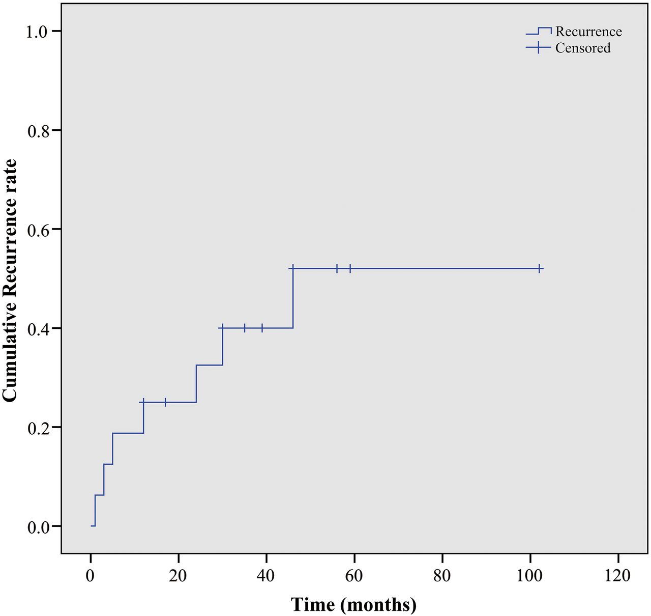
Cumulative recurrence curve of 16 patients.
As showed in figure 2, compared with controls matched for gender and age, patients showed strong in situ expressions of IL-21 and pERK1/2, respectively (p<0.05). Distribution of clinicopathological outcomes was compared with IL-21 and pERK1/2 expressions to assess the potential prognostic variables. Patients with strong IL-21 staining intensity had a lower recurrence rate than those with moderate staining intensity (p=0.049). Patients with overexpression of pERK1/2 had a lower recurrence rate than those with low expression (p=0.019). However, differences were not statistically significant by gender, age, eosinophils, location, multiplicity, laterality, size, duration and primary outbreak (table 1).

Expressions of pERK1/2 in Kimura disease (KD) (A) and controls (B) as well as expressions of IL-21 in KD (C) and normal controls (D). Arrow: note the positivity pattern (×100 and×400). IL, interleukin.
Univariate analysis showed that age, expressions of IL-21 and pERK1/2 were associated with prognosis. To adjust for those variables, the parameters listed in table 2 were calculated in a multivariate analysis. Statistically, pERK1/2 was an independent prognostic factor (p=0.020), while age, gender, eosinophils, multiplicity, laterality, size, duration, primary outbreak and expression of IL-21 were not.
Univariate and multivariate variable Cox regression model analysis of parameters for time to recurrence in Kimura disease
Discussion
KD is a benign, chronic inflammatory disorder of vague aetiology. Kim first reported on the disease in 1937 and Kimura provided more detail in 1948.17 ,18 About 400 patients have been reported on in English publications.10 It is endemic in Oriental male subjects.1 KD must be distinguished from other diseases, such as angiolymphoid hyperplasia with eosinophilia (ALHE), neoplasms from the parotid gland, Sjögren's syndrome, etc. Although ALHE and KD have some similarities, the former is characterised by absent fibrosis and exuberant angiomatoid proliferation pathologically; additionally, peripheral eosinophils and IgE concentrations are typically not elevated clinically.12 ,13
In this study, age varied from 11 to 61 years and peaked at the second and fourth decades, which is similar to previous reports that male patients aged between 20 and 40 years were most affected.19 The lesion was commonly seen in the head and neck region, and the parotid region and cervical lymphadenopathy were the most dominant sites of involvement. Epiglottis, larynx, median nerve and chest wall are less often affected.11 ,20–22
Scholars compared three modalities for KD, and this meta-analysis found that surgery combined with radiotherapy leads to the lowest recurrence rate.2 Radiotherapy dosage from 20 to 44 Grays has been advocated.3 ,23 Radiation of 36–40 Gray were delivered, which was a moderate dose, and no secondary malignant transformation was observed in the follow-up. After an average follow-up period of 32.1 months, the disease recurred in 44% (7/16) of our patients. The high recurrence rate may stem from (1) a different sample size and duration of follow-up; (2) chief complaint was included as one of the criteria for recurrence; (3) diagnosis was based on excisional biopsy rather than fine needle aspiration biopsy as used elsewhere.
The aetiology of KD is unknown, but impairment of immune regulation may play a part. The presence of an increased level of IL-4 and IL-5 expressed by mast cells, activated eosinophils and T cells was validated in KD.24 ,25 OKT4-positive granuloma T cells were shown to be responsible for the production of two natural mediators for tissue eosinophilia in KD.26 With polarisation of Th2 and Tc1, the number of Th2 and Tc1 cells rather than Th1 and Tc2 cells was elevated.5
Elevation of IL-4, IL-5, IL-6, IL-10, IL-13, IL-22, granulocyte-macrophage colony-stimulating factor (GM-CSF), thymic stromal lymphopoietin (TSL), TSL receptor, IL-25 receptor, eotaxin, regulated on activation normal T cell expressed and secreted (RANTES), CC-chemokine receptor 3 (CCR3), tumour necrosis factor-α, eosinophil cationic protein, major basic protein, sIL-2 receptor, thymus and activation regulated chemokine (TARC/CCL17), eotaxin-3/CCL26, vascular endothelial growth factor and prostaglandin D2,25 ,27–41 and reduction of IL-1727 were seen in KD, whereas IL-4, IL-5, IL-6, IL-10, IL-13, IL-17, IL-25, IL-33, IL-33 receptor, interferon-γ, GM-CSF27 ,29 ,30 ,33 ,35 ,37 ,42 were within the normal ranges. Although there were several inconsistencies, an overwhelming majority of researchers, in our opinion, agree that Th2 polarisation and the increased syntheses of Th2-type cytokines may play crucial roles in the pathogenesis of Kimura disease. These processes may be related to the pathogenesis of this disorder.
Immunostaining for IL-21 and pERK1/2 in patients was stronger than in controls, and in both cases was linked with absence of disease-free. Additionally, pERK1/2 is a prognostic factor for longer DFS. As a pleiotropic cytokine, IL-21 plays a part in lymphocyte activation, proliferation, differentiation and survival.6 It functions through a heterodimeric receptor comprising the unique IL-21R and the common gamma-chain of the IL-2R. IL-21R is widely expressed on cells—either adaptive and innate immune cells or non-immune cells.43–45 Elevation of IL-21, therefore, has been subsequently confirmed in a number of autoimmune diseases—for example, systemic lupus erythematosus, inflammatory bowel disease, atopic dermatitis.46–49 IL-21 stimulation increased interferon-γ production and curbed the development of Th2 cells and molecules.50 Meanwhile, IL-21 downregulated the secretion of IgE.51 Numerous reports have shown that IL-21 functions via many signal transduction pathways, including downstream effectors such as ERK1/2.43–45 ERK1/2 takes part in translating extracellular stimuli—cytokines, chemokines and so forth—into an intracellular process leading to cell proliferation, differentiation, migration, inflammation and survival.52–56 In addition, ERK1/2 signalling plays a critical role in T cell activation and Th1/Th2 polarisation. It has been reported that ERK1/2 has an immunosuppressive role.57–59 Meanwhile, inhibition of ERK1/2 led to Th2 polarisation.60 It was proved that the ERK1/2 signalling pathway plays a negative role in IL-4, inducing Th2 development and secretion of IL-4 in Th2 cells. This is consistent with the demonstration that ERK1/2 has an immunosuppressive role.
In conclusion, overexpression of IL-21 and pERK1/2 in KD, and upregulation of IL-21 and pERK1/2 is associated with freedom from disease. This study suggests that the IL-21/pERK1/2 pathway is activated in KD. As KD is a rare disease, a further multicenter study with a large sample size should be conducted.
Take home messages
Kimura disease (KD) is a rare, benign inflammatory disorder of unknown aetiology and pathogenesis.
We demonstrated overexpression of interleukin (IL)-21 and pERK1/2 in a series of 18 patients with KD compared with age- and gender-matched controls.
IL-21 and -ERK1/2 may be involved in the pathogenesis of KD, and pERK1/2 might be a potential prognostic indicator in KD.
References
Supplementary materials
Abstract in Chinese
This web only file has been produced by the BMJ Publishing Group from an electronic file supplied by the author(s) and has not been edited for content.
- Abstract in Chinese - Online abstract
Footnotes
Handling editor Cheok Soon Lee
Contributors All authors contributed to the design of the study, writing, or critical review of the manuscript, patient care and analysis and interpretation of data. All agreed to submission of the manuscript.
Competing interests None declared.
Patient consent Obtained.
Provenance and peer review Not commissioned; externally peer reviewed.