Article Text

Abstract
This is a review of the morphological spectrum of fatty tumours containing a component of spindle cells, highlighting the immunohistochemical and cytogenetic workup that is now mandatory for accurate diagnosis, with the goal of providing a practical approach for practising surgical pathologists. There have been significant advances in recent years in classifying and understanding the pathogenesis of fatty tumours with spindle cells, based on the correlation of histological, immunohistochemical and cytogenetic/molecular findings. In spite of this, morphological diagnosis and accurate classification of fatty tumours with spindle cells can be challenging to diagnostic pathologists. A group of three lesions: spindle cell lipoma, mammary-type myofibroblastoma and cellular angiofibroma share morphological features and are united by retinoblastoma protein (pRb) loss. Closely allied to these lesions, especially spindle cell lipoma is the newly designated atypical spindle cell lipomatous tumour, which shares morphological, immunohistochemical and cytogenetic features with the trio of tumours lacking nuclear pRb. All of these lesions lack MDM2 and CDK4 amplification as well and separation is based on clinical features, principally location. Atypical lipomatous tumour or well-differentiated liposarcoma shows retention of pRb but overexpression and amplification of MDM2. Fatty tumours with spindle cells need to be extensively sampled, with careful attention paid to cellular atypia and location, and they need to have immunohistochemical workup with pRb, MDM2, desmin, CD34 and p16. In addition, cytogenetic analysis for MDM2 and CDK4 amplification has become crucial for the proper identification of these lesions.
- Spindle cell fatty tumours
- atypical lipomatous tumour
- atypical spindle cell lipomatous tumour
- cellular angiofibroma
- liposarcoma
- mammary-type myofibroblastoma
- spindle cell lipoma.
Statistics from Altmetric.com
- Spindle cell fatty tumours
- atypical lipomatous tumour
- atypical spindle cell lipomatous tumour
- cellular angiofibroma
- liposarcoma
- mammary-type myofibroblastoma
- spindle cell lipoma.
Introduction
Soft tissue sarcomas are diverse mesenchymal malignancies that account for approximately 1% of adult solid tumours.1 Despite their rarity, they hold a diagnostic and intellectual mystique that transcends their relative infrequency under a general diagnostic pathologist’s microscope. Adipocytic tumours represent the most common type of mesenchymal tumours.2 Awareness and classification of atypical and malignant adipocytic tumours have evolved rapidly in the last few years due to a better biological understanding from large series, and with new entities being described by virtue of the application of ancillary techniques such as cytogenetics, molecular genetics and immunohistochemistry. With this has come a level of morphological complexity that necessitates a more than desultory examination of fatty tumours microscopically.
The morphological, and often the immunohistochemical, similarities between fatty tumours can cause diagnostic challenges for even experienced surgical pathologists. We present a synthesis of recently described fatty lesions with a spindle cell component, the differential diagnosis and a review of the currently known morphological, immunohistochemical and molecular features of these tumours. The dilemma confronting the non-expert pathologist dealing with a fatty tumour with spindle cells is nomenclature. Calling a lesion a liposarcoma triggers a particular management algorithm together with labelling the patient as having a sarcoma. Failing to recognise a lesion as potentially locally recurring is the other side of the diagnostic quandary. Thus, the goal of this overview is to highlight key histological features, provide diagnostic pointers to help categorise these lesions as accurately as possible and separate them from other key histological mimics enabling pathologists to make informed and appropriate comments regarding behaviour and management. It is not meant to be a comprehensive expose on each entity, many of which are dealt with in specialised soft tissue textbooks, but a practical perspective when confronted by lesions composed of fat and spindle cells (table 1).
Fatty tumours with spindle cells
Spindle cell lipoma
Nomenclature
Spindle cell and pleomorphic lipomas (SC/PL) are characterised by a morphological continuum and have identical cytogenetics, are considered a single entity.3
Clinical features
They are relatively uncommon, comprising approximately 1.5% of adipocytic neoplasms and being 60 times less frequently encountered than simple lipomas.4 SC/PLs are solitary lesions that grow slowly in a very restricted anatomical distribution, namely, the posterior neck, shoulder and back. They occur most frequently (90%) in adult males, in their 40s–60s and usually measure 3–5 cm in diameter.5 Thus, the clinical feature of note aiding the diagnosis of SC/PL is location.
Microscopic findings
SC/PLs are well-circumscribed, often encapsulated tumours, located in the dermis and subcutis, composed of a mixture of mature adipocytes and spindle cells, frequently set in a myxoid matrix (figure 1A). The spindle cells are cytologically bland, arranged in short fascicles in a ‘school of fish’ pattern.6 Ropey stromal collagen (thick, refractile, eosinophilic collagen) is a characteristic feature (figure 1B). The ratio of the mature fat to the spindle cells can vary significantly. Infrequently, fat is present in a small amount in ‘low-fat’ and ‘fat-free’ spindle cell lipomas (SCLs), which can cause diagnostic challenges.5 Vessels may be prominent, and may be arborising and thick-walled. Occasionally, mast cells are numerous.
- Download figure
- Open in new tab
- Download powerpoint
Spindle cell lipoma (SCL) consists of an admixture of fat and spindle cells which are usually uniform and bland (A). The stromal collagen is disposed as birefringent, so-called ropey appearing strands (B). The stroma can be myxoid and occasionally the spindle cells manifest mild atypia (C). SCL are strongly CD34 positive (D).
Atypical cells range from rare to frequent and are characterised by hyperchromatic, enlarged nuclei, located in both fatty and non-fatty stromal areas (figure 1C). These atypical cells may form a semi-circle of nuclei, designated as ‘floret cells’.
It is important to note that lipoblasts, which are cells with hyperchromatic nuclei that are indented or sharply scalloped due to lipid-rich monovacuolated or multivacuolated cytoplasmic droplets, may be seen occasionally in otherwise morphologically and cytogenetically typical examples of SC/PL, particularly in pleomorphic lipomas.6 7
Immunohistochemistry and cytogenetics
The spindle cells are CD34 positive (figure 1D), while CD99 and bcl-2 are usually positive too. S100 stains fat cells only. Nuclear retinoblastoma protein (pRb) is lost in SC/PL and typically, p16 immunoexpression is not seen in the adipocytes or the spindle-shaped tumour cell nuclei, although up to 20% of cases can also be positive.8 Both MDM2 and CDK4 are usually negative, but again a small proportion of cases can show immunopositivity.
The tumour suppressor gene, RB1, encoding the pRb located at 13q14, is deleted in these lesions,9 explaining the loss of expression of immunohistochemical marker pRb.8 SC/PL lacks amplification of MDM2 and CDK4.
Treatment and prognosis
Local excision is almost always curative and metastases have never been reported.
Mammary-type myofibroblastoma
Clinical features
Mammary-type myofibroblastoma (MTMF) is a benign mesenchymal lesion, first described in the breast, which is now known to have a wide anatomical distribution, most commonly occurring in the groin, inguinal area, trunk and lower extremity.10 11 They are well-circumscribed lesions, with a median size of 5–7 cm, occurring twice as commonly in males in their 50s.
The degree of morphological overlap between MTMF, SCL and cellular angiofibroma has raised the question whether these benign tumours are truly distinct entities or represent points along a single spectrum of genetically related (all have a deletion or rearrangement of 13q14 with subsequent loss of nuclear pRb) tumours.10
Microscopic findings
MTMF is a well-circumscribed, bland, spindle cell proliferation, reminiscent of SCL, with a variably prominent lipomatous component (figure 2A). The tumour cells generally show no atypia, and the adipocytes show minimal variation in size and shape. The stroma is typically collagenous, and occasionally hyalinised or myxoid (figure 2B). Morphological heterogeneity has also been described including cellular, epithelioid and neurilemmoma-like variants.12–15
- Download figure
- Open in new tab
- Download powerpoint
Mammary-type myofibroblastoma (MTMF) also is composed of variable proportions of fat and uniform spindle cells (A) set within a stroma that can be myxoid (B). MTMF are desmin (C) and CD34 positive (D).
Immunohistochemistry and cytogenetics
Immunohistochemically, in most cases (up to 90%), the lesional cells express desmin and CD34 diffusely (figure 2C,D).10 Approximately 40% also are smooth muscle actin (SMA) and epithelial membrane antigen positive. Very occasional cases are S100, MDM2 and CDK4 positive too. Most MTMFs show loss of pRb nuclear expression as a result of 13q14 abnormalities.16
Differential diagnosis
The differential diagnosis of MTMF can be broad and somewhat dependent on the anatomic location and/or the presence of unusual morphological features.10 However, in most situations the differential diagnosis is limited and most notably includes other tumours in the 13q/Rb family, namely SC/PL and cellular angiofibroma.16–19
SCL occurs almost exclusively in the posterior shoulder, neck or upper back region, has more consistently present ropey, refractile collagen bundles and, in contrast to MTMF, is negative for desmin while being positive for CD34.4 10
Cellular angiofibroma is generally limited to the inguinal or groin region and contains wispy rather than ropey or thick collagen, usually has a more limited adipocytic element and has a prominent component of rounded stromal vessels, often with perivascular fibrosis or hyalinization.18 20 See below for fuller discourse on cellular angiofibroma.
Myolipomas/lipoleiomyomas are also benign tumours and may be distinguished from MTMF by more conspicuous smooth muscle morphology with tighter fascicles. In addition, they do not share the cytogenetics of MTMF and do not show loss of pRb.
Other entities with more clinical significance that are occasionally considered in the differential diagnosis of MTMF include atypical lipomatous tumour (ALT), desmoid tumour (fibromatosis) and solitary fibrous tumour (SFT). It is important to distinguish these from MTMF as these tumours have a significant risk for recurrence and, in rare cases (most notably in SFT), metastasis.10 21 22
ALT: MTMF can be distinguished from ALT by the lack of significant nuclear atypia, the lack of variation in adipocyte size and the absence of lipoblasts. By immunohistochemistry, ALTs are positive for MDM2 and CDK4, reflecting amplification of 12q13-15, which can also be demonstrated using fluorescence in situ hybridisation (FISH).23 Nuclear pRb expression is retained in ALT, which can serve as another helpful marker in this distinction.16
Desmoid tumour/fibromatosis: unlike MTMF, desmoid tumours comprise long fascicles of spindle cells and frequently demonstrate nuclear expression of β-catenin.24 They are not well circumscribed and are desmin negative.
SFT may occasionally enter the differential diagnosis of MTMF, particularly the ‘fat-forming’ variant of SFT. These two entities share a CD34-positive immunophenotype, but SFTs have a patternless architecture and are not characterised by loss of pRb (immunohistochemically and cytogenetically), and do not express desmin. Instead, SFT harbours recurrent gene rearrangements resulting in overexpression of STAT6, a sensitive and specific marker for SFT.25–27
Treatment and prognosis
MTMFs do not recur, even if incompletely excised, and have no potential for metastasis.10
Cellular angiofibroma
Clinical features
Cellular angiofibroma is an uncommon benign mesenchymal tumour, occurring mainly in the genital region of both females and males.18 20 Extragenital localisations have also been rarely reported, including in the retroperitoneum, pelvic and lumbar regions, anus, urethra, trunk and oral mucosa.18 20 28–30
Microscopic findings
Cellular angiofibromas are typically well-circumscribed lesions, localised to the superficial soft tissue. They are composed of bland spindle-shaped (sometimes epithelioid) cells, arranged in no particular pattern, in an occasionally myxoid stroma with wispy collagen (figure 3A). Numerous small-sized to medium-sized, thick-walled blood vessels with mural hyalinization are typically present (figure 3B).18 20 Vessels may contain fibrin thrombi, vessel wall inflammation and degenerative changes with hemosiderin. A mature adipocytic component is variably seen.18 20
- Download figure
- Open in new tab
- Download powerpoint
Cellular angiofibroma consists of uniform cells often set within a myxoid stroma with less prominent stromal collagen (‘wispy’) compared with spindle cell lipoma (A). Characteristically, but not exclusively, vessels of varying calibre are encountered (B).
Immunohistochemistry and cytogenetics
Cellular angiofibromas are usually positive for CD34 (30%–60%), progesterone receptor and oestrogen receptor (over 90%) and negative for desmin.18 Loss of nuclear pRb staining is a characteristic finding. In keeping with this immunohistochemical pattern, essentially all cases show a loss of genetic material from the 13q14 region, as indicated by monoallelic deletion of RB1 and FOXO1.19
Differential diagnosis
SFT: see ’differential diagnosis' subsection of the ’mammary-type myofibroblastoma' section above.
Angiomyofibroblastoma could potentially be confused with cellular angiofibroma, due to its localisation to the genital region.31 32 Angiomyofibroblastomas are composed of more epithelioid, and often plasmacytoid, cells, arranged in cords and nests, preferentially located around blood vessels. The tumour cells express desmin but are only rarely positive for CD34.18 Nuclear pRb immunopositivity is usual.
Deep (‘aggressive’) angiomyxoma also involves the pelvic region and tends to be quite large, displaying an infiltrative growth pattern with entrapment of mucosal glands, fat, muscle and nerves. The prominent vascular component shows hyalinization. However, myoid bundles, mostly adjacent to medium-sized blood vessels, are a typical finding.18 There is variable positivity for desmin, smooth muscle actin and CD3431 32 and retention of nuclear pRb.
Prognosis and treatment
There are a few reported cases in the literature of cellular angiofibroma with atypia or sarcomatous features. The sarcomatous component described shows variable features, including ALT, pleomorphic liposarcoma and pleomorphic sarcoma not otherwise specified.18 20 29 33 34 Although the follow-up of all of these reported cases did not disclose any local recurrence or metastases,33 the biological significance of atypia and sarcomatous transformation in cellular angiofibromas remains uncertain.
Atypical spindle cell lipomatous tumour
Nomenclature
This category of fatty tumours is a repository for tumours composed of atypical spindle cells and a variable fat component. Initially, cases were called ALTs or well-differentiated liposarcoma (WDL). However, it soon became apparent that a subset of spindle cell fatty tumours had distinct immunohistochemical and cytogenetic features and included entities such as spindle cell liposarcoma, atypical SCL and fibrosarcoma-like lipomatous tumour.8 This entity should not be confused with ALT, which is also known as WDL.
Clinical features
Atypical spindle cell lipomatous tumour (ASCLT) constitutes a distinct category of adipocytic neoplasms of intermediate biological potential, which most frequently affects adult males in their 50s, has a median size of 5 cm and has a predilection for the limbs and limb girdles.35
These neoplasms were first described by Dei Tos et al 36 in 1994, when they characterised a series of six cases that they designated ‘spindle cell liposarcoma’, which they considered to be variants of WDL.
Microscopic findings
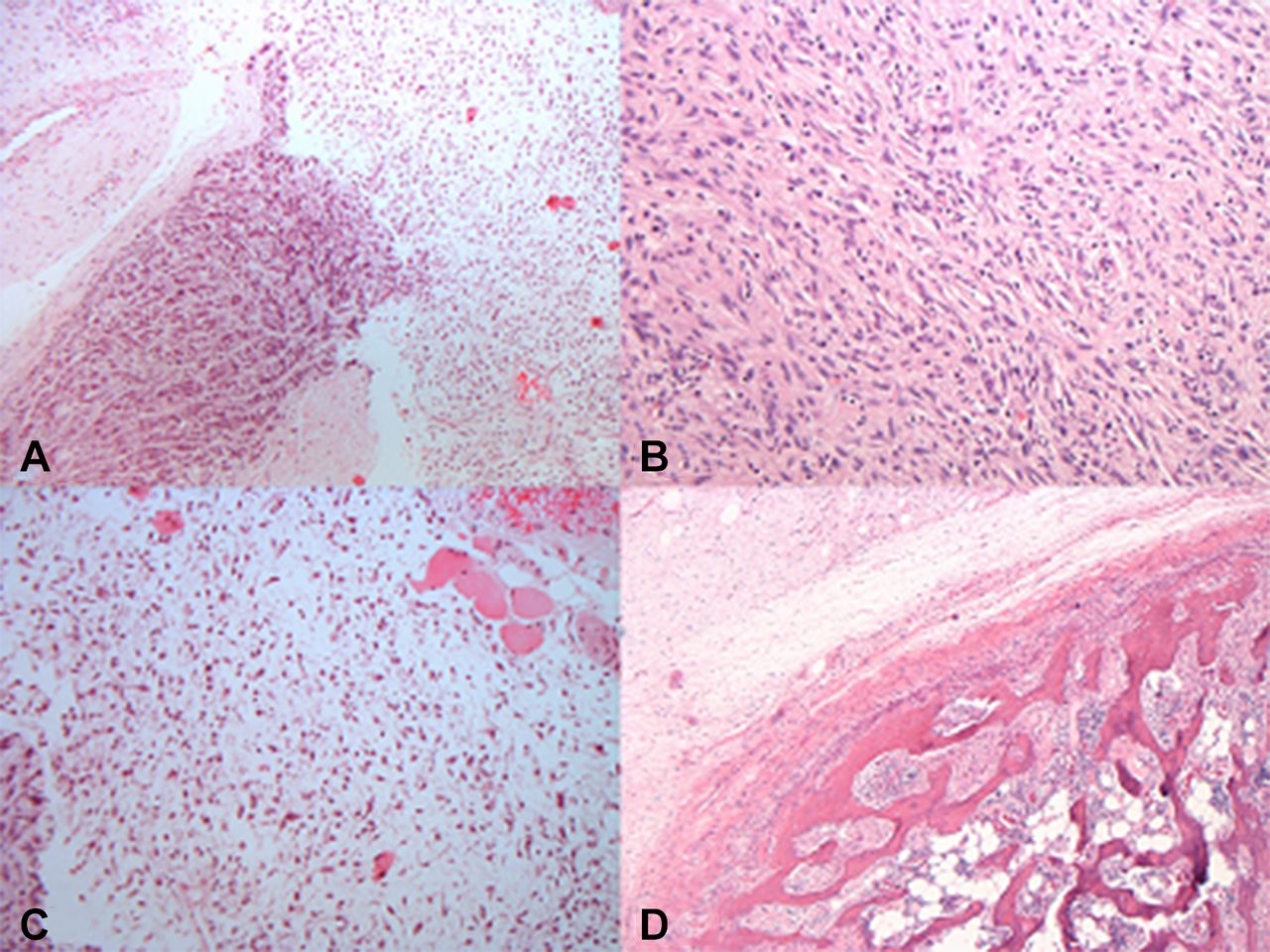
Microscopically, ASCLTs have an ill-defined fibrous pseudo-capsule (figure 4A), and are composed of a vaguely lobular neoplastic proliferation of relatively uniform spindle cells, admixed with a variably prominent adipocytic component, set in a fibrous, collagenous or myxoid stroma (figure 4B). The proportions of spindle cells, adipocytes and extracellular matrix varies, resulting in a broad range of microscopic appearances.35
- Download figure
- Open in new tab
- Download powerpoint
Atypical spindle cell lipomatous tumour has a lobular appearance with a fibrous pseudocapsule (A) and is made up of uniform spindle cells, admixed with a variably prominent adipocytic component, set in a fibrous, collagenous or myxoid stroma (B). Spindle cells have ovoid nuclei with variably hyperchromatic chromatin and generally regular nuclear contours (C). The fatty component shows variation in fat cell size (D).
The neoplastic spindle cells are elongated, with indistinct, pale, eosinophilic cytoplasm, and ovoid nuclei with variably hyperchromatic chromatin and generally regular nuclear contours (figure 4C). The degree of nuclear atypia and hyperchromasia of these spindle cells varies between tumours, with most tumours showing only focal and/or mild atypia. However, prominent and/or diffuse atypia can be seen in approximately one-third of cases.35
The adipocytic component predominantly comprises mature-appearing adipocytes, but variation in adipocyte size can be seen (figure 4D).
Univacuolated or multivacuolated lipoblasts are present in almost half of cases, but usually in small numbers (figure 5A). Lipoblasts range from small, univacuolated or bivacuolated to larger, multivacuolated cells, with hyperchromatic scalloped nuclei and sharply punched-out vacuoles. In addition, in the majority of cases, bizarre, hyperchromatic, often multinucleated cells are scattered throughout the tumour, within the adipocytic or spindle cell components (figure 5B).35

Univacuolated and multivacuolated lipoblastic cells are present in atypical spindle cell lipomatous tumour (A). Also bizarre, hyperchromatic, often multinucleated cells are encountered in either the adipocytic or spindle cell components (B).
Extracellular matrix is abundant in most tumours, varying widely from purely myxoid to predominantly collagenous. The amount of collagen present varies. The presence of bundles of ropey collagen is a rare finding.35
Thus, due to the extremely variable proportions of spindle cells, adipocytes, lipoblasts and extracellular matrix, as well as the variable quality of the extracellular matrix, there is a wide range of microscopic appearances of ASCLTs. These tumours can be paucicellular, with few, cytologically bland, spindle cells with minimal nuclear atypia, set in an abundant myxoid matrix with scattered mature adipocytes. They can bear a close resemblance to an SCL. At the other extreme, these tumours may be significantly more cellular, composed of numerous spindle cells showing diffuse, mild-to-moderate nuclear atypia, with variable presence of lipoblasts and less extracellular matrix. It is therefore extremely important to sample these fatty lesions with spindle cells thoroughly, if not in total.
At present, there are no reproducible morphological, immunohistochemical or molecular criteria to accurately subclassify atypical spindle cell lipomatous neoplasms into clinically meaningful subsets.
Immunohistochemistry and cytogenetics
The neoplastic cells express CD34 (65%), S100 (40%) and desmin (20%). Loss of nuclear pRb expression is encountered in around 50%–60% of cases, correlating with RB1 heterozygous deletion. However, it is important to note that either retention or loss of nuclear pRb staining is still compatible with the diagnosis of ASCLT. Weak and/or focal expression of MDM2 (5%) or CDK4 (5%) can be seen, but always in the absence of corresponding gene amplification.35 The combination of MDM2 and CDK4 positivity together is not encountered in ASCLT. Typically, no nuclear p16 expression is seen in the adipocytes or the spindle-shaped tumour cells of ASCLT.8
Alterations such as 13q deletion with loss of pRb, and monosomy 7 have been shown to be recurrent findings in these lesions.8 37 38 FISH analysis does not demonstrate MDM2 or CD4K amplification.35 This latter finding separates ASCLT from liposarcoma.
Differential diagnosis
The morphological differential diagnosis of ASCLT is broad, and includes benign and malignant lesions (including low-grade malignant peripheral nerve sheath tumour, dermatofibrosarcoma protuberans, ALT/WDL, low-grade dedifferentiated liposarcoma (DDL)).35
Spindle cell/pleomorphic lipomas as noted above can have slight atypia of the spindle cell component or bizarre cells in the pleomorphic variant. Both location and the loss of pRb are strongly in favour of the diagnosis of SC/PL (bearing in mind that approximately half of ASCLTs can also show loss of pRb). Given that MDM2 is also not amplified in ASCLT, separation from SCL on morphological, immunohistochemical and cytogenetic grounds can be extremely difficult. If the lesion is cytologically bland and located in the neck/upper back region, then SCL is most likely. If located in the limbs and one is considering a diagnosis of SCL, then caution should be exercised, multiple sections examined for atypia and a diagnosis of ASCLT should be rendered.
Diffuse neurofibromas are often located within the dermis and subcutis of young patients, and are composed of a monomorphic proliferation of S100-positive spindle cells, with wavy/buckled nuclei. Adipocytic differentiation is rare, but entrapment of adjacent structures, including fat, may occur thus simulating a spindle cell and fat lesion. The detection of hyperplastic nerve bundles and Meissnerian corpuscles can aid in correctly identifying diffuse neurofibromas.35
Fat-forming SFT generally occurs in deep soft tissue and has a variably prominent adipocytic component.39 It has a ‘patternless’ architecture, characteristically variable cellularity and branching blood vessels, and is composed of bland spindle cells embedded in a collagenised stroma. Virtually, all SFTs demonstrate nuclear expression of STAT6, CD34, CD99 and bcl-2 by immunohistochemistry.26
Low-grade malignant peripheral nerve sheath tumours (MPNSTs) are usually better circumscribed and more cellular than ASCLTs. They are composed of slender spindle cells, with tapering/wavy nuclei exhibiting mild nuclear atypia, growing in fascicles and sheets, with characteristic perivascular accentuation. The lesional cells show variable S100 protein and SOX-10 positivity.40
Dermatofibrosarcoma protuberans (DFSP) arising in skin may show marked infiltration of underlying subcutaneous adipose tissue, mimicking an adipocytic neoplasm with a spindle cell component. The spindle cells of DFSP are typically monotonous, growing in a characteristic storiform pattern. The adipose tissue infiltrated by DFSP is benign and therefore, shows no cytological atypia and lacks variation in adipocyte size and shape.35 Up to 85% of DFSPs harbour the t(q21;q13)17 22 translocation.
Low-grade DDLs: a subset of DDLs morphologically may be indistinguishable from ASCLT on the cellular end of the spectrum.41 Morphological features favouring DDL are the presence of an abrupt transition from a well-differentiated adipocytic component into non-lipogenic spindle cell areas, and/or the presence of higher-grade areas and widespread cytological atypia. In contrast, the presence of lipogenesis intimately admixed with, rather than sharply demarcated from, the spindle cell component is characteristic of ASCLTs. DDL can be ruled out by demonstrating absence of MDM2 and CDK4 expression by immunohistochemistry, and lack of MDM2 amplification by FISH testing. This is a critical distinction, given the metastatic potential of DDLs.
Treatment and prognosis
ASCLT is a clinically low-grade lesion, which requires adequate surgical excision with clear margins due to its infiltrative growth pattern that can result in a low, but non-negligible, tendency for local recurrence, with reported rates of about 12% for incompletely removed lesions.35 36 The risk for dedifferentiation and/or metastasis is minimal.35 36
Liposarcoma
Liposarcomas are now grouped into several categories, namely: ALT or WDL, DDL, myxoid/round cell liposarcoma and pleomorphic liposarcoma.
Several fatty tumours with spindle cells, such as SCL, MTMF, cellular angiofibroma, ASCLT and WDL, all show considerable morphological overlap, thus warranting an overview attempting to delineate these entities.
Atypical lipomatous tumour/well-differentiated liposarcoma
Nomenclature
The term ALT is preferred in superficial soft tissues that are amenable to wide local excision. Where curative resection cannot be achieved, the term WDL is used for the same lesion.11
WHO classifies WDL into three main subtypes: adipocytic, sclerosing and inflammatory. Adipocytic or lipoma-like liposarcoma consists of mature adipocytes, exhibiting variation in cell size and focal nuclear atypia and hyperchromasia.42 The sclerosing subtype comprises scattered distinctive bizarre stromal cells, associated with rare multivacuolated lipoblasts set in a fibrillar collagenous background. The inflammatory subtype contains a polyphenotypic lymphoplasmacytic infiltrate, with a predominance of B lymphocytes.42–44
Clinical features
ALT/WDL is a locally aggressive mesenchymal neoplasm occurring in deep-seated locations, such as in the retroperitoneum and mediastinum.
Microscopic findings
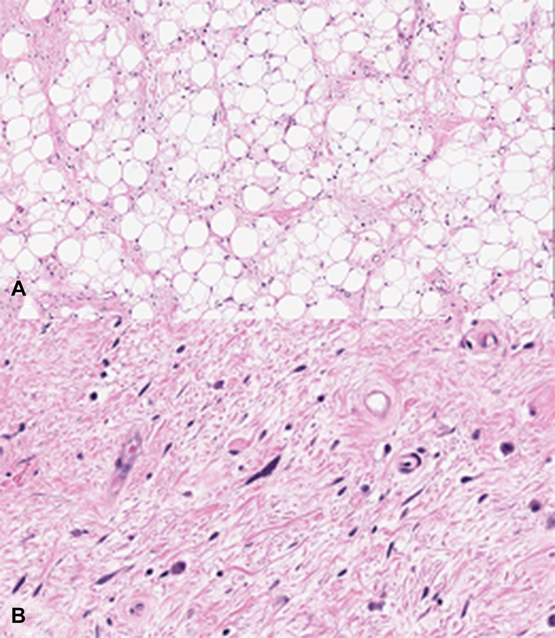
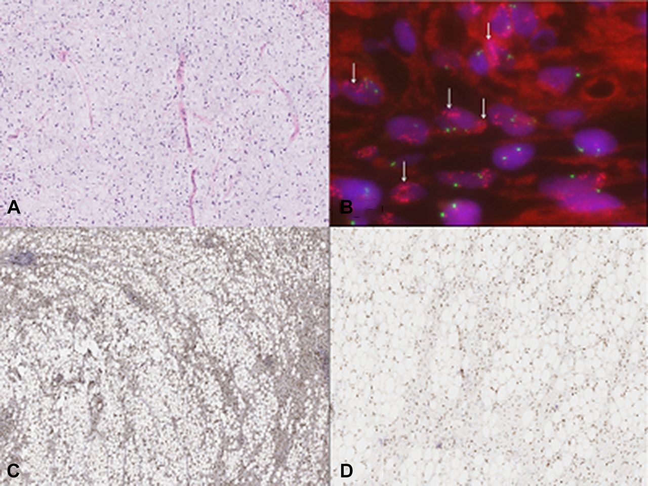
ALT/WDL is composed, entirely or in part, of an adipocytic proliferation with significant variation in adipocyte size (figure 6A) and a variable number of atypical, enlarged, hyperchromatic (‘inky’ nuclei) stromal cells, present in fibrous septae (figure 6B). Lipoblasts may or may not be present in ALT/WDL, and this is reflected in the current WHO diagnostic criteria for ALT/WDL, where lipoblasts are considered a helpful clue, but not a prerequisite, for the diagnosis.7 Myxoid change is well recognised in ALT/WDL, and when extensive, such cases may be mistaken for a myxoid liposarcoma (figure 7A). Various combinations of these features may occur and there are no significant clinicopathological, molecular or prognostic differences.45

Atypical lipomatous tumour or well-differentiated liposarcoma contains adipocytes displaying significant variation in adipocyte size (A) with variable numbers of atypical, enlarged, hyperchromatic (‘inky’) nuclei stromal cells, often present in fibrous septae (B).

Myxoid stromal change is frequently encountered in atypical lipomatous tumour/well-differentiated liposarcoma, and can be extensive enough to resemble myxoid liposarcoma (A). Of diagnostic importance is the demonstration of MDM2 amplification by fluorescence in situ hybridisation (see arrows) (B). These lesions are p16 positive (C) and show retention of nuclear retinoblastoma protein staining (D).
Immunohistochemistry and cytogenetics
ALT/WDLs are characterised by supernumerary ring chromosomes or giant marker chromosomes that demonstrate amplifications in the chromosomal region 12q13-15 (figure 7B)46; these amplifications constantly affect MDM2 (100%), and frequently affect CDK4 (90%) and HMGA2, genes. Thus, ALT/WDLs demonstrate MDM2 and CDK4 overexpression by immunohistochemistry. However, MDM2 staining tends to be weak and scattered in ALT/WDLs. In addition, MDM2 staining may be difficult to interpret, especially if macrophages are present, as they can stain positively. This can be a pitfall in lipomas with secondary degenerative changes, where inflammatory cells and macrophages are positive for MDM2.23 CDK4 immunohistochemistry has a low sensitivity and approximately 10% of ALT/WDLs do not possess amplification of CDK4.47
Amplification of MDM2 and CDK4 detection using FISH is the recommended gold standard methodology.48 49
p16Ink4A (p16) is a transcript of the cyclin-dependent kinase inhibitor 2A (CDKN2A) gene and it inhibits cell cycle progression by binding to CDK4.50 It has been shown to be overexpressed in ALT-WDL by both quantitative reverse-transcription PCR and immunohistochemistry (figure 7C), and consequently, p16 has been proposed as a diagnostic marker.51 Pathologists should be aware of, and disregard, cytoplasmic staining that may be observed in endothelial and inflammatory cells.52
Of note, nuclear pRb expression is typically intact in ALT/WDL (figure 7D).
Differential diagnosis
From a practical and patient management point of view, the most important differential diagnosis for ALT/WDL is a benign fatty tumour. The distinction between ALT/WDLs and benign adipocytic tumours is crucial because of differences in prognosis and treatment. In ALT/WDL, the atypical stromal cells may be poorly represented or absent and lipoblasts may not be seen. In lipomas, fibrosis, necrosis, inflammatory cells and pseudolipoblasts may be present. MDM2 gene amplification by FISH is an essential adjunct to prevent both underdiagnosis and overdiagnosis of ALT/WDL.
Clay et al suggest that FISH should be performed in the following scenarios pertaining to problematic lipomatous tumours: in recurrent ‘lipomas’; in cases with equivocal cytological atypia; in lesions of the retroperitoneum, pelvis or abdomen; in any deep-seated tumour of the extremity measuring >10 cm in a patient over the age of 50 years and in special clinical situations as directed by treating clinicians.53
Treatment and prognosis
ALT/WDL has no metastatic potential and therefore such tumours are better classified as being of intermediate (but locally aggressive) biological potential. However, the overwhelming majority of retroperitoneal and mediastinal WDLs are ultimately associated with frequent local recurrence and significant mortality, even in the absence of dedifferentiation.
Dedifferentiation occurs in up to 10% of ALT/WDLs, with the risk of dedifferentiation being much higher in the retroperitoneum than elsewhere, almost certainly as a consequence of the longer lesional duration and larger tumour size at this site.11 54
Dedifferentiated liposarcoma
Clinical features
ALT/WDL and DDL form the largest subgroup of liposarcomas, and represent a morphological and behavioural spectrum representing one disease entity.55 56 The classic definition of DDL states that it is a ‘non-lipogenic sarcoma’ arising in association with a WDL/an ALT.
DDLs present most frequently in middle-aged and older adults, with an equal gender distribution. They are typically large (>10 cm), multinodular masses.54 The retroperitoneum is considered the most common site of DDLs (where a spindle cell sarcoma is a DDL until proven otherwise), but they also occur in other locations, including the extremities, the paratesticular region, and, more rarely, the trunk (including mediastinum and thorax), head and neck.54 57–59 DDLs typically surround adjacent visceral structures but frank invasion of these structures is rare.54
Up to 90% of DDLs present as de novo neoplasms while the remainder occur as a recurrence of a pre-existing ALT/WDL, after an average interval of 7.7 years.41 Dedifferentiation can occur in up to 10% of ALT/WDLs at any site,54 with the risk being greater in tumours that are deep-seated, particularly those in the retroperitoneum, where this risk is approximately 28%.54 59 60
Microscopic findings
DDLs show a spectrum of morphological appearances, although the majority show features of undifferentiated pleomorphic sarcoma or spindle cell sarcoma not otherwise specified,54 with high-to-moderate cellularity, and with cells disposed in loose fascicles, patternless distributions or sometimes with a storiform architecture, within variably fibrous stroma. This is sharply demarcated from the well-differentiated component (figure 8A). Prominent cellularity with nuclear pleomorphism andmoderate to marked cytological atypia are seen, the mitotic index is variable, and necrosis can also be seen (figure 8B).61 62
Dedifferentiated liposarcoma (DDL) is characterised by a sharp demarcation between the well-differentiated and dedifferentiated components (A). The dedifferentiated component is made up of spindle cells with mild-to-moderate atypia in fascicles resembling a spindle sarcoma (B), set within a myxoid stroma (C). Unusual histological accompaniments include metaplastic bone (D).
Prominent myxoid stroma can be a feature of DDLs (figure 8C). When present, atypical spindle or ovoid cells with a myxofibrosarcoma-like morphology are present within the myxoid stroma. In addition, a prominent coarse plexiform vascular pattern (similar to that seen in myxofibrosarcoma), and sometimes a ‘pulmonary oedema-like’ pattern (similar to that of myxoid liposarcoma) may be seen.63
Metaplastic bone, meningothelial-like whorls and metaplastic cartilage may be seen rarely (figure 8D).64–66
DDLs may exhibit heterologous differentiation towards other mesenchymal lineages, including chondroid, osteoid, myoid (both smooth and skeletal muscle differentiation).41 67 68 Rarely, angiosarcomatous differentiation has been described.69
A minority of DDLs are of low cellularity and comprise only histologically ‘low-grade’ areas resembling fibromatosis or low-grade fibromyxoid sarcoma.41 70 71 Low cellularity DDLs are composed of sparsely to moderately cellular proliferations comprising loose fascicles or patternless distributions of fibroblast-like spindle cells with mild nuclear atypia and low mitotic activity.54
A small subset of cases may demonstrate lipoblastic differentiation within the dedifferentiated component (ie, rare cases may be ‘lipogenic’).72 This finding is referred to as DDL with ‘homologous lipoblastic differentiation’ or ‘pleomorphic liposarcoma-like features’.67 73
Immunohistochemistry and cytogenetics
DDLs can show variable expression of CD34, with focal positivity for smooth muscle actin and desmin, while S100 protein is absent in non-lipogenic areas of DDLs.54 Most DDLs (>90%) show immunohistochemical expression of MDM2 and CDK4 and there is strong correlation between expression of these immunohistochemical stains and their gene amplification status.23 74 The combination of p16 with CDK4 and MDM2 is useful in distinguishing DDLs from other adipocytic neoplasms, and is more sensitive than the use of CDK4 and MDM2 alone. Approximately 93% of DDLs express at least two antigens of this triad of markers, and p16 has been shown to be the most sensitive and specific marker for detecting DDLs.74
The assessment of MDM2 gene amplification by FISH is a highly useful adjunctive diagnostic tool for the diagnosis of DDL.75
A recent paper details the results of whole-exome sequencing performed on 206 sarcomas, including 50 DDLs, all of which were defined by 12q13-15 amplifications, including highly recurrent copy-number gains or amplification of MDM2 (100% of samples), CDK4 (92%) and HMGA2 (76%), as well as FRS2 (96%) and NAV3 (60%).1 Other frequent somatic copy-number alterations involved genes reported to inhibit adipocyte differentiation, namely PTPRQ (46%), JUN (42%), DDIT3 (32%), CEBPA (24%) and YAP1 (16%). Recurrent deletions of ATRX (30%), NF1 (28%) and CDKN2A (44%) were also detected.1
Differential diagnosis
The diverse histological patterns of DDL can lead to difficulty in distinguishing it from other soft tissue sarcomas, particularly in patients without an antecedent history of ALT/WDL, and in tumours in which a component of ALT/WDL is lacking.54
Morphological clues to identify DDLs are the presence of an abrupt, sharp transition from a well-differentiated adipocytic component into non-lipogenic spindle cell areas, and/or the presence of higher-grade areas, widespread atypia and varied morphology or cellular pleomorphism.54 Essentially all neoplasms in the differential diagnosis of DDL will lack any adjacent WDL, and will tend to lack expression of p16, CDK4 and MDM2 by immunohistochemistry and lack evidence of MDM2 amplification by FISH.54
It is worth bearing in mind that it has been shown that most neoplasms arising in the retroperitoneum that are diagnosed as undifferentiated pleomorphic sarcoma (so-called ‘malignant fibrous histiocytoma’), in fact represent DDLs.76
Treatment and prognosis
The diagnosis of DDL is prognostically significant, as it has a lower tendency towards local recurrence and metastasis, when compared with some other liposarcomas and with morphologically similar pleomorphic soft tissue sarcomas such as leiomyosarcomas or undifferentiated pleomorphic sarcomas.62
Radical surgical excision remains the mainstay of treatment, although radiotherapy is considered a valuable treatment option in retroperitoneal DDLs.54
DDL behaves in a more aggressive manner than ALT/WDL, with a greater propensity for local recurrence (in approximately 41% of cases) as well as the capacity to metastasize (in 15%–30% of cases), but it tends to behave less aggressively than other pleomorphic sarcomas.41 54 62
Macroscopic tumour clearance at any site has been shown to be significantly associated with reduced local recurrence and improved survival.77 A recent study has shown that retroperitoneal liposarcomas with myogenic differentiation, and particularly those with a rhabdomyoblastic component, have a significantly poorer outcome.78 The most important adverse prognostic factor has been reported to be a location in the retroperitoneum, with tumours here showing significantly worse survival than those at other anatomic sites.41
Even histologically, low-grade DDLs have been shown to have the capacity to metastasize and to behave like traditional DDLs, rather than like WDLs.41 79
Take home messages
Fatty tumours with a spindle cell component encompass both benign and malignant tumours.
Immunohistochemistry and/or cytogenetic analysis is critical for the correct diagnosis of spindle cell fatty tumours.
Spindle cell lipoma/mammary-type myofibroblastoma/cellular angiofibroma/atypical spindle cell lipomatous tumours (ASCLT) are lesions that are characterised by spindle cells (often bland) and fat, nuclear retinoblastoma protein loss and absence of amplification of MDM2 and CDK4.
ASCLT is a newly created group of tumours incorporating spindle cell liposarcoma.
ASCLT can mimic spindle cell lipoma morphologically, immunohistochemically and cytogenetically. Such cases are separated by location of the lesion.
Thorough sampling of fatty lesions with spindle cells is advocated in view of the variable and overlapping features.
References
Footnotes
Handling editor Cheok Soon Lee.
Contributors AJM and RC contributed equally to the conceptualising and writing of the manuscript.
Funding This research received no specific grant from any funding agency in the public, commercial or not-for-profit sectors.
Competing interests None declared.
Provenance and peer review Commissioned; internally peer reviewed.
Data sharing statement None.