Article Text

Abstract
Aims To demonstrate clinical application of a mesodissection platform that was developed to combine advantages of laser-based instrumentation with the speed/ease of manual dissection for automated dissection of tissue off standard glass slides.
Methods Genomic analysis for KRAS gene mutation was performed on formalin fixed paraffin embedded (FFPE) cancer patient tissue that was dissected using the mesodissection platform. Selected reaction monitoring proteomic analysis for quantitative Her2 protein expression was performed on FFPE patient tumour tissue dissected by a laser-based instrument and the MilliSect instrument.
Results Genomic analysis demonstrates highly confident detection of KRAS mutation specifically in lung cancer cells and not the surrounding benign, non-tumour tissue. Proteomic analysis demonstrates Her2 quantitative protein expression in breast cancer cells dissected manually, by laser-based instrumentation and by MilliSect instrumentation (mesodissection).
Conclusions Slide-mounted tissue dissection is commonly performed using laser-based instruments or manually scraping tissue by scalpel. Here we demonstrate that the mesodissection platform as performed by the MilliSect instrument for tissue dissection is cost-effective; it functions comparably to laser-based dissection and which can be adopted into a clinical diagnostic workflow.
- DIAGNOSTICS
- CANCER GENETICS
- PROTEINS
This is an Open Access article distributed in accordance with the Creative Commons Attribution Non Commercial (CC BY-NC 4.0) license, which permits others to distribute, remix, adapt, build upon this work non-commercially, and license their derivative works on different terms, provided the original work is properly cited and the use is non-commercial. See: http://creativecommons.org/licenses/by-nc/4.0/
Statistics from Altmetric.com
Introduction
Knowledge of the mutation status of cancer-related genes and the quantitative levels of drug target proteins in tumour cells can assist in selection of targeted cancer therapy.1–3 Genomic-based technologies are becoming the benchmark for mutation detection in formalin fixed paraffin embedded (FFPE) patient tissue, while mass spectrometry is a robust quantitative approach to measuring protein levels in FFPE patient tissue.4–7 Genomic and proteomic analysis of patient tissue depends on the percentage of tumour cells in the sample, making it imperative that highly enriched tumour cell populations be procured from the heterogeneous tissue microenvironment using tissue dissection methodology.8–10
Tissue dissection in the clinical molecular diagnostics laboratory is often performed by manually scraping tissue (via scalpel) directly off standard glass slides. This is performed at a very low cost but with little resolution in light of tissue heterogeneity. Laser microdissection instrumentation was developed to address lack of resolution, yet these instruments are expensive, labour intensive, and often rely on special slides or photoactivation film.
Here we demonstrate the application of mesodissection that incorporates into a single platform advantages of laser microdissection and manual dissection, while improving upon their individual disadvantages.11 Mutation detection and quantitative protein analysis of mesodissected tumour tissue demonstrate application to a clinical cancer diagnostic laboratory workflow in an economical, automated and robust platform.
Materials and methods
Genomics
Multiple serial sections (5 µM thick) from a lung cancer block known to harbour a KRAS point mutation (p.G12C_c.34G>T) were cut onto standard glass slides at ARUP Laboratories (Salt Lake City, Utah, USA) under strict Internal Review Board regulations. Images from an H&E section were pre-marked to identify areas of pure tumour cells, then used to guide dissection using the 2iD software on the MilliSect mesodissection instrument (avanscibio.com; AvanSci Bio, Salt Lake City, Utah, USA). For dissection, each pre-marked image was aligned with the corresponding live image and areas of tumour and non-tumour cells dissected separately with either 200 µM or 400 µM xScisors (avanscibio.com; AvanSciBio, Salt Lake City, Utah, USA) using a low detergent milling buffer (2 mM TRIS (pH 8.5), 0.2 mM EDTA, 0.1% TWEEN-20). For DNA preparation, the recovered tissue was centrifuged (2000g) to a pellet, and all but 20 μl of the supernatant was discarded. An equal volume of light mineral oil was added, followed by heating to 92°C for 1 h under constant shaking (1500 rpm). Proteinase K was added to 0.5 µg/µL and tissue heated to 56°C for 1 h under constant shaking (1500 rpm). The enzyme was heat inactivated for 15 min at 92°C, followed by removal of the oil with AvanSciBio Wicking Strips (avanscibio.com; AvanSci Bio, Salt Lake City, Utah, USA). Total DNA was quantified using PicoGreen (Life Technologies, Grand Island, New York, USA).
Proteomics
A Her2+ breast cancer tissue block (IHC 3+) was obtained from Asterand (Detroit, Michigan, USA) and de-identified prior to shipment. A single section (10 µM) was cut onto a DIRECTOR slide (OncoPlex Diagnostics, Rockville, Maryland, USA) and multiple serial sections cut onto standard glass slides. Pre-marked areas of tumour cells were used to guide dissection using the 2iD software on the MilliSect instrument. A Leica LMD6000 microdissection instrument was used to dissect correlative areas of tumour cells off the DIRECTOR slide. An entire section was scraped into a tube using a scalpel for manual dissection. Four additional sections on glass slides were used for mesodissection of the marked tumour cell areas using the MilliSect instrument as directed by the pre-marked images. Tissue was dissected with Liquid Tissue buffer using 200 μM xScisors. Liquid Tissue lysates were prepared from the laser dissected, manual dissected and mesodissected tissue according to manufacturer's recommendations (OncoPlex Diagnostics, Rockville, Maryland, USA). Total protein was quantified by a modified microBCA assay (Pierce, Rockford, Illinois, USA)
Results and discussion
Figure 1A shows an example (section 7) of pre-marked areas of tumour and non-tumour cells (pre-dissection and post-dissection) for mesodissection and subsequent DNA mutation analysis. Each dissection took approximately 5 min, which is comparable with performing dissection with a scalpel. DNA amounts ranged from 54 to 129 ng (average 19 ng/mm2) in the dissected tumour cell populations, and 50–78 ng (average 5.6 ng/mm2) in the dissected non-tumour benign tissue (table 1). Less DNA/mm2 was recovered from the non-tumour tissue likely due to the lower cellularity of this area. Crude DNA lysates were used with the Sequenome MassArray platform (Agena Biosciences) for KRAS mutation (p.G12C_c.34G>T) detection. Data in table 1 indicate an average allele frequency (AF) of 61% with a tight range of 58.8%–63.1% in the tumour cell populations across all serial sections. A Z-score of 10 was achieved for each analysis, indicating high confidence data. The mutation was detected in three of the seven benign tissue dissections with an average allele frequency of 5.6% and low Z-scores. Results indicate highly consistent and reliable mutation detection in tumour cell populations collected by mesodissection across serial sections of the same tumour block. Analysis of non-tumour benign tissue resulted in either no detection of the mutation or clinically unreliable data (low allele frequency/low Z score).

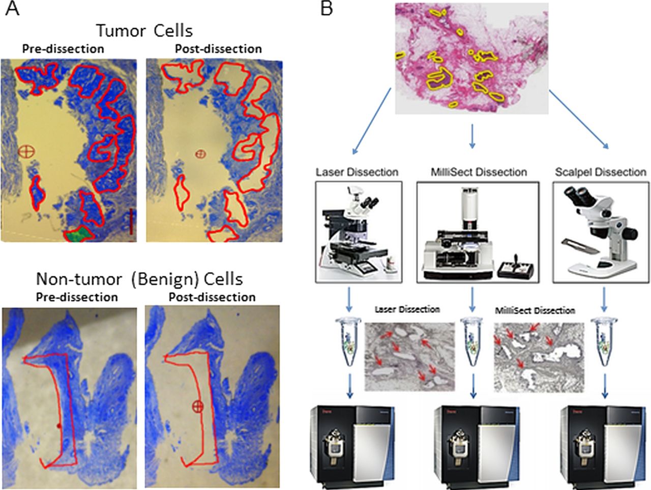
(A) Genomic analysis of mesodissected KRAS+ lung cancer tissue. An example of pre-dissection and post-dissection of tumour cells and neighbouring non-tumour benign tissue from formalin fixed paraffin embedded tissue sections off standard glass slides using the MilliSect instrument (http://www.youtube.com/watch?v=U6C5wVOkH3I). Dissection guidance was achieved using the 2iD software (http://www.youtube.com/watch?v=rBgMhHlSyl4). Tissue was collected by mesodissection mediated by xScisor technology (http://www.youtube.com/watch?v=XJ6yADqdVtM&feature=youtu.be). (B) Proteomic analysis of mesodissected Her2+ breast cancer tissue. Example mark-up of a section showing the regions of tumour cells to be dissected. Much of the section is non-tumour benign tissue consisting of non-cellular material. One section was manually dissected, another section dissected using laser instrumentation and four additional serial sections mesodissected using the MilliSect instrument. Examples of post-dissection are shown. Liquid Tissue lysates were prepared and protein quantitation performed by selected reaction monitoring.
Results of KRAS mutation detection using the Sequenome MassArray technology
Proteomic analysis strategy and examples of pre-dissection and post-dissection are shown in figure 1B. Once dissected, Liquid Tissue lysate was prepared and total protein of each lysate ranged from 5.6 to 11.13 µg (average 0.97 µg/mm2) in tumour cell populations mesodissected with the MilliSect instrument (table 2). Total protein for the laser-based microdissection was 6.7 µg (0.83 µg/mm2) and 8.2 µg (0.47 µg/mm2) in the scalpel dissection (table 2). Each Liquid Tissue lysate was interrogated in triplicate by selected reaction monitoring (SRM) on a Quantiva triple quadrupole mass spectrometer (ThermoScientific, San Jose, California, USA). Quantitation of a specific Her2 tryptic peptide (ELVSEFSR) by SRM-mass spectrometry was performed as described, and the levels are reflected in amol of peptide per microgram of total protein analysed.7 Likewise, the β-actin (AVFPSIVGR) and tubulin (YLTVAAVFR) tryptic peptides were quantified in triplicate to indicate the cellularity of the dissection and levels are reflected as femtomole of the peptide per microgram total protein analysed.7 Coefficient of variations (CVs) of <9% were obtained for all SRM analyses indicating high assay reproducibility (table 2).
Results of Her2, β-actin and tubulin SRM assays performed in triplicate are shown
Laser instrumentation provides the highest resolution for tumour cell purity (based on single cell resolution) and results indicate a benchmark Her2 peptide level of 597.5 amol/µg (CV=2.26%). Manual scalpel dissection of the entire tissue section provides the lowest resolution and demonstrates Her2 peptide level of 168.7 amol/µg. Cell populations collected by MilliSect mesodissection consistently showed comparable levels of Her2 peptide as the laser-dissected cells (table 2). Quantitation of β-actin and tubulin indicates that all sections dissected by the MilliSect and the LMD6000 contain high peptide levels, also indicating a high degree of cellularity. In contrast, β-actin and tubulin levels in the manual dissections show remarkably lower levels, reflecting the fact that much of the scraped tissue consists of extracellular matrix (reduced cellularity).
This study demonstrates highly selective, specific and efficient collection of tumour cells directly from FFPE tissue sections mounted on plain glass slides using the mesodissection platform that functions comparably to laser dissection instrumentation. In addition, mesodissection provides higher dissection resolution than scalpel dissection, resulting in highly confident molecular diagnostic results as reflected in the reduction of false positives/negatives. The simplicity and much-reduced cost of the MilliSect instrument compared with laser-based instruments, and the fact that manual scraping does not provide purified tumour cell populations, demonstrates that mesodissection can become a technological cornerstone in the cancer diagnostic laboratory for molecular analysis of FFPE patient tissue.
Take home messages
-
A novel tissue mesodissection platform was used to procure highly enriched populations of tumour cells from formalin fixed paraffin embedded (FFPE) cancer tissue sections directly off standard glass slides. KRAS mutation detection and Her2 quantitative selected reaction monitoring protein analysis indicate the specificity/resolution of tumour cell dissection with clear application to clinical diagnostic analysis of FFPE patient tumour tissue.
Acknowledgments
We thank Dr Katherine Geiersbach for ARUP Laboratories for directing digital annotation and Noah Welker from ARUP Laboratories for performing the Sequenome MassArray experiments, and Dr Sheeno Thyparambil and Dr Wei-Li Lao for proteomic expertise.
References
Footnotes
-
Contributors DK designed and supervised the proteomic experiments and wrote the manuscript. NA and RP designed, supervised and performed the genomic dissection and sample preparation experiments.
-
Funding US Department of Health and Human Services-National Institutes of Health 2R44GM100645-02.
-
Competing interests NA and RP are employees at AvanSci Bio, which has developed and markets the MilliSect. DK provides consulting services to AvanSci Bio.
-
Provenance and peer review Not commissioned; externally peer reviewed.