Article Text

Abstract
Aims Recent reports have identified recurrent MED12 somatic mutations in fibroadenomas and phyllodes tumours. The frequency and type of somatic mutations were noted to be similar to those of uterine leiomyomas. We aimed to investigate protein expression of MED12, correlating it to MED12 mutational status and expression of oestrogen receptors (ER).
Methods Immunohistochemistry was performed on a total of 232 fibroepithelial lesions (100 fibroadenomas, 132 phyllodes tumours) diagnosed at the Department of Pathology, Singapore General Hospital using MED12, ERα and ERβ antibodies. Expressions were evaluated in both stroma and epithelium, and correlated with MED12 mutational status.
Results MED12 mutation was significantly associated with high MED12 protein expression (H-score >150) in the stroma (p=0.029), but not in the epithelium. It was not associated with ERα and ERβ protein expression in both stroma and epithelium. MED12 protein expression was significantly correlated with ERα in epithelial (p=0.007) and ERβ in stromal (p=0.049) components. MED12 was not significantly different between fibroadenomas and phyllodes tumours. Epithelial expression of ERα was significantly higher in fibroadenomas (p<0.001) than in phyllodes tumours. Conversely, both epithelial and stromal expression of ERβ was significantly higher in phyllodes tumours (p<0.001).
Conclusions Positive associations observed between MED12 and ERα, ERβ immunohistochemical expression suggest a biological interplay between the proteins. The lack of significant association of MED12 mutation with ER protein expression indicates a need to further explore the functional impact of MED12 mutations on the ER signalling pathway in breast fibroepithelial lesions.
- BREAST
- IMMUNOHISTOCHEMISTRY
- HISTOPATHOLOGY
Statistics from Altmetric.com
Introduction
Breast fibroepithelial lesions are biphasic neoplasms comprising a proliferation of both epithelial and stromal components. Fibroadenomas and phyllodes tumours are the two major entities of fibroepithelial lesions. Our recent studies reported frequent somatic MED12 mutations in both fibroadenomas (59%) and phyllodes tumours (62.5%).1 ,2 MED12 encodes for the mediator complex subunit 12 (MED12) protein, which assembles with other subunits to form a large protein complex known as the mediator complex. The evolutionarily conserved 26-subunit mediator complex plays a central role in regulating transcriptional processes, which are vital for protein coding.3 Besides, the mediator complex is also an essential coactivator for a broad range of nuclear hormone receptors.4 Prenzel et al5 reported a decrease in oestrogen receptor α (ERα) protein levels and a significant impairment to oestrogen-regulated transcriptome following knockdown of the MED12 gene in MCF7 breast cancer cell lines.5 Kang et al6 demonstrated that the mediator complex interacts directly with ERα and ERβ and enhances receptor function in vitro. Together with our previous finding of MED12 mutations being associated with dysregulated oestrogen signalling,1 we set out to investigate the protein expression of MED12 in breast fibroepithelial lesions, correlating the findings with MED12 mutational status and ER expression.
Materials and methods
Samples
A total of 232 fibroepithelial lesions was included in this study, of which 100 and 132 cases were fibroadenomas and phyllodes tumours (77 benign, 41 borderline, 14 malignant), respectively. These cases were partly derived from our previous study cohorts.1 ,2 Samples were obtained from the archives of the Department of Pathology, Singapore General Hospital with ethics approval from the Centralized Institutional Review Board (CIRB 2005/002/F). Cases were reviewed and subtyped using criteria recommended by the WHO classification of breast tumours.7 Briefly, phyllodes tumours are diagnosed when there is presence of fronded architecture accompanied by increased stromal cellularity. The benign phyllodes tumour has mild stromal cellularity and nuclear atypia, pushing borders, without stromal overgrowth and four or less mitoses per 10 high-power fields. The malignant phyllodes tumour is diagnosed when there is marked stromal cellularity and nuclear atypia, presence of stromal overgrowth, permeative margins and mitoses of 10 or more per 10 high-power fields. The borderline phyllodes tumour has intermediate features between benign and malignant tumours. Fibroadenomas are fibroepithelial neoplasms without the presence of fronded architecture and increased stromal cellularity.
Immunohistochemistry and evaluation
Whole sections of 4 μm thickness were cut from formalin-fixed, paraffin embedded (FFPE) tissue blocks onto charged slides (Leica Biosystems, Richmond, IL, USA). Sections were deparaffinised with two changes of xylene and graded alcohols. Then, tissues were pretreated with heat-induced epitope retrieval method before immunohistochemistry was performed. Briefly, endogenous peroxidase activity was blocked with hydrogen peroxide followed by primary antibody incubation with optimised dilution and time as shown in table 1. Breast carcinoma acted as positive control for all antibodies, while negative control was achieved by omission of primary antibody on breast carcinoma tissue.
Details and optimised conditions for MED12, ERα and ERβ antibodies
Slides were scanned with IntelliSite Pathology Ultra Fast Scanner (Philips Digital Pathology Solutions, The Netherlands) and viewed with IMS Viewer (Philips, The Netherlands). Staining was evaluated by two independent observers blinded to clinical data and mutation status. MED12 and ERα expression was assessed in the nucleus, while ERβ expression was assessed in the nucleus and cytoplasm separately. Epithelial and stromal expression was examined separately. Intensity and percentage of tumour cells stained were assessed. A four-tiered staining intensity was employed—no expression, weak expression, moderate expression and strong expression. Semiquantitative H-score8 was derived, taking into account both staining intensity and percentage of tumour cells stained, with the following equation: H-score=(1×% of tumour cells with weak staining)+(2×% of tumour cells with moderate staining)+(3×% of tumour cells with strong staining). Associations between MED12 and ERα, ERβ immunohistochemical expression were assessed using H-score as a continuous variable to determine the strength of relationship. For comparisons of immunohistochemical expression between wild-type and mutant MED12 tumours, categorical data were employed. High MED12 protein expression was defined as H-score 150 and above using a systematic exploration of thresholds in this study. ERα and ERβ positivity was defined by at least 1% immunoreactive nuclei.9
Immunohistochemistry for caldesmon on a case of fibroadenoma that expressed ERα in the stroma was performed using the BOND-MAX (Leica Biosystems, Germany) automated system. Caldesmon (Dako M3557) antibody was applied in a dilution of 1:70 for 20 min after the tissue section was pretreated with ER1 pH 6.0 solution at 100°C for 20 min. Staining was visualised using the Bond Polymer Refine Detection system (Leica Biosystems).
Mutation analysis of MED12 exon 2
Samples were interrogated for mutational status of MED12, specifically targeting exon 2 as previously reported.1 ,2 Briefly, DNA extraction was performed with the QIAamp DNA FFPE Tissue Kit (Qiagen, The Netherlands) according to manufacturer's protocol. Amplicons were generated and pooled with the Access Array System (Fluidigm, San Francisco, California, USA). Ultradeep targeted sequencing was performed on the MiSeq instrument (Illumina, San Diego, CA, USA) using MiSeq Reagent Kit V3 (Illumina, USA). Resulting reads were aligned against the reference human genome (hg19) with the Burrows–Wheeler Alignment tool (V.0.6.2). Variant allele frequencies exceeding a threshold of 5% were called.
Statistical analysis
Mann–Whitney U test (H-score as continuous data), χ2 and Fisher's exact tests (H-score as categorical data) were performed to evaluate differences between MED12 protein expression of wild-type and mutant MED12 tumours. Associations between MED12, ERα and ERβ protein expression were analysed with Spearman's correlation test. Differential expression of MED12, ERα and ERβ between fibroadenomas and phyllodes tumours was assessed with Fisher's exact test. All analyses were performed with PASW Statistics for Windows, V.18.0 (SPSS).
Results
Characteristics of study cohort
Median age of the study cohort was 39 years, ranging from 15 to 79 years. Patients with fibroadenomas were significantly younger than patients with phyllodes tumours (p<0.001). No significant differences were observed between ethnic distribution and frequency of MED12 mutations of fibroadenomas and phyllodes tumours. As a whole, MED12 mutations were not associated with age. Detailed characteristics of the study cohort are shown in Table 2. However, there was a trend of younger age associating with mutant MED12 (median age 30) compared with wild-type MED12 (median age 38) among patients diagnosed with fibroadenomas (p=0.061). Among patients with phyllodes tumours, there were no differences in age at diagnosis between patients harbouring wild-type MED12 and mutant MED12 tumours.
Characteristics of study cohort
MED12 protein immunohistochemical expression in MED12 wild-type and mutant tumours
MED12 protein expression between MED12 wild-type and MED12 mutant tumours was not significantly different in the stroma, quantified with H-score as a continuous variable. We further explored a systematic selection of a threshold (H-score 50, 100, 150, 200 and 250) to define high MED12 protein expression in the stroma in association with mutation status. At a H-score threshold of 150, MED12 mutant tumours were significantly associated with high MED12 protein expression in the stroma (p=0.029). No significant associations were observed with other thresholds. A stratified analysis of MED12 mutant tumours into different types of mutations (table 3) revealed high MED12 protein expression in the stroma was associated with MED12 missense mutations (p=0.023) as shown in figure 1A. Low MED12 protein expression in the stroma was associated with MED12 indel mutations and splice site mutations (figure 1B).
Protein expression of MED12 and ER in tumours with wild-type MED12 and mutant MED12

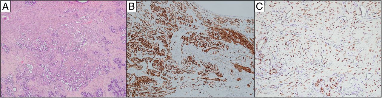
Immunohistochemical protein expression of MED12. (A) MED12 staining was intense in both epithelium and stroma of this benign phyllodes tumour harbouring a point mutation at codon 44. (B) An area of stromal overgrowth in a malignant phyllodes tumour harbouring an indel mutation expressing low MED12 protein expression (H-score below 150) in the stroma.
MED12 immunoexpression was also observed in the epithelium of fibroepithelial lesions (figure 1A), with no obvious differences observed between wild-type MED12 and mutant MED12 tumours, respectively (p=0.729).
Associations of ERα, ERβ immunohistochemical expression with MED12 mutation status and MED12 protein expression
A subset of 184 cases was available for ERα and ERβ protein expression assessment. ERα was typically expressed in the epithelium, but was absent in the stroma (figure 2A) except for one fibroadenoma harbouring wild-type MED12 (figure 3A). Upon reviewing the case and performing immunohistochemistry for caldesmon (figure 3B), a smooth muscle component was noted in the stroma that expressed ERα (figure 3C). Further analysis for stromal ERα expression was excluded as it was not statistically viable. On the contrary, ERβ was noted in the stroma as well as in the epithelium, with expression observed in both nuclei and cytoplasm of fibroepithelial lesions (figure 2B). The highest ERβ expression was observed in epithelial nuclei, followed by epithelial cytoplasm, stromal nuclei and stromal cytoplasm, with mean H-scores of 22, 6, 3 and 1, respectively.

Immunohistochemical protein expression of oestrogen receptor α (ERα) and β (ERβ). (A) ERα is seen in the epithelium, but not in the stroma in this example of a benign phyllodes tumour. Other parts of the tumour disclosed typical phyllodal fronds with stromal hypercellularity. (B) Nuclear expression with cytoplasmic decoration of ERβ was observed in the epithelium of a fibroadenoma.
A case of fibroadenoma with smooth muscle metaplasia. (A) The fibroadenoma shows bundles of smooth muscle fibres amid and extending beyond sclerosing adenosis. (B) Immunohistochemistry for caldesmon decorates the smooth muscle fibres. (C) Oestrogen receptor α immunohistochemistry shows nuclear positivity in the smooth muscle fibres as well as in the nuclei of ductular epithelium.
ERα epithelial expression was positively correlated with MED12 epithelial expression with Spearman's coefficient of 0.198 (p=0.007). This association was stronger in fibroadenomas than in phyllodes tumours (table 4). A significant positive correlation between cytoplasmic ERβ expression and MED12 nuclear expression was observed in the stroma among fibroadenomas with Spearman's coefficient of 0.198 (p=0.049). Conversely in phyllodes tumours, a negative correlation was observed despite not being significant statistically.
Spearman's correlation analysis for associations between MED12 and ER protein expression*
Among the 184 cases, 168 cases had MED12 mutation status available. For correlation with mutational status, ERα and ERβ expression was classified into two categories employing the threshold recommended by the American Society of Clinical Oncology/College of American Pathologists (ASCO/CAP), where positivity was defined by at least 1% immunoreactive nuclei.9 Results are shown in table 3. Both ERα and ERβ expression was not significantly different between MED12 wild-type and MED12 mutant tumours. However, it was noted that among the MED12 mutant tumours, ERβ stromal cytoplasmic positivity was associated with indel mutations (p=0.039).
Differential immunohistochemical expression of ERα, ERβ and MED12 in fibroadenomas and phyllodes tumours
Expression of stromal and epithelial MED12 was not significantly different between fibroadenomas and phyllodes tumours (table 5). However, MED12 expression in the epithelium was inversely associated with tumour grade among phyllodes tumours (p=0.01).
Expression of MED12, ERα and ERβ in fibroadenomas and phyllodes tumours
ERα was positive in all fibroadenomas, while only 86.9% of phyllodes tumours had positive ERα in the epithelium (p<0.001). Of the 11 ERα-negative cases, 6 (12.5%) were benign, 3 (10.7%) were borderline and 2 (25%) were malignant tumours. The proportions of such negative cases in each grade were, however, not significantly different (p=0.563).
Expression of ERβ was significantly higher in phyllodes tumours than fibroadenomas in all components (table 5). Within phyllodes tumours, expression in the stromal cytoplasm was significantly higher in the malignant group (62.5%), compared with benign (14.6%) and borderline (35.7%) tumours (p=0.006).
Discussion
The discovery of highly recurrent MED12 somatic mutations in breast fibroadenomas1 has led to a surge of reports published recently on MED12 somatic mutations in fibroadenomas and the closely related phyllodes tumours (table 6). The frequency of MED12 somatic mutations reported ranges from 45% to 80%, not significantly different between fibroadenomas and phyllodes tumours, although some authors report a lower frequency in malignant phyllodes tumours10 ,11 while other studies show no significant difference among phyllodes tumours of different grades.2 ,12 In uterine leiomyomas, mutations of MED12 are common, but are rare in its malignant counterpart the leiomyosarcoma.13–15 The composition of MED12 mutations in breast fibroepithelial tumours across different studies are comparable, with missense mutations (single nucleotide change resulting in an altered amino acid in the protein) in codon 44 as the most frequently encountered alteration, similar to what was found in uterine leiomyomas.15
Reports of MED12 somatic mutations in fibroadenomas (FA) and phyllodes tumours (PT)
MED12 protein was highly expressed in the stroma with H-score above 150 in the majority (91%) of cases. An increased percentage of cases with high MED12 stromal expression was observed in MED12 mutant tumours despite the narrow differences observed (p=0.029). Specifically, high MED12 stromal protein expression was associated with MED12 missense mutation. This finding contrasts with those in studies by Ravegnini et al16 and Yoon et al,17 where MED12 protein expression was not correlated with MED12 mutation. Ravegnini et al16 reported a small number of missense mutants in their study cohort, while Yoon et al17 employed a scoring method assessing staining intensity only. We also observed an association between low MED12 protein expression with MED12 indel and splice site mutations, corroborating findings by Bertsch et al,13 where complex MED12 mutations had significant lower immunoreactivity for MED12 in leiomyomas. The current antibody employed targets a specific region near the C-terminus (between 2150 and C-terminus) of the protein, a region not translated from the exon 2 mutation sites. Indel mutations result in a shift of amino acids and may lead to reduced detection sensitivity by the antibody, possibly explaining the association with low MED12 protein expression. Missense mutations at exon 2, however, may not affect antibody detection of the antigen, which is located near the C-terminus site. The mechanism of MED12 protein regulation is currently not well studied. Mutations in MED12 may alter regulation of the protein similar to the situation with c-myc gene, where mutations around Thr-58 may result in protein stabilisation preventing degradation and hence augmenting c-myc protein levels.18 The development of an antibody specifically targeting the protein translated from exon 2 will be useful in investigating the effects of altered protein products resulting from the mutations.
We previously showed through expression profiling that genes upregulated in MED12 exon 2 mutant fibroadenomas were associated with dysregulated oestrogen signalling.1 ERs, represented by ERα and ERβ, are important transcription factors for oestrogen signalling in cells vital for development and maintenance of reproductive functions.19 To our knowledge, this is the first report investigating ER protein expression in relation to MED12 protein expression and mutation status in fibroepithelial lesions. We observed no significant differences in both ERα and ERβ expression between MED12 wild-type and MED12 mutant tumours, which suggests that MED12 mutations may not directly affect ER protein expression despite associations observed in gene expression profiling. The molecular mechanism of MED12 exon 2 mutations affecting the multifaceted oestrogen signalling pathway is still largely unknown despite a recent report of MED12 G44D mutant cells demonstrating a significant loss of mediator-associated cyclin-dependant kinase (CDK) activity relative to MED12 wild-type cells.20 MED12, CDK8, cyclin C and MED13 form the well-known ‘CDK8 module’ within the mediator complex.3 This four-subunit module acts as a molecular switch, which controls the coactivator function of the mediator complex.21 ,22 Future studies in elucidating the direct and functional relationship between MED12 exon 2 mutations and the oestrogen signalling pathway will be important to understand the effects of such mutations on the oestrogen signalling pathway.
Despite no significant differences in ERα protein expression in wild-type versus mutant MED12 tumours, we observed a significant positive association between MED12 protein and ERα expression in the epithelium, where ERα expression increases with increasing MED12 expression. This is consistent with findings by Prenzel et al,5 where ERα expression was impaired when MED12 was knocked down in breast cancer cell lines. However, in the stromal component, we observed no ERα expression in all cases except for the single fibroadenoma with smooth muscle metaplasia, even if the tumours expressed MED12 protein. Our observation of ERα negativity in the stroma of fibroepithelial lesions corroborates that of Sapino et al,23 who also noted that ERα was not detected in the stroma of both fibroadenomas and phyllodes tumours. The disparity in results between epithelial and stromal components resonates well with the notion that the epithelium and stroma have distinct behaviour24 and are likely regulated differently. In addition, the regulation of ERα expression is complex with a plethora of factors involved, such as estradiol ligand, SET7 methyltransferase and human progesterone receptor isoform hPR-A.25 ,26
The relationship between fibroadenomas and phyllodes tumours has been an intriguing topic, with our group recently describing the genomic profiles of fibroepithelial neoplasms.31 They have overlapping histological properties despite potentially divergent outcomes. Fibroadenomas are benign and biologically innocuous, while phyllodes tumours have a tendency to recur. Table 6 shows the findings of various groups denoting a similar frequency of MED12 mutations in fibroadenomas and phyllodes tumours, corroborating our findings and underscoring their biological similarity. On the contrary, we noted a significant difference in ERα and ERβ expression between fibroadenomas and phyllodes tumours. These opposing observations speak to the complexity and heterogeneity of fibroepithelial lesions, requiring further interrogation of molecular pathogenesis to enhance understanding of the relationship between the two entities.
A limitation of this study is the small number of non-missense mutations observed, hampering meaningful interpretation despite statistically significant differences. Our attempt of stratifying fibroepithelial lesions into fibroadenomas and phyllodes tumours to analyse their individual associations of MED12 mutation status with MED12 and ER immunohistochemical expression also yielded no significant findings (results not shown). The prognostic impact of MED12 and ER expression was not demonstrated in this study as our aim was to determine the relevance of MED12 mutations in correlation with protein expression, as well as their association with ERα and ERβ expression. Nonetheless, we had previously shown that phyllodes tumours harbouring MED12 mutations augured a better recurrence-free survival.2
In conclusion, positive associations observed between MED12 and ERα, ERβ immunohistochemical expression suggests a biological interplay between the proteins. No significant differences in ER protein expression were observed between wild-type and mutant MED12 tumours. Future studies to understand the relationship between MED12 and ER will be important to elucidate the functional effects of MED12 mutations in the oestrogen signalling pathway.
Take home messages
MED12 mutation was associated with high MED12 protein expression in the stroma of fibroepithelial lesions.
MED12 protein expression correlated with oestrogen receptor expression.
MED12 protein expression was not significantly different between fibroadenomas and phyllodes tumours.
References
Footnotes
Handling editor Cheok Soon Lee
Contributors WJT performed experiments, evaluated IHC expression, analysed statistical data and wrote the manuscript. JYC evaluated IHC expression and reviewed the manuscript. AAT selected cases, validated IHC expression and reviewed the manuscript. JCTL, NDMN and JSYT carried out immunohistochemistry staining and reviewed the manuscript. VCYK collated clinical data and reviewed the manuscript. WKL, JT, CCYN, VR and SN performed mutation analysis and reviewed the manuscript. BHB contributed to the scientific content of the study. BTT and PHT initiated and supervised the study, wrote and revised the manuscript. All authors read and approved the final manuscript.
Funding This study was supported by the SGH Research Grant SRG-S/01/2014.
Competing interests None declared.
Ethics approval Centralized Institutional Review Board (CIRB 2005/002/F).
Provenance and peer review Not commissioned; externally peer reviewed.