Article Text

Abstract
Background: Steatosis occurs in more than 50% of patients with chronic hepatitis C and is associated with increased hepatic fibrosis. In many of these patients the pathogenesis of steatosis appears to be the same as for patients with non-alcoholic fatty liver disease—that is, related to visceral adiposity and obesity.
Methods: The effect of a three month weight reduction programme on liver biochemistry and metabolic parameters was examined in 19 subjects with steatosis and chronic hepatitis C. Paired liver biopsies were performed in 10 subjects, prior to and 3–6 months following the intervention, to determine the effect of weight loss on liver histology.
Results: There was a mean weight loss of 5.9 (3.2) kg and a mean reduction in waist circumference of 9.0 (5.0) cm. In 16 of the 19 patients, serum alanine aminotransferase levels fell progressively with weight loss. Mean fasting insulin fell from 16 (7) to 11 (4) mmol/l (p<0.002). Nine of 10 patients with paired liver biopsies had a reduction in steatosis irrespective of viral genotype. In these subjects the median modified Knodell fibrosis score decreased from 3 to 1 (p=0.04) and activated stellate cells significantly decreased (p<0.004).
Conclusions: Weight loss in patients with chronic hepatitis C may be associated with a reduction in steatosis and abnormal liver enzymes and an improvement in fibrosis, despite the persistence of the virus. Weight reduction may provide an important adjunct treatment strategy for patients with chronic hepatitis C.
- fatty liver
- calorie restriction
- fibrosis
- stellate cells
- weight loss
- HCV, hepatitis C virus
- BMI, body mass index
- ALT, alanine aminotransferase
- α-SMA, α-smooth muscle actin
- HOMA, homeostasis model of assessment
Statistics from Altmetric.com
- HCV, hepatitis C virus
- BMI, body mass index
- ALT, alanine aminotransferase
- α-SMA, α-smooth muscle actin
- HOMA, homeostasis model of assessment
Hepatitis C virus (HCV) is a major cause of chronic liver disease with an estimated 170 million chronic carriers worldwide. Most infections are persistent and the majority of cases develop chronic liver disease with various outcomes, ranging from an asymptomatic carrier state to chronic hepatitis, cirrhosis, and hepatocellular carcinoma. Although the therapy of chronic HCV has improved substantially during the last decade, complete recovery occurs in less than 50% of treated patients. With the standard regimen of interferon alpha-2b in combination with ribavirin for 24–48 weeks, a sustained virological response is achievable in only 30–60% of treated patients.1–3 In clinical practice, many patients with chronic HCV ask about lifestyle modification, including dietary manipulations, to improve their liver disease. Apart from avoiding alcohol, there is a paucity of data regarding lifestyle changes that are associated with amelioration of liver injury.
Steatosis is a common histological finding, occurring in more than 50% of patients with chronic hepatitis C.4 In patients with viral genotype 3, steatosis may be a cytopathic effect of the virus.5 However, in at least a proportion of patients with HCV, the pathogenesis of steatosis appears to be the same as for patients with non-alcoholic fatty liver disease—that is, related to visceral adiposity and elevated circulating insulin levels.6 In a previous study we demonstrated a highly significant relationship between steatosis and body mass index (BMI) in patients with untreated chronic HCV4 and similar results have now been noted in other centres.7 These studies also revealed a significant relationship between steatosis and hepatic fibrosis, suggesting that in chronic HCV, steatosis may play a role in disease progression.4, 7, 8
We hypothesised that weight reduction in patients with chronic HCV would be accompanied by a decrease in the degree of hepatic steatosis and a reduction in circulating insulin levels. Our second hypothesis was that a reduction in steatosis would be associated with an improvement in hepatic inflammation and fibrosis. To investigate these hypotheses, we studied the effect of a three month weight reduction programme on liver biochemistry and metabolic parameters in 19 subjects with chronic HCV. To investigate the effect of weight loss on liver histology, paired liver biopsies were performed in 10 subjects, prior to and 3–6 months following the intervention.
MATERIALS AND METHODS
Selection of patients
Twenty three subjects with steatosis and chronic HCV seen in the Hepatitis Management Clinics at the Princess Alexandra and Royal Brisbane Hospitals between July 1999 and August 2000 were invited to participate in the study. Informed consent was obtained from each patient and the study protocol was approved by the hospital research ethics committees. Criteria required for entrance into the study were: (a) chronic HCV with circulating HCV RNA (detected by Amplicor HCV Monitor assay; Roche, New Jersey, USA) and abnormal serum aminotransferase levels for at least 6 months; (b) liver biopsy demonstrating ≥grade 1 steatosis; (c) alcohol consumption <70 g/week; (d) ineligibility or unwilling for current antiviral treatment; and (e) residence within metropolitan Brisbane to enable weekly attendance at clinic during the three month weight reduction programme.
Four subjects withdrew in the first four weeks due to exacerbation of schizophrenia (n=1), excessive alcohol consumption (n=2), or an increase in work commitments preventing clinic attendance (n=1). In 13 subjects a liver biopsy was performed within a 12 month period prior to commencement of the weight reduction programme. Ten of these patients consented to a repeat liver biopsy 3–6 months following completion of the intervention.
Weight reduction programme
During the 12 week intervention period, subjects were counselled on an individualised energy restriction and exercise regimen to promote a goal of 0.5 kg weight loss per week. In order to expedite weight loss, dietary restriction and exercise were initiated simultaneously. It was recommended that diets be composed of nutrients contributing to total energy as 55% carbohydrate, 15% protein, and 30% fat. No vitamin, mineral, or herbal preparations were taken during the intervention. Subjects were seen weekly by the dietitian during the intervention and monthly during the 12 month maintenance period. Weight and waist circumference measurements and details on alcohol, exercise, and diet history were recorded at each visit. A seven day diet diary was completed once per month during the intervention period. The macronutrient content of the diets was analysed using Foodworks version 2.10.133 (Xyris Software 1998–2000, Australia). Subjects were required to limit alcohol to <20 g/week during the intervention period. Alcohol intake was averaged and recorded as grams per week.
Liver histology
Liver biopsy specimens were fixed in 10% neutral buffered formalin and embedded in paraffin. Steatosis was graded as follows: 0; 1 (<30% of hepatocytes affected); 2 (30–70% of hepatocytes affected); or 3 (>70% of hepatocytes affected). The degree of inflammation and fibrosis was assessed and graded using the modified Knodell scoring system of Ishak and colleagues.9 The sections were analysed by an experienced hepatopathologist (AC) who was blinded to patient identity and time of biopsy.
The extent of α smooth muscle actin (α-SMA) staining was determined as a marker of stellate cell activation in the paired liver biopsy samples. Immunohistochemistry was performed on sections of formalin fixed paraffin embedded liver using a monoclonal antibody for α-SMA (1:120, clone 1A4; Dako, Denmark) as previously described.10 Cells that were α-SMA positive were counted in 10 separate areas using an ocular grid and the number of positive cells calculated per mm2. Portal and acinar areas were assessed separately.
Biochemical, virological, and metabolic studies
Blood samples for measurement of cholesterol, triglycerides, glucose, and insulin were obtained after an overnight fast prior to and at completion of the 12 week intervention. Liver enzymes were assayed monthly during the intervention and routine biochemical tests were performed using a Hitachi 747-100 Analyser (Roche, Australia). Viral genotyping was performed using the Inno-Lipa HCV II assay (Innogenetics, Zwijnaarde, Belgium). Viral load quantitation was not available for this patient group. Insulin resistance was determined using the homeostasis model of assessment (HOMA).11
Statistical analysis
Continuous normally distributed variables (such as weight, BMI, waist circumference, and insulin and macronutrient intake) were summarised as mean (SD) (mean (SEM) for graphical presentation). Alcohol intake, degree of steatosis, stage of fibrosis, and inflammation scores, all being either skewed or ordinal, were summarised using the median. The degree of association between continuous normally distributed variables (such as between BMI and insulin) was assessed using Pearson's correlation coefficient (r). The degree of association between any non-normal variables (such as between changes in steatosis and weight loss) was measured using Spearman's non-parametric correlation coefficient (rs).
The significance of the effect of the intervention, assessed in terms of changes in weight, BMI, waist circumference, as well as biochemical changes, was assessed using paired t tests. Changes in the degree of steatosis, stage of fibrosis, or grade of inflammation over the intervention period were assessed using the Wilcoxon signed rank test. All analysis was done using SPSS version 10.0. Statistical significance was taken at the 5% level.
RESULTS
Study patients
Demographic information for those 19 patients who completed the programme is detailed in table 1. The age of the patients ranged from 29 to 67 years (mean 43 (8.2)). All subjects were HCV RNA positive with viral genotype 1 (n=11), 2 (n=1), or 3 (n=7). Mean BMI of patients prior to intervention was 30.2 (5.0) kg/m2 (range 21–40). Mean waist circumference was 102 (7) cm in males and 98 (14) cm in females (range 74–119). Median prior alcohol intake was 0 g/day. At entry, 12 of the 19 subjects had grade 2 or 3 steatosis, and seven had moderate to severe fibrosis with a Knodell fibrosis score of 4–6. Mean fasting cholesterol, triglycerides, and glucose levels were within the normal range. Fasting insulin levels were mildly elevated and were significantly associated with BMI (r=0.58, p=0.02) (fig 1).
Demographic, histological, and metabolic parameters in the 19 subjects undergoing weight reduction

Correlation between fasting serum insulin and body mass index prior to weight reduction (r=0.58, p=0.02).
Twelve patients had received prior antiviral treatment, either with a single course of interferon monotherapy (n=6) or a combination of interferon and ribavirin (n=2), or repeated courses of both monotherapy and combination therapy (n=4). In the 10 subjects with paired liver biopsy samples before and after weight reduction who had previous antiviral treatment (n=5), the “pre” liver biopsy was taken at least six months after completion of antiviral therapy. These details have been documented in table 2. There was no difference in demographic, biochemical, histological, or dietary parameters between those patients with and without a repeat liver biopsy. However, there was a greater number of subjects with viral genotype 3 in the group that was rebiopsied (p=0.04).
Histological parameters in patients with paired liver biopsies before (pre) and after (post) weight reduction
Response to 12 week weight reduction programme
Compliance
The 19 subjects (11 males, eight females) who completed the programme all lost weight, with a mean weight loss of 5.9 (3.2) kg (range 1.6–11.9), resulting in a mean decrease in BMI of 2 (1) kg/m2 (p<0.0001) and a mean reduction in waist circumference of 10.4 (3.6) cm in males and 7.0 (6.3) cm in females (range 1.5–19.5) (p<0.0001, table 1). Prior to the study, all subjects had a sedentary lifestyle with no regular exercise. During the intervention period, all subjects recorded an increase in activity to an average of 30 minutes aerobic exercise per day (data not shown).
Diet diaries revealed a significant reduction in energy intake with mean change from 11 500 to 6800 kJ/day (2740 to 1620 calories/day) (p=0.002) and reduction in total fat content with a mean change from 94 to 52 g/day (p=0.003). At enrolment, dietary composition was 50% carbohydrate, 20% protein, and 30% fat. At completion, there was a tendency to increase per cent carbohydrate and decrease per cent fat, although this was not statistically significant (p=0.07 and 0.39, respectively).
Serum ALT and metabolic parameters
In 16 of the 19 patients, serum alanine aminotransferase (ALT) levels fell progressively with weight loss. Overall, the mean ALT level decreased from 137 (80) to 94 (67) U/l at week 12 (p=0.002, fig 2).

Decrease in weight and serum alanine aminotransferase (ALT) levels during the 12 week intervention period (mean (SEM), n=19).
Mean fasting insulin fell from 16 (7) to 11 (4) mmol/l (p<0.002). Females had a significantly higher initial fasting insulin level than males (p=0.026) but after weight reduction there was no significant difference. Mean HOMA score decreased from 4.2 (3.3) to 2.5 (1.1) (p=0.03). There was a general downward trend in cholesterol and triglyceride levels pre and post weight reduction but these were not significant.
Liver histology
Paired pre and post treatment liver biopsy specimens were available from 10 patients (table 2). Nine of the 10 patients had a reduction in steatosis (median reduction of one grade) irrespective of viral genotype (p<0.005, fig 3) Representative photomicrographs of liver biopsies pre and post weight reduction are shown in fig 4. The degree of reduction in steatosis was significantly associated with percentage weight loss (rs=0.81, p=0.005).
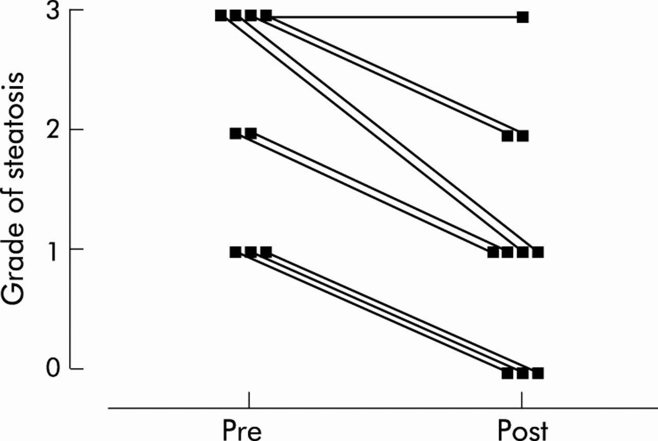
Grade of hepatic steatosis in patients before (Pre) and after (Post) weight reduction (median pre=2; post=1, p<0.005).
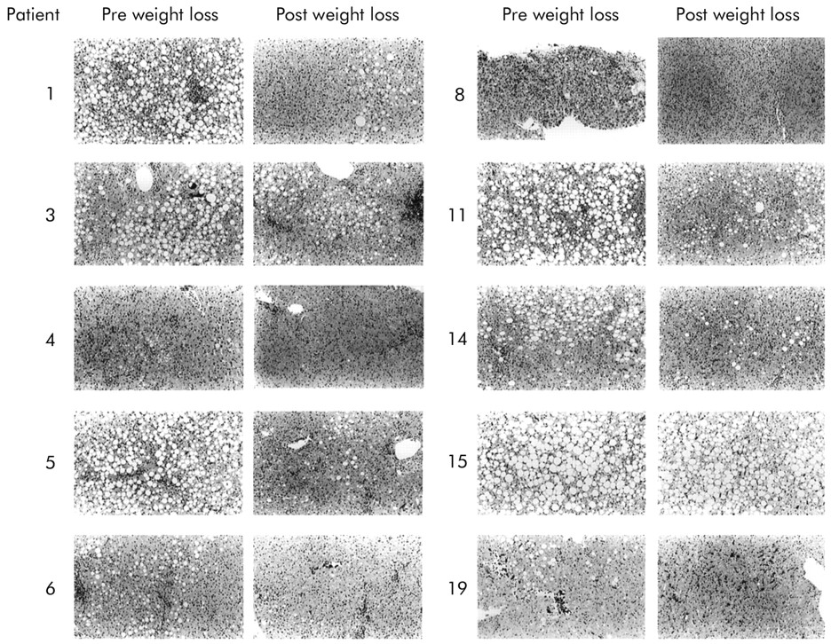
Representative photomicrographs of each paired liver biopsy before (Pre) and after (Post) weight reduction (haematoxylin and eosin; original magnification×100).
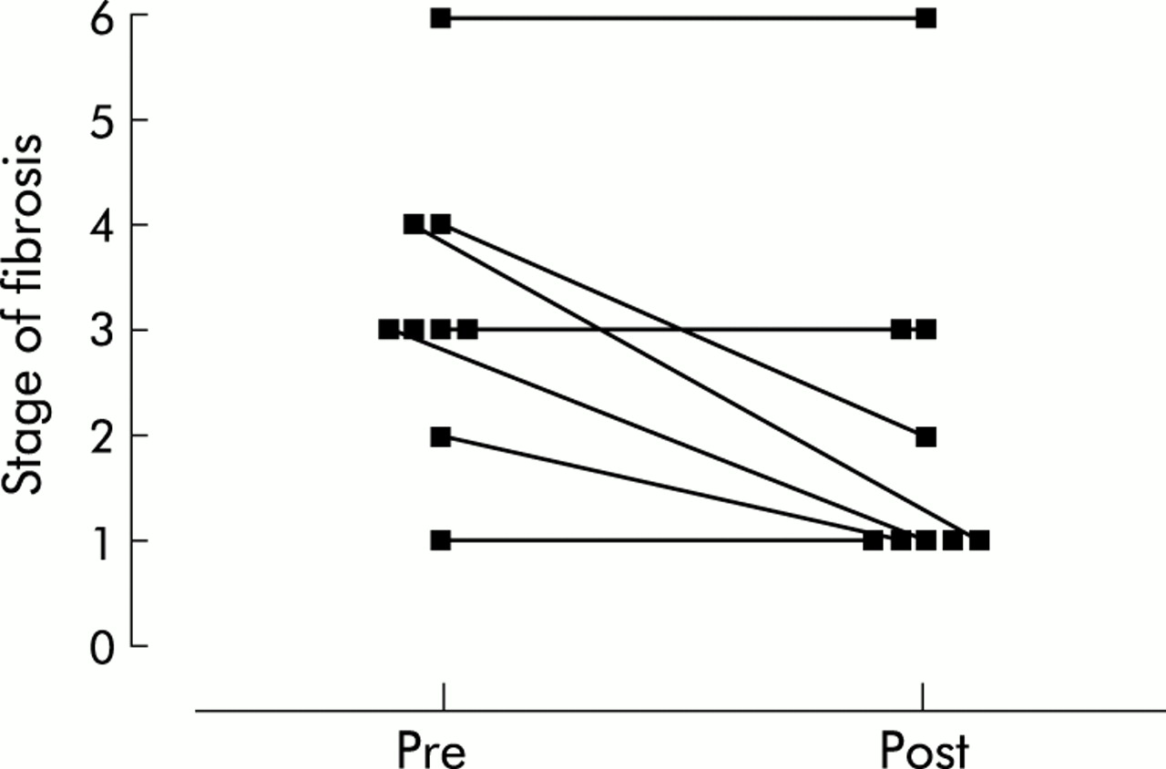
Five of the nine subjects with a reduction in steatosis demonstrated a decrease in fibrosis while in four subjects fibrosis was unchanged (p=0.04, fig 5). In the subject with persistent severe steatosis, fibrosis increased. Deposition of mild fine perisinusoidal collagen in the acini was seen in 9/10 patients initially but disappeared on repeat biopsy in three of these cases. Activated stellate cells, detected by α–SMA staining, were reduced in both portal tracts and acini in 8/10 patients following dietary intervention. The median number of α–SMA positive cells in the portal tracts and acini decreased from 333 (322) to 149 (200) cells/mm2 and 32 (28) to 3 (16) cells/mm2, respectively (p=0.004, table 2).

Modified Knodell fibrosis score in patients before (Pre) and after (Post) weight reduction, in those patients with a reduction in steatosis following weight loss (median pre=3; post=1, p=0.04)
The median histological activity index for all 10 subjects did not significantly decrease (p=0.14) although the reduction in acinar inflammation approached significance (p=0.08, data not shown).
Maintenance of weight loss
To date, eight subjects have had at least six months of follow up after completion of the dietary intervention (range 6–18 months). Three of these subjects have regained >25% of their lost body weight (28%, 48%, and 85%).
DISCUSSION
These results demonstrate for the first time that weight reduction in patients with chronic HCV may reduce hepatic steatosis, irrespective of viral genotype. The reduction in steatosis may be accompanied by a significant decrease in stellate cell activation and in some cases (5/9) regression of hepatic fibrosis was demonstrated.
Most patients with chronic HCV and steatosis were in the overweight or obese weight range (26–35 kg/m2) and had significant visceral adiposity, as demonstrated by an elevated waist circumference (>94 cm in males and >80 cm in females).12, 13 In these patients, loss of as little as 2.6% body weight resulted in a reduction in steatosis, with a significant association between the amount of body weight lost and the degree of change in steatosis. However, at least two patients with HCV genotype 3 were not overweight, suggesting that their steatosis may be due to a cytopathic effect of the virus, as previously described.5, 14–17 Interestingly, even in these lean patients with HCV genotype 3, weight loss was accompanied by a significant reduction in steatosis.
Steatosis was accompanied by mild fine subsinusoidal fibrosis with a chicken wire appearance similar to that seen in steatohepatitis. We have previously shown in chronic HCV that the extent of α-SMA staining (as a marker of stellate cell activation) is correlated with the degree of portal inflammation and the stage of portal fibrosis, but not with the grade of hepatic steatosis.18 In the current study, we again demonstrated a wide variation between individuals in the extent of α-SMA staining. This suggests that in clinical samples (as opposed to rodent models) there may be some dissociation between stellate cell activation and collagen deposition, probably reflecting differences between individuals and also in collagen turnover. However, following weight loss, there was a striking reduction in stellate cell activation in some patients and this was seen in both the acini and portal tracts. Resolution of fibrosis following removal of injurious agents is thought to occur via apoptosis of hepatic stellate cells.19
Although weight loss is recommended in the management of obese patients with fatty liver disease, there has been little evidence to support a beneficial effect on the progression of liver disease. Our study is the first to demonstrate histological improvement following weight loss in patients with fatty liver as a cofactor associated with a primary liver disease. A previous study has shown a decline in ALT and an improvement in symptoms and physical findings following ≥10% weight loss in a small group of overweight patients with chronic hepatitis.20 In a study of obese patients with primary fatty liver disease, repeat liver biopsy after a three month trial of dietary restriction and exercise showed a reduction in steatosis, and a trend towards a reduction in inflammation and fibrosis that was not statistically significant.21
The mechanisms by which weight loss and a reduction in steatosis improve portal inflammation and fibrosis in chronic HCV remain to be determined. Steatosis is associated with an increased production of reactive oxygen species which initiate lipid peroxidation, resulting in activation of hepatic stellate cells. The mechanism for this is not precisely known although recently steatosis has been shown to alter the expression of mitochondrial membrane proteins, including uncoupling protein 2.22 This protein has been implicated in the generation of reactive oxygen species and lipid metabolism.23 Irrespective of its effect on steatosis, energy restriction may also have a direct effect on the inflammatory response associated with HCV. In various rodent models, calorie restriction has been shown to reduce the intensity of inflammation and levels of proinflammatory cytokines.24, 25 In our patients, energy restriction was modest, and the effect of the dietary intervention in the absence of steatosis was not addressed.
Despite the overall decrease in ALT with weight loss, this was not correlated to the reduction in steatosis in our patients. In these patients with steatosis and chronic HCV, fasting serum insulin levels were mildly elevated and significantly associated with BMI. Fasting hyperinsulinaemia and HOMA score are commonly used as surrogate markers for insulin resistance and occur frequently in overweight individuals. It has been suggested that insulin resistance may contribute to hepatic steatosis by a number of mechanisms. In lean individuals, insulin inhibits hormone sensitive lipase, providing control over the release of free fatty acids from adipocytes. In contrast, in obesity the loss of insulin sensitivity at the level of the adipocyte results in increased movement of fatty acids from peripheral stores to the liver. In addition, one of the actions of insulin in the liver is to increase the transcription of genes involved in fatty acid synthesis.26 This stimulation may be responsible for the increase in triglyceride levels in the liver and plasma that accompany hyperinsulinaemia.
Compliance in this cohort of patients was very good, with a mean weight loss of 5.9 kg over a 12 week period. This was achieved by a combination of intensive weekly intervention by a dietitian, restriction in energy intake, and an increase in daily activity. The contribution of each component to the change in liver biochemistry and histology could not be differentiated in our patients but will be of great interest in future studies. Maintenance of this weight loss may be challenging, as in a recent review of long term outcomes after lifestyle interventions alone, individuals regained around 60% of the weight they had originally lost after one year.27 However, our follow up to date has been encouraging, and perhaps the motivation of patients with a chronic disease and no other immediate treatment options may improve compliance with dietary and lifestyle modifications.
This study has demonstrated that weight loss in patients with chronic HCV is achievable and may result in a reduction in hepatic steatosis, irrespective of viral genotype. A small amount of weight loss may be associated with a reduction in abnormal liver enzymes and an improvement in fibrosis, despite the persistence of the virus. We believe that weight reduction may provide an important new adjunct treatment strategy for patients with chronic HCV.
Acknowledgments
The Princess Alexandra Hospital Research and Development Foundation and the Department of Nutrition and Operational Support Services, Princess Alexandra Hospital are gratefully acknowledged.
REFERENCES
Linked Articles
- Commentary