Article Text

Abstract
Aims: As part of the UK antimicrobial resistance strategy and action plan, the Public Health Laboratory Service (PHLS) is required to collect antibiotic susceptibility data so that resistance trends and patterns can be monitored. Most laboratories report urine Gram negative isolates, as “coliforms” according to morphological appearance, but without an acceptable identification system the antimicrobial surveillance data will be meaningless. Commercially available identification systems tend to be expensive and time consuming. Chromogenic agars, which claim to improve the detection of mixed cultures and identification of organisms from urine, have now become available and may provide a cost effective alternative. The primary aim of this study was to compare the performance of cystine lactose electrolyte deficient (CLED) agar with a chromogenic agar (Oxoid urinary tract infection medium; CUTI) in terms of isolation rates and ability to detect mixed cultures. Secondary aims were to evaluate the correlation of “presumptive” identification of isolates from chromogenic media with that of two commercial identification systems and to appraise the sensitivity of the semiquantitative loop and filter paper strip culture techniques.
Method: One thousand, four hundred and sixty six urine samples were examined in four laboratories using the semiquantitative culture methods of 1 μl loop and filter paper strip. The degree of accuracy of organism identification was measured by comparing the presumptive identification using colony colour supplemented with simple bench tests, with identification obtained from two more complex commercial systems.
Results: There was no significant difference between the performance of the loop and filter paper strip methods on the CLED agar, but the CUTI agar performed significantly better than the CLED agar for the detection of significant isolates and mixed cultures. This difference was greater using the loop method. Identification of the organisms using the commercial systems gave > 99% agreement and was therefore considered suitable as a standard against which to compare the presumptive CUTI identification. Using the manufacturer's colony colour criteria in combination with a bench indole test, the CUTI medium was 99% specific for Escherichia coli, although this was reduced to 97% if the indole test was omitted. Citrobacter spp were the most commonly misidentified organisms, giving false presumptive identification as E coli. By testing oxidase activity to differentiate Pseudomonas spp and the absence of indole production to support the identification of Proteus mirabilis, the CUTI medium provided a suitable identification for 86.8% of Gram negative isolates. The remaining 13.2% would require further identification.
Conclusion: CUTI medium improves the detection of mixed cultures, thereby improving the reliability of reporting of significant isolates when compared with CLED agar. When supplemented with simple bench tests it provides an identification system capable of speciating 86.8% of Gram negative isolates and providing a valuable cost effective mechanism for antimicrobial resistance surveillance.
- Gram negative organisms
- identification
- detection
- cystine lactose electrolyte deficient agar
- chromogenic urinary tract infection medium
- BSOP 41, PHLS standard operating procedure
- cfu, colony forming units
- CLED, cystine lactose electrolyte deficient agar
- CUTI, chromogenic urinary tract infection medium
- PHLS, Public Health Laboratory Service
- PYR, l-pyrrolidonyl-β-napthylamide
- UTI, urinary tract infection
- WBC, white blood cell count
Statistics from Altmetric.com
- Gram negative organisms
- identification
- detection
- cystine lactose electrolyte deficient agar
- chromogenic urinary tract infection medium
- BSOP 41, PHLS standard operating procedure
- cfu, colony forming units
- CLED, cystine lactose electrolyte deficient agar
- CUTI, chromogenic urinary tract infection medium
- PHLS, Public Health Laboratory Service
- PYR, l-pyrrolidonyl-β-napthylamide
- UTI, urinary tract infection
- WBC, white blood cell count
Over recent years, the systematic identification of all Enterobacteriacae from urine isolates has been progressively reduced in the UK. Most laboratories now use the generic term “coliform” for Enterobacteriaceae from routine urine samples to improve cost efficiency. As part of the UK antimicrobial resistance strategy and action plan,1 the Public Health Laboratory Service (PHLS) is now required to collect antibiotic susceptibility data to aid the monitoring of antibiotic resistance trends and patterns2 within the UK.
Intrinsic resistance patterns of genera and in some cases species of Enterobacteriaceae vary. Outbreaks of particular Enterobacteriaceae are regularly described in hospital acquired infection and the interpretation of the added burden of extrinsic resistance, as distinct from the selection of genera and species with intrinsic resistance, cannot be assessed without improved identification. In addition, the BSAC sensitivity test method3 sometimes interprets zone sizes differently by genera or species of Enterobacteriaceae.
A completed audit performed by one laboratory within the Midlands group indicated that 60% of all Gram negative laboratory isolates deemed potentially clinically relevant, and reported with antimicrobial susceptibilities, originated from urine samples and that approximately 30% of urine specimens yielded a significant isolate. There are few data on systematic variation of species and antibiotic resistance by anatomical site of infection or the effects of sampling selection for chemotherapeutic failure or recurrent infection. Nonetheless, because they are so common, urine samples would provide a suitable sample population for antibiotic surveillance. Without reliable identification methods, the collection of antibiotic surveillance data would not offer insights into the added burden of intrinsic resistance, the source and origin of resistant clones, or any potential method of intervention.
As described in the PHLS standard operating procedure (BSOP 41),4 acute uncomplicated urinary tract infections (UTIs) are usually caused by a single bacterial species, of which Escherichia coli is the most common.
The confirmation of UTI generally requires the demonstration of significant bacteriuria by culture. Several methods of semiquantifying bacteria in urine have been shown to provide varying degrees of sensitivity. These include the calibrated loop technique, the filter paper strip method, and the use of multipoint technology.5–7 From an ad hoc survey within the PHLS, the use of a 1 μl loop inoculated on to CLED medium is the most widely recognised method, with lower limits of detection of 106 colony forming units (cfu)/litre. However, for ease of use and economy of media utilisation, many laboratories use the inoculated filter paper strip method, which will detect as few as 107 cfu/litre, or multipoint inoculation.
“Without reliable identification methods, the collection of antibiotic surveillance data would not offer insights into the added burden of intrinsic resistance, the source and origin of resistant clones, or any potential method of intervention”
Initial studies by Kass8 suggested that bacterial counts of ≥105 cfu/litre were indicative of infection and that counts below this usually indicated contamination. Conflicting studies have suggested that the quantitative criteria for the definition of significant bacteriuria will be variable for different patient populations and specimen types.9 Similarly, although it is accepted that in most cases a single causative organism will be responsible for infection, the literature search revealed conflicting reports. BSOP 41 recommends the reporting of up to two colony types if pyuria is present,4 and this criterion was adopted for the purpose of this study.
CLED medium, first described by Sandys10 and later modified by Mackey and Sandys,11 is generally used in the UK for diagnostic routine urinary bacteriology as a non-selective medium capable of supporting the growth of most urinary pathogens and giving good colonial differentiation without spread of Proteus spp. A recent development has seen the basal CLED medium being combined with chromogenic substrates to detect the production of β-d-galactosidase, β-glucosidase, and tryptophan deaminase to produce a chromogenic urinary tract infection medium (CUTI).12 It is claimed that when used it will support the presumptive identification of urinary isolates and enable mixed cultures to be detected more easily. Identification on this medium can be supplemented with simple rapid tests such as indole production. If these claims were substantiated, this medium would provide a cost effective mechanism of facilitating the collection of antibiotic surveillance data in the routine diagnostic laboratory.
An evaluation of this medium was undertaken in four laboratories, adopting a standardised protocol to compare the isolation rates and detection of mixed cultures with CLED agar using the 1 μl loop and filter paper strip methods. Correlation of presumptive identification of isolates to identification using commercial kits was used to evaluate specificity.
METHODS AND MATERIALS
Media and reagents
CLED agar (CM301; Oxoid, Basingstoke, UK) and CUTI medium (CM949; Oxoid) were produced according to the manufacturer's instructions. All plates were produced at one site and subjected to full quality control procedures before distribution. All plates were used within 14 days of preparation on the basis of pre-trial shelf life testing, which showed no decline in recovery rates or colony size over this period.
Indole reagent (p-dimethylaminocinnamaldehyde; Sigma, Poole, Dorset, UK) was produced as a 1% wt/vol solution in 10% hydrochloric acid. This was quality controlled using positive control E coli (NCTC 9001) and negative control Enterobacter cloacae (NCTC 11936).
The following reagents were also used: filter paper strips (BTR 1; Mast, Bootle, Merseyside, UK), API 20 E and 20NE (Bio-Merieux, Basingstoke, UK), BBL Crystal (Becton-Dickinson, Oxford, UK), MicrobankTM Cryo beads (PL. 160; Prolab, Wirral, UK), cephalexin 30 μg disks (CL30; Oxoid), and the OBIS PYR test (ID580M; Oxoid). These were all used according to manufacturers' instructions.
Samples
Samples were selected on the basis of a white blood cell count (WBC) of > 100 × 106/litre. A total of 1466 routine samples received during a two week period in January and February 2000, from both hospital and general practice, were included in our study.
Media inoculation and incubation
Using a 1 μl loop, all 1466 samples containing > 100 × 106/litre WBC were inoculated on to ¼ plate CLED and CUTI medium. In addition, all the samples were inoculated on to square CLED and CUTI agar plates using the filter paper strip method. All plates were incubated in air at 37°C ± 1°C for 18–24 hours.
Plate reading
The staff of the laboratories previously inexperienced with the use of CUTI medium were given two weeks of training before the study to ensure familiarity with colony appearances. CLED and CUTI media plates were read independently and by different members of staff.
Data on bacterial growth and significant isolates were recorded at the time of reading and discrepancies verified daily by a separate senior member of staff. Bacterial growth was recorded semiquantitatively on the basis of colony counting. For loop inoculated plates, one colony equated to 106 cfu/litre, increasing to > 108 cfu/litre for more than 100 colonies. For filter paper strip cultures, between one and five colonies equated to 107 cfu/litre, increasing to 108 cfu/litre for 20 colonies.
For the purpose of our study, samples containing no more than two organism types were considered significant. Samples containing three or more organism types were classified as mixed growths and no further investigations were performed.
Isolates from CLED agar were recorded according to generic classification with bench tests performed as applicable—that is, the staphylococcal latex test for coagulase clumping and/or protein A, oxidase, catalase test, and novobiocin sensitivity. Isolates from CUTI agar were recorded according to colour as described in the manufacturers' instructions. As recommended, spot indole using DMACA reagent (Sigma) was performed on all pink colonies.
Significant Gram negative bacilli isolates from the loop inoculated CLED and CUTI medium were saved on to Microbank Cryo beads (Prolab) and stored at −70°C for subsequent identification.
Identification
All Gram negative isolates were identified using two commercial systems, Bio-Merieux API (20E and 20NE) and Becton-Dickinson Crystal (E/NF). Isolates giving discrepant results were submitted to the Identification Services, Central Public Health Laboratory, Colindale, London, for identification by additional methods.
Supplementary tests
All organisms identified as Citrobacter spp and a random 4% selection of E coli were further tested for cephalexin susceptibility and l-pyrrolidonyl-β-napthylamide (PYR) activity. All organisms identified presumptively as Proteus spp were tested for indole production.
Statistical methods
Data were collated centrally and entered into an Excel spreadsheet. Differences between laboratories were compared using the χ2 test and differences between methods using McNemar's test.
RESULTS
Method/media comparisons
One thousand, four hundred and sixty six urine samples were examined from the four laboratories. Statistical analysis indicated no significant difference between the results submitted from the four sites and therefore the amalgamated results are presented here.
Tables 1– 4 and figs 1–4 summarise the isolation results and the detection of mixed cultures.
Isolation rates (%) from 1466 samples using the 1 μl loop method
Isolation rates (%) from 1466 samples using the filter paper strip method
Comparison of isolation rates (%) from 1466 samples using loop inoculated and filter paper strip methods for cystine lactose electrolyte deficient agar
Comparison of isolation rates (%) from 1466 samples using loop inoculated and filter paper strip methods for chromogenic urinary tract infection medium agar
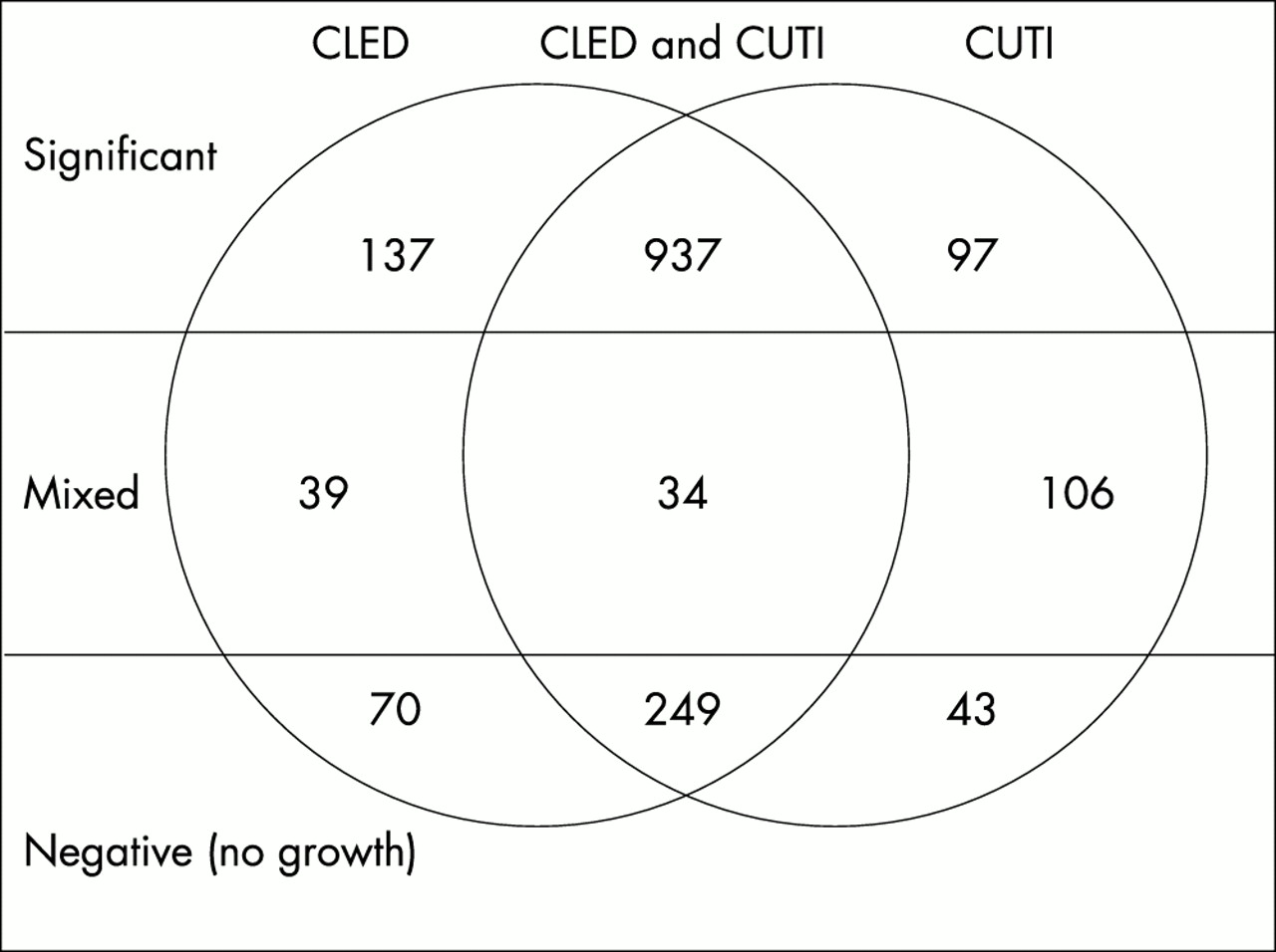
Distribution of isolates using the 1 μl loop method. CLED, cystine lactose electrolyte deficient agar; CUTI, chromogenic urinary tract infection medium.
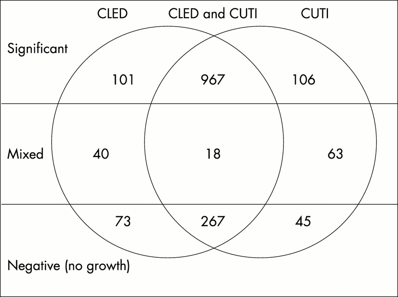
Distribution of isolates using the filter paper strip method. CLED, cystine lactose electrolyte deficient agar; CUTI, chromogenic urinary tract infection medium.

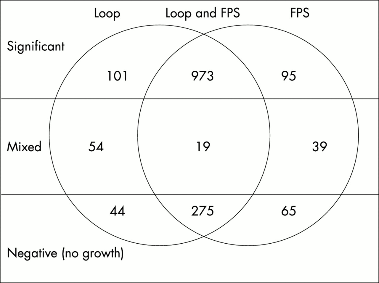
Comparison of isolation rates using loop inoculated and filter paper strip (FPS) methods for cystine lactose electrolyte deficient agar.

Comparison of isolation rates using loop inoculated and filter paper strip (FPS) methods on chromogenic urinary tract infection agar.
The comparison of CLED with CUTI for the detection of mixed cultures showed a highly significant improvement in detection by both the loop (p < 0.001) and filter paper strip (p = 0.030) methods using CUTI medium (tables 1,2; figs 1, 2). It can be seen that for both methods CUTI gave significantly fewer samples with no growth (p = 0.014 and p = 0.013), indicating that CUTI supported the growth of more urinary isolates than CLED.
A direct comparison of the loop and filter paper strip methods (table 4; fig 4) confirmed the superiority of the loop method in detecting mixed cultures on CUTI medium (p < 0.001), but there was no significant difference between the inoculation methods on CLED medium (p = 0.14; table 3; fig 3)
Overall, the CUTI loop inoculated method identified more mixed growths than the filter paper strip methods (p < 0.001) and hence fewer significant isolates (p = 0.012). Fewer plates were recorded as no growth by this method (p = 0.077; table 4; fig 4).
Identification
Using the CUTI loop inoculated results, of the 1466 samples tested, 973 samples (66.4%) gave significant isolates (≤ 2 isolates/sample), resulting in 676 saved Gram negative isolates, with the remaining isolates consisting of a variety of organisms including staphylococci, streptococci, yeasts, and lactobacilli.
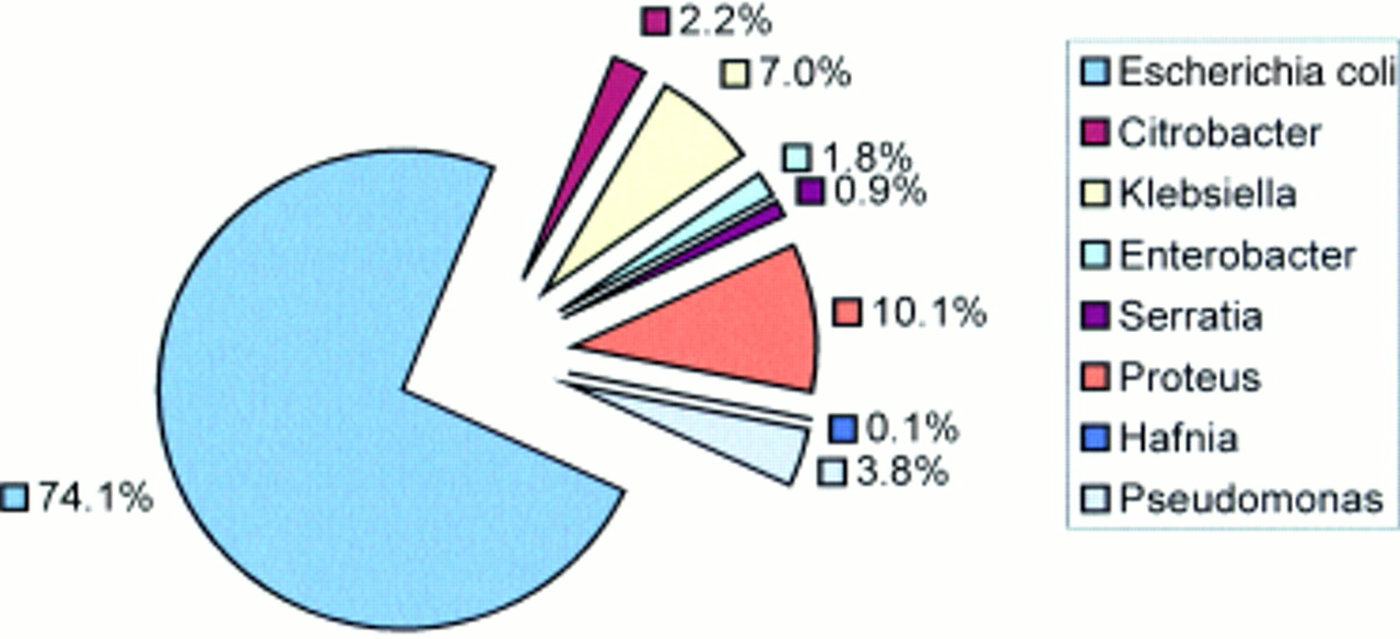
Identification of the Gram negative isolates using Bio-Merieux API 20E/NE and Becton and Dickinson Crystal E/NF gave > 99% (672 of 676) agreement and therefore the amalgamated results are presented here (see fig 5 for a summary identification of isolates).
Identification of isolates from chromogenic urinary tract infection medium.
Using the colour criteria provided by Oxoid, the correlation of presumptive identification from CUTI medium with confirmed identification using API/BBL is summarised in table 5.
Isolate identification from chromogenic urinary tract infection medium using commercial identification system
It can be seen that the CUTI medium in combination with the bench indole test was 99% specific for E coli but this was reduced to 97% if the indole test was omitted because 11 of 15 Citrobacter spp gave false presumptive E coli identification (pink colonies). Although the indole test eliminated all Citrobacter freundii isolates (seven of the 11 Citrobacter spp), it was not sufficient to eliminate the misidentification of Citrobacter koseri. To perform indole tests to eliminate the misidentification of seven of 15 Citrobacter spp would require the testing of all pink colonies (74.3% of all isolates). It might be considered that this specificity gain is not sufficient to warrant the large number of indole tests that would need to be performed.
It was anticipated that the misidentification of Citrobacter spp as E coli would affect the apparent prevalence of cephalosporin resistance as a result of chromosomal β-lactamases, which would affect surveillance data. On the evidence of previous studies, it was expected that 80% of C freundii would be resistant to cephalexin,13 and therefore cephalexin resistance could be used as an alternative marker of a need for further identification. However, supplementary testing of the pink citrobacter isolates (one isolate of C koseri and 10 isolates of C freundii) demonstrated that approximately 50% were resistant (six isolates of C freundii) to cephalexin. These results demonstrate that reliance on cephalexin resistance as a marker in the absence of an indole test would still misidentify half of the citrobacter isolates as E coli.
If cephalexin sensitivity is used to increase the specificity of E coli identification of pink colonies, a PYR test would be required to differentiate the cephalexin resistant isolates as E coli or C freundii. Further testing of the E coli and citrobacter isolates from the study confirmed that all the E coli isolates tested were PYR negative and citrobacter isolates were PYR positive.
Using the PYR test and cephalexin susceptibility, the sensitivity and specificity achieved for E coli identification were 97.2% (487 of 501) and 98.2% (487 of 496), respectively.
The chromogenic medium did not differentiate between isolates presenting as blue/purple colonies. These included species of klebsiella, enterobacter, and serratia and further identification tests would be required.
One hundred per cent sensitivity and specificity for Pseudomonas spp were achieved using the criteria of isolates being colourless and oxidase positive. Ninety four per cent of the brown colonies were species of proteus/morganella. Negative indole tests accurately distinguished Proteus mirabilis, which comprised 2% of the isolates.
Overall, 86.2% of isolates could be identified using the chromogenic medium, supplemented by cephalexin susceptibility and oxidase tests. Table 6 summarises these data. The remaining 13.8% of isolates would require further identification.
CUTI presumptive identification (ID) summary
DISCUSSION
Semiquantitative plating of urine samples on to CLED agar has been an integral part of the microbiological investigation of UTI for many years. Previous comparisons of methods comparing loop inoculation with filter paper strips have indicated that both are suitable as a means of identifying infection. Our study has confirmed that for the detection of significant isolates there was no significant difference between loop and filter paper strip inoculation methods when using CLED agar.
Conversely for CUTI medium, there were significant differences in the detection of mixtures. A higher proportion of significant isolates by the filter paper strip method corresponded to a higher proportion of mixed cultures by the loop method, confirming the superiority of the loop method to detect mixed cultures.
If loop inoculation is used, the impact of changing from CLED to CUTI is to decrease the number of significant isolates from 1074 to 1034 (p = 0.011). This decrease of 40 is the result of an increase in the detection of mixed cultures. If the filter paper strip method is used the impact of changing from CLED to CUTI is to decrease the number of significant isolates from 1073 to 1068, but this result was not significant.
The increased number of mixed cultures detected on CUTI medium was largely accounted for by the presence of Gram positive organisms, especially enterococci. Because the main objective of the research was to validate the performance of CUTI medium for Gram negative identification this was not formally assessed as part of the trial.
“Our study has confirmed that for the detection of significant isolates there was no significant difference between loop and filter paper strip inoculation methods when using cystine lactose electrolyte deficient agar”
Commercial identification systems (Bio-Merieux API 20 E and Becton-Dickinson BBL Crystal) performed equally well for the identification of Gram negative isolates.
Using the supplementary bench tests, 86% of Gram negative isolates could be identified on CUTI medium. The identification of E coli, Pseudomonas spp, and P mirabilis could be confirmed by simple spot tests.
Citrobacter spp were commonly misidentified as E coli and, as described by Chagla et al,14 the bench indole test failed to differentiate the Citrobacter spp from E coli. Improved differentiation between Citrobacter spp and E coli has been demonstrated previously by Chagla et al using a PYR hydrolysis test.14 Supplementary testing of the saved isolates using the Oxoid PYR kit confirmed that all of the Citrobacter spp isolated were PYR positive compared with the negative PYR results obtained from the E coli isolates. However, to undertake a PYR test on all pink colonies would be much more expensive than performing an indole test in a routine laboratory. It had been suggested that by screening for cephalexin susceptibility and performing a PYR test on resistant isolates, the misidentification of Citrobacter spp as E coli would be resolved, but this was found to be only partially correct. Of the 11 pink Citrobacter spp isolated, six were found to be cephalexin resistant and could therefore be correctly identified with a supplementary PYR test. The remaining five cephalexin sensitive, pink Citrobacter spp would be falsely identified as E coli, which resulted in an E coli specificity of 98.2%.
Further work to develop a reliable and inexpensive identification system for the remaining 13.2% of Gram-negative isolates is required.
Take home messages
-
Although more expensive than cystine lactose electrolyte deficient (CLED) medium, Oxoid chromogenic urinary tract infection (CUTI) medium improves the detection of mixed cultures, thereby improving the reliability of reporting of significant isolates
-
In addition, the loop method is superior to the filter paper strip method for detecting mixed cultures
-
When supplemented with simple bench tests, the CUTI loop method provides an identification system capable of speciating 86.8% of Gram negative isolates
-
This method would provide a valuable cost effective mechanism for antimicrobial resistance surveillance
The cost implications of using pre-poured plates of CUTI medium as a direct replacement for CLED medium into a routine laboratory testing 50 000 samples each year would be an additional £3750. However, this needs to be assessed against the cost of undertaking less susceptibility testing (more mixed growths) and the added value of gathering reliable antibiotic surveillance data.
In summary, CUTI medium although more expensive than CLED, improves the detection of mixed cultures and when supplemented with simple bench tests provides an identification system that is capable of speciating 86.8% of Gram negative isolates, facilitating antimicrobial resistance surveillance for E coli and P mirabilis.
Acknowledgments
We thank the staff of the Midlands Public Health Laboratories for their technical support and Oxoid for supplying the media.
Footnotes
-
The authors present this study on behalf of the PHLS (Midlands) bacterial methods evaluation group.