Article Text

Abstract
The designation NESTIN refers to a member of the family of intermediate filaments and comes from the fact that this protein is expressed mainly in neuroepithelial stem cells. However, nestin is not expressed in mature elements and terminal cell differentiation is associated with loss of immunoreactivity to this protein. Therefore, immunohistochemical assessment of nestin expression might be useful to differentiate between mature and immature elements. The aim of this study was to analyse nestin expression in various tumours of neuroectodermal and vascular origin and to determine whether its detection has practical relevance. The results indicate that the immunohistochemical detection of nestin expression could be useful in astrocytomas and malignant melanomas, where it could be used as an auxiliary indicator of dedifferentiation and progression. Because of the weak and heterogeneous expression of nestin in neurinomas, phaeochromocytomas, and carcinoids, nestin detection in these lesions is of little practical use.
- HGA, high grade astrocytoma
- LGA, low grade astrocytoma
Statistics from Altmetric.com
The designation NESTIN refers to a member of the family of intermediate filaments and comes from the fact that this protein is expressed mainly in neuroepithelial stem cells.1–3 However, nestin is not expressed in mature elements and terminal cell differentiation is associated with loss of immunoreactivity to this protein. Therefore, immunohistochemical assessment of nestin expression might be useful to differentiate between mature and immature elements. The aim of our study was to analyse nestin expression in various tumours of neuroectodermal and vascular origin, and to determine whether its detection has practical relevance.
MATERIALS AND METHODS
Formalin fixed, paraffin wax embedded specimens from 30 cases of low grade astrocytoma (LGA) and 40 cases of high grade astrocytoma (HGA) were used (table 1). Moreover, 16 cases of benign melanocytic naevi, nine malignant melanomas, 11 capillary and 12 cavernous haemangiomas, 10 schwannomas, 11 phaeochromocytomas, and nine carcinoid tumours were included. We used an indirect immunohistochemical technique with primary anti-nestin antibody (clone 5326; Chemicon, Chandler’s Ford, Hampshire, UK). The results of staining were assessed semiquantitatively by estimation of the “histoscore” (percentage of positive cells × intensity of staining).
Low grade and high grade astrocytomas
RESULTS
We found significantly higher expression of nestin in the cytoplasm of neoplastic cells in HGA compared with LGA. The mean histoscores were 1.6 in LGA and 3.9 in HGA (p ⩽ 0.001; fig 1). Nestin expression was also seen in tumour endothelial cells, but there was no significant difference between the two grades of tumour. In contrast to benign melanocytic naevi, which were negative for nestin, strong positivity for nestin was seen in all cases of malignant melanoma (p ⩽ 0.0001). We also found nestin expression in endothelial cells and pericytes in capillary haemangiomas; however, these cells were nestin negative in cavernous haemangiomas and also in cavernous parts of capillary haemangiomas (p ⩽ 0.003). The expression of nestin in neurinomas, phaeochromocytomas, and carcinoids was weak and heterogeneous.
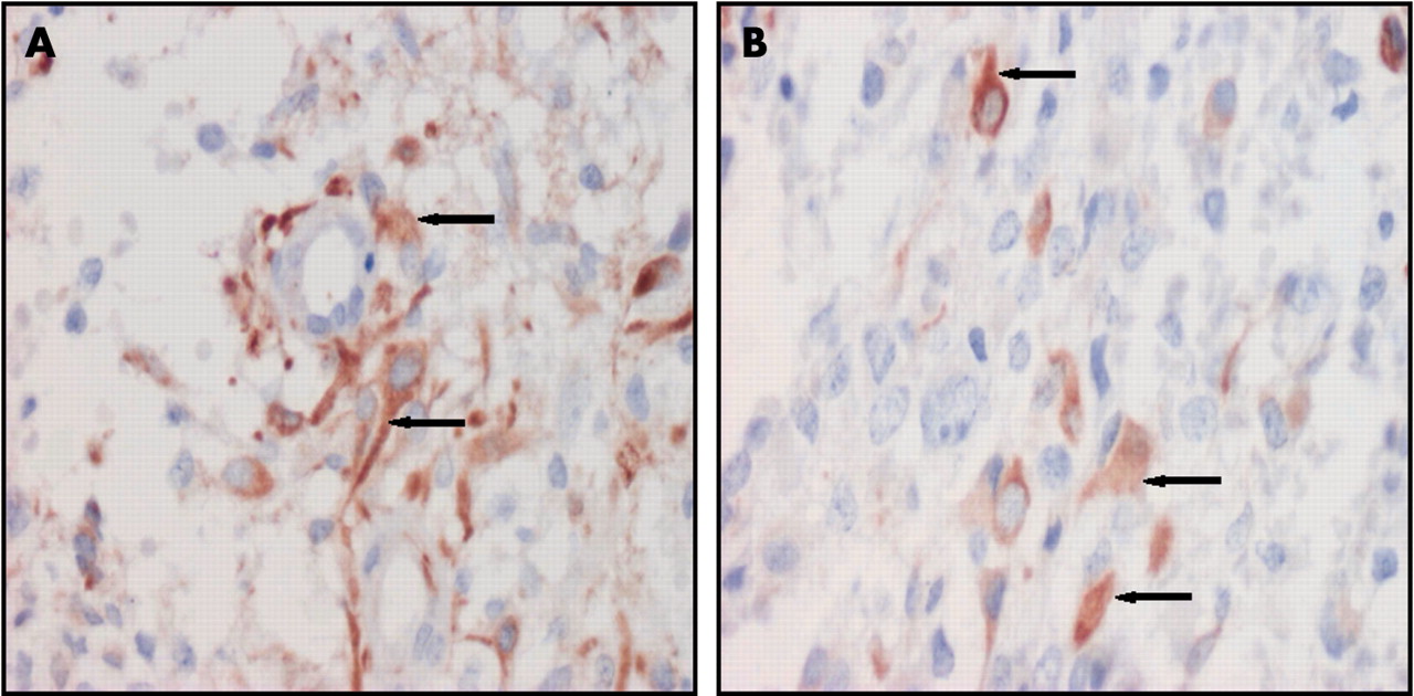
(A, B) Positive nestin expression in the cytoplasm of glioblastoma grade IV tumour cells (arrows).
DISCUSSION
Based on our results, we anticipate that the immunohistochemical detection of nestin expression may be a useful tool for the grading of astroglial tumours.4 The presence of nestin in HGA is probably a sign of immaturity. Similarly, nestin positive cells in LGA may be a sign of later dedifferentiation/progression to HGA.
The pattern of nestin expression in pigmented lesions was similar to that seen in astrogliomas. Nestin positivity was seen in immature malignant cells in malignant melanomas, whereas differentiated melanocytes in benign melanocytic naevi were nestin negative. Therefore, the presence of nestin may indicate dedifferentiation in these tumours also.5
We have no explanation for the difference between nestin expression in capillary and cavernous haemangiomas. It is possible that endothelial cells in cavernous haemangiomas are in a differentiated, mature state and are therefore nestin negative. Obviously, further research is necessary in this field.
Because of the weak and heterogeneous expression of nestin in neurinomas, phaeochromocytomas, and carcinoids, nestin detection in these lesions is of little practical use. Taken together, the results of our study indicate that the immunohistochemical detection of nestin expression has significant value in astrocytomas and malignant melanomas, where it could be used as an auxiliary indicator of dedifferentiation and progression.
Take home messages
-
The immunohistochemical detection of nestin expression could be useful in astrocytomas and malignant melanomas, where it could be used as an auxiliary indicator of dedifferentiation and progression
-
Because of the weak and heterogeneous expression of nestin in neurinomas, phaeochromocytomas, and carcinoids, nestin detection in these lesions is of little practical use
Acknowledgments
This study was supported by grants MSM 1510001 and IGA MZ CR NK/6727-3