Article Text

Abstract
Taking a kidney biopsy is not a trivial procedure. The sample is almost invariably smaller than the pathologist would like. Investigation usually requires division into even smaller samples to permit the application of specialist techniques. In some cases the biopsy is taken not for diagnosis, but to assess the extent of tissue damage. The clinical need is sometimes extremely urgent. These features all underline the crucial importance of collaboration between pathologist and nephrologist if maximum benefit is to be obtained from such very small samples. Consequently, in deciding what to do with a renal biopsy, flexibility and thought are required rather than a single prescribed list of procedures. This article, written after extensive international consultation, represents an attempt to define current best practice in the laboratory handling of renal biopsy specimens, while not neglecting the need to tailor processing to the individual needs of each case.J Clin Pathol
- renal biopsy
- immunofluorescence
- immunohistochemistry
- electron microscopy
Statistics from Altmetric.com
The purpose of a renal biopsy varies with each case, but is likely to include one or all of the following, with varying priority:
-
To establish a tissue diagnosis, or at least to exclude other diagnostic possibilities that could have a similar clinical presentation.
-
To assess the severity and activity of the lesion (“grade”).
-
To assess the amount of irreversible scarring (“stage”).
This variation in clinical needs underlies the requirement for close clinicopathological cooperation in the evaluation of renal biopsy specimens.
Taking the biopsy
In experienced hands, renal biopsy has an extremely low mortality, but it is rarely a painless procedure. The renal cortex is a very vascular tissue; perinephric haematoma is a common consequence and considerable morbidity is not rare. Consequently, renal biopsy is not undertaken lightly, and the subsequent handling and processing of the extremely small specimens should also be done with great care. It is questionable whether renal biopsy should be performed outside specialist renal units where the necessary clinical skills can be acquired and maintained. Similarly, it is questionable whether the histological evaluation of renal biopsies should be undertaken by laboratories on an occasional basis. If a laboratory that is not accustomed to handling renal biopsy specimens is called upon to do so, the pathologists concerned should have no hesitation in seeking advice and assistance from specialist centres.
Biopsies can be taken by nephrologists or radiologists, according to local practice. To ensure that adequate clinical information is available it might be necessary to insist that the nephrologist fills in the biopsy request form before asking the radiologist to perform the biopsy.
Renal biopsies are usually obtained using a Tru-cut needle or spring loaded biopsy gun, under local anaesthetic cover, and with ultrasound guidance. Biopsy is undertaken as a sterile procedure and the person who takes the biopsy should be able to pass the tissue to an assistant for confirmation that renal cortex has been obtained, without compromising sterility.
This assistant will commonly be another physician, a nurse, or a biomedical scientist rather than a histopathologist. Nevertheless, because staff rotate it might on occasion be necessary for a pathologist to train a new member of staff in recognition and division of the sample.
The appropriate size of biopsy needle is contentious1; assessment of what constitutes an “adequate sample” depends heavily on the condition to be sampled, which, of course, is usually unknown. With wider biopsies it might be possible to divide the specimen along its length to provide material for freezing and electron microscopy. This is not possible with an 18 gauge needle, which is a minimum, and at that calibre at least two cores should be requested.
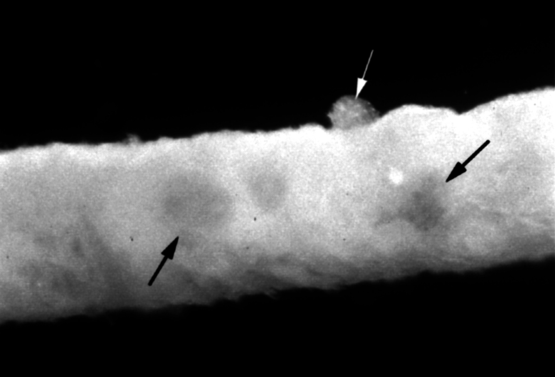
The biopsy should be placed in a drop of saline immediately after removal from the patient and examined under a good dissecting microscope with an adequate light source. With appropriate magnification it is easy to recognise normal renal cortex and to identify glomeruli on the surface (fig 1). This might be more difficult with chronic disease and fibrosis, which might cause the tissue to be uniformly pale, or with acute disease where hypercellular, bloodless glomeruli may take on the appearance of surrounding tubulo-interstitial tissues.

Fresh renal cortical biopsy, taken with an 18 gauge needle, under a dissection microscope. Glomeruli marked by arrows, on the surface (white arrow) and within the biopsy (black arrows).
Some cases of acute renal failure and of suspected allograft rejection require urgent processing. Waiting until the next morning is rarely detrimental, but waiting over a weekend can result in loss of renal function. Therefore, the renal pathology laboratory must have an on call service.
Dividing the biopsy
Division of the biopsy should be done within minutes of removal from the patient so that artefactual ultrastructural changes do not develop. Guidelines for division of the specimen will vary with the type of biopsy, the clinical question being asked, the urgency of that question, and the approach to subsequent investigations taken in the individual laboratory.
In most cases, native kidney biopsies will need to be divided into three parts for conventional light microscopy, electron microscopy, and immunofluorescence studies.
In theory, a single glomerulus each could be adequate for electron microscopy and immunofluorescence studies. Therefore, where possible, the bulk of the specimen should be submitted for light microscopy so that variations in the morphology of disease between glomeruli can best be assessed. However, in practice, handling samples less than 1 mm in diameter can be difficult, especially for immunofluorescence. Therefore, if the needle biopsy contains over 1 cm of cortex it is reasonable to take 1 mm for electron microscopy and 2 or 3 mm of cortex for immunofluorescence, assuming that the presence of at least one glomerulus in each sample can be guaranteed.
If the sample is more limited, division will depend on the clinical question. It might be appropriate to omit the immunofluorescence specimen, especially if immunoperoxidase methods are available in the laboratory concerned. If the sample is tiny and the clinical question is not urgent, it might be appropriate to process the entire sample for electron microscopy. This will permit conventional light microscopy using resin sections and ultrastructural studies of the same material. Only the immunological studies will be lost, and much can be inferred from the distribution of any electron dense deposits. Alternatively, some authorities argue that (in the absence of immunoperoxidase methods) a frozen sample can be reprocessed for light and even electron microscopy. If this is to be attempted, snap freezing in freezing isopentane is probably needed to minimise crystal artefact. However, if the clinical question is urgent there might be a more pressing need for rapid paraffin wax processing.
This need to assess each case on its merits, rather than following rigid rules, highlights the importance of collaboration between renal pathologists and renal physicians. If the biopsy is taken and divided by a radiologist, it is important that the radiologist is aware of the subtleties of the clinical problem and of the potential for pathological investigation.
Handling the unfixed biopsy can result in artefacts, of which the pathologist must be aware. “Holes” can result from the use of synthetic sponges, and bizarre cytoplasmic vacuolation can occur as a result of the accidental substitution of water for saline.
It has recently been observed that if the biopsy needle is washed after the procedure, several glomeruli are often found to be adherent to the needle—even in some cases where renal tissue has not been retrieved. Studying isolated glomeruli is difficult and time consuming, but they can be used for some processes, especially electron microscopy, if more solid tissue is in very limited supply.2
Fixation and processing
The most widely used fixative for light microscopy of renal biopsy specimens is neutral buffered formaldehyde. This has the advantage of being in general use within the laboratory for most specimens. It also permits immunohistochemical studies on the paraffin wax processed specimen. Bouin's fluid, mercury based fixatives, and Karnowsky's fixative have been advocated, and certainly give better preservation of some aspects of morphology, especially the tubules. However, some of these fixatives render subsequent immunohistochemical studies difficult or impossible.
For biopsies where an extremely urgent clinical answer is required, microwave fixation might be of assistance; we have found microwave fixation in isolation to be unsatisfactory, but it does provide a convenient way by which formaldehyde can be heated, resulting in adequate fixation within minutes. Precautions are required to avoid inhalation of formaldehyde vapour.
Paraffin wax processing of renal biopsy specimens can be carried out with the routine diagnostic workload, although a rapid process schedule should be available for urgent specimens.
Several resin impregnation techniques have been advocated for light microscopy of renal biopsies because they permit thinner sections to be cut. This facilitates the use of high magnification, which is commonly necessary in evaluating glomerular disease.3 The images obtained are certainly aesthetically superior, but it can be questioned whether the diagnostic information gained is greatly improved. The approach certainly does not render electron microscopy unnecessary. The disadvantages are potentially increased processing times, additional laboratory staff labour, and sacrifice of the possibility of immunohistochemical studies.
Sectioning and staining
High magnification light microscopy is crucial to evaluating glomerular disease, so thin sections are essential. Thick sections artefactually increase the apparent cellularity of glomeruli and can result in erroneous diagnoses. Given the small size of a needle biopsy specimen, it should be possible to cut paraffin wax sections at 3 μm thickness or less. Thicker sections may be necessary for some stains and investigations, such as Congo red or immunoperoxidase studies.
There seems to be a wide variation in the number of levels at which sections are cut and examined. Again, good practice depends on the nature of the question. With a good sized sample and a straightforward diagnosis, one might be sufficient. If it becomes necessary to identify small segmental lesions (and their location in the glomerulus), serial sections might be needed.
As a minimum, sections should be routinely stained by a technique that highlights cells, such as haematoxylin and eosin, and one that highlights the basement membranes and connective tissue matrix, such as methenamine silver. Some advocate using a periodic acid Schiff (PAS) stain to achieve both goals. This will certainly provide good cellular detail, but the resolution of basement membranes is superior with a methenamine silver stain. Unfortunately, methenamine silver staining is technically more demanding, because the process is analogous to the development of a monochrome film. Overdevelopment or underdevelopment can result in a stain that is of no value. An optimally stained methenamine silver of a normal glomerulus should make peripheral basement membranes appear as thin, waving ribbons that are continuous, but that nevertheless permit the transmission of some light. There has been a fashion in recent years to use haematoxylin and eosin as a counterstain to the methenamine silver stain, rather than the conventional neutral green. This certainly facilitates the examination of the relations between glomerular cells and matrix. Alternatively, a trichrome counterstain gives a pleasing result.
A variety of other special stains might be called for in specific circumstances. A Congo red stain for amyloid is usually a wise precaution whenever there is heavy proteinuria or a systemic disease that predisposes to amyloid; extremely small quantities of amyloid that are quite undetectable on haematoxylin and eosin can have a devastating functional effect on the glomerular basement membrane. An elastin stain might be of assistance in evaluating vascular disease, but rarely adds further information to that available from a good methenamine silver. A variety of trichrome stains have been used in the past to delineate glomerular deposits of immunoglobulins and fibrin, but have been largely superseded by immunohistochemical studies.
In renal transplant pathology, the development of the Banff classification4,5 has laid great emphasis on the use of the PAS stain to delineate tubular basement membranes and to identify tubulitis, the principal marker of acute rejection. For this purpose PAS staining is certainly equal to methenamine silver, and is technically easier. However, pathologists and technical staff should be aware that to demonstrate basement membranes clearly in thin sections, a PAS stain optimised for detecting mucus or fungi is insufficient. The times in periodic acid and in Schiff's reagent should both be doubled,6 to 10 and 20 minutes, respectively, with fresh solutions prepared as needed.
Electron microscopy
Electron microscopy has become less used in recent years in many aspects of diagnostic pathology, but has retained its importance in renal pathology. A recent questionnaire survey indicated that 60% of UK laboratories handling renal biopsies perform ultrastructural studies on all renal biopsies where appropriate material is available.7 It can be argued that this is a luxury, and that it is possible in a proportion of cases to identify the pathological process from light microscopy alone with sufficient confidence to permit the omission of electron microscopy. This approach is taken by most of the remaining laboratories. This is a question of balancing the use of resources with the benefits obtained. Electron microscopy will occasionally produce unexpected second diagnoses. It may refine apparently obvious diagnoses; for example, the finding of mesangial electron dense deposits in a case of membranous glomerulonephritis suggests that the glomerulonephritis is secondary, probably to lupus, rather than idiopathic. It might also detect unsuspected method failures, as when electron microscopy shows electron dense deposits in a case with apparently negative immunohistochemical results. The balance will be influenced by the opinion and experience of the pathologist and the available resources.
In renal transplant pathology, electron microscopy has frequently been omitted unless there is a suspicion of recurrent or de novo glomerular disease. However, there is evidence that ultrastructural examination of the basement membranes of peritubular capillaries for splitting gives a relatively specific marker of chronic rejection,8 so this might change in the future.
Fixation of electron microscopy specimens conventionally uses glutaraldehyde, or a glutaraldehyde/formaldehyde mixture such as Karnowsky's fixative, with postfixation using osmium tetroxide. Processing is conventionally into an epoxy resin. Semi-thin sections for block selection are stained using toluidine blue; this is frequently a neglected resource, because light microscopy of these sections can provide much information about the underlying disease process. Ultrathin sections are stained with uranyl acetate and lead citrate9 before examination under the electron microscope.
In cases of haematuria, ultrastructural assessment should include measurement of basement membrane thickness, to exclude thin membrane nephropathy. Accurate morphometric assessment of basement membrane thickness is labourious and is probably beyond the resources of most units. However, a more limited measurement of basement membrane thickness can yield useful information, especially if the approach is standardised for each case.10 Basement membrane thickness seems to be influenced heavily by fixation and processing as well as measurement schedules, so each electron microscopy unit needs to assess its own “normal range” for this measurement.
In specialist units, some variations on this approach may be undertaken, especially if immunoelectron microscopy is to be performed. This typically requires fixation with freshly prepared paraformaldehyde because glutaraldehyde destroys the antigenicity of most epitopes. Processing is usually into a proprietary resin with low temperature polymerisation. This approach permits immunoelectron microscopy for a restricted range of antigens and might, if carefully performed, cause little loss in ultrastructural morphology.11 Nevertheless, it is not an approach that is widely taken.
In institutions that regard themselves as centres of excellence, it seems reasonable to argue that electron microscopy should be performed on all biopsies of native kidney. In a review of laboratory practice in renal pathology, many respondents expressed the opinion that to carry out evaluation of renal biopsy specimens without at least having the availability of electron microscopy is negligent.7 As a minimum, electron microscopy must be performed where light microscopy does not provide a diagnosis that adequately explains the observed clinical features. Ideally, the renal pathologist should examine the specimen directly, but samples for electron microscopy can be sent by post if the laboratory does not have its own facilities; photographs taken by a suitably trained laboratory scientist are usually adequate. If an appropriately fixed sample is not available, useful results can often be obtained by reprocessing material out of a paraffin wax block, or from a frozen sample. Cellular detail is largely destroyed, but basement membranes and electron dense deposits are surprisingly well preserved.
Immunohistochemistry
Immunohistochemical investigations should be a routine part of the investigation of native renal biopsies. A basic panel of antibodies should be used for the detection of tissue deposits of IgG, IgA, IgM, and complement (usually C3). This represents a minimum. Additional antibodies can be used at the pathologist's discretion. Widely used antibodies include those directed against:
-
C4 and/or C1q: to indicate classical pathway complement activation.
-
C5b–9: to detect membrane attack complex deposition.
-
Fibrin: to detect fibrinoid necrosis in vessels and glomeruli, and fibrin in crescents.
-
Both κ and λ light chains: not only for myeloma kidney, but also the more subtle changes of light chain nephropathy.
The list of antibodies that might be used in special cases is extensive. A selection includes those directed against:
-
Specific amyloid types.
-
Specific α chains of type 4 collagen (Alport's syndrome).
-
Fibronectin (fibronectin glomerulopathy).
-
Type 3 collagen (collagenous glomerulopathy).
-
Viral antibodies.
-
Tamm-Horsfall protein.
METHODS OF DETECTION
The detection of immunoglobulins in glomeruli is rendered difficult by the presence of abundant plasma proteins, which must be removed to avoid non-specific staining. This has resulted in a continuing popularity of frozen sections and immunofluorescence techniques in renal pathology, where they have been superseded in most other branches of immunohistochemistry by methods that are applicable to paraffin wax sections. In paraffin wax sections, the plasma proteins have been fixed in the tissues and must be removed by a digestion process rather than simple washing.
Immunofluorescence
This approach has the advantage of simplicity and reliability because plasma proteins can be removed from frozen sections simply by washing. The essential panel of antibodies against immunoglobulins and complement can be used with a direct immunofluorescence method, which is extremely simple and quick. In some hospitals this investigation is carried out in the immunology or microbiology department. There can be no justification for this practice. The reason is purely historical; the method is not difficult and it is surely preferable for the morphology of the immunofluorescence sections to be read by the person who is reporting the rest of the morphological appearances.
Immunofluorescence has several disadvantages. A separate frozen specimen must be taken at the time of biopsy. A cryostat and epifluorescence microscope are required. The preparations must be mounted in aqueous media and they are not permanent; exposure to light causes bleaching. This can be reduced but not eliminated by using specialist mounting media and storing the sections in the dark in a refrigerator. Ideally, relevant images should be stored, photographically or digitally.
IMMUNOHISTOCHEMISTRY USING PARAFFIN WAX SECTIONS
A detection method applied to sections from the block that is re-used for routine microscopy has obvious advantages; it allows correlation with morphology and results in a permanent preparation that does not require special equipment or a separate sample for its production or examination. However, immunofluorescence remains the method of choice in 83% of UK laboratories because of the technical difficulties of the alternative methods.7
Renal tissue contains large quantities of alkaline phosphatase and peroxidase; to block peroxidase completely is easier than blocking alkaline phosphatase, and most laboratories choose a peroxidase based detection method.
After formaldehyde fixation, proteolytic digestion is essential to remove plasma proteins and to “unmask” epitopes in glomerular immunoglobulin deposits. Unfortunately, excessive digestion will remove deposits as well as plasma. Different biopsies seem to require different digestion times, even if they are fixed and processed to a standardised protocol. Three approaches have been taken to overcome this.
-
Trial and error: for each biopsy, sections are incubated with a proteolytic enzyme (usually trypsin) for variable periods. They are all then stained with a single antibody (anti-albumin or anti-IgG). The trypsinisation time that is just long enough to remove plasma from capillary loops is then selected for a subsequent run using all the antibodies required.7
-
Direct observation: under phase contrast microscopy, it is possible (with suitable experience) to see directly when plasma has been completely digested from the glomerular capillary loops.12
-
Washing: it is reported that washing the biopsy in saline or tissue culture medium for one hour before fixation and paraffin wax processing makes digestion of plasma from the capillary loops much easier and less crucial. This approach has the disadvantage that if material is subsequently reprocessed from the paraffin wax block for electron microscopy, the morphology will be more severely disrupted.13
All these approaches require that all the sections used are cut to a consistent thickness; 3 μm is commonly used.
With renal transplant biopsies, immunohistochemical studies are usually omitted, but are mandatory where there are any unusual features or if there is suspicion of recurrent or de novo glomerular disease. Immunohistochemistry is of surprisingly low sensitivity in the detection of antibody mediated rejection.
Reporting
In renal pathology, interpretation of the morphological appearances often requires detailed clinical information, and it mighty not be obvious to the pathologist exactly what sort of information the clinician requires. For this reason, it is important that the pathologist should discuss renal biopsies with the clinical team using a multiheaded microscope or video microscope system.
Because the special investigations might take some time, it is often appropriate to issue a preliminary report.
A final renal biopsy report should typically contain the following components.
-
Identification of material submitted. Proportion of cortex, proportion of medulla, and other tissues included. Can juxtamedullary cortex be specifically identified? How many glomeruli are available? This is best expressed as “at least”, meaning the largest number seen in any single section.
-
Changes in glomeruli. Cellularity, sclerosis, necrosis, crescents, numbers of glomeruli affected, segmental, or global changes. With focal changes, the proportion of glomeruli affected should be indicated.
-
Tubules. Evidence of acute damage, extent of atrophy (as an estimated percentage), casts, crystals, inclusions, etc.
-
Interstitium. Lymphocytic infiltrates, fibrosis, patchy, or diffuse changes. An estimate of these features often gives a more meaningful assessment of “activity” and “chronicity” than the glomerular changes.
-
Blood vessels. Number and size present; any age related changes, any acute changes.
-
Immunohistochemical results.
-
Electron microscopic results.
-
Conclusions.
In drawing conclusions, it might be necessary to provide a differential diagnosis, with arguments for and against each possibility. In this case, it is usually helpful to give some estimate of the pathologist's view of the probability of each diagnosis proffered.
The nomenclature of renal disease contains some terms that are purely descriptive (for example, focal segmental proliferative glomerulonephritis) and terms that, as far as present understanding permits, are thought to represent specific disease processes (for example, IgA nephropathy). As far as possible, the distinction should be made clear—for example: “The pattern is of a focal segmental proliferative glomerulonephritis. Special investigations show that this is the result of IgA nephropathy.”
It is usually helpful to give an estimate of the activity and chronicity (grade and stage, respectively); specific treatments for renal disease are still relatively few and clinically the most important part of the report is often an estimate of the reversibility of the lesion rather than a specific diagnosis.
Classification systems have been devised for use in some renal diseases, but these should be used with care. The problems with such schemes are well illustrated by the WHO classification for lupus nephritis. This followed several earlier contentious attempts, and the WHO classification itself has been through three revisions, the most recent in 1995.14 All are still in use, usually without an explanation of which is being used. Furthermore, interobserver variation in the application of the WHO classification is considerable. The use of such schemes at the end of a report might be helpful, and should certainly be included if the nephrologists request it, but it is essential to be sure that the recipient understands the meaning. They are not a substitute for careful description. The best route to conveying the severity of a continuous variable such as inflammation or fibrosis is to discuss the images with the responsible clinicians over a video microscope.
For renal transplant biopsies, the pattern of reporting should be essentially the same, although immunohistochemistry and electron microscopy are commonly omitted, especially in grafts under six months of age, unless there is a suspicion of recurrent or de novo glomerular disease. Electron microscopy has been advocated for the detection of peritubular capillary basement membrane splitting in chronic rejection, but its use is not yet widespread. The conclusion of a transplant biopsy report should probably include the Banff classification of renal transplant pathology, or at least use terms (such as a specific grade of tubulitis) that permit translation to a specific Banff category. However, as noted above, there is interobserver variation in the application of the classification, and two slightly different versions (Banff 93–954 and Banff 975) are in existence. In biopsies taken for the diagnosis or exclusion of acute rejection, it is often helpful if the pathologist also integrates the morphological appearances with the clinical information provided, and suggests whether treatment for an episode of acute rejection is likely to be justified or not.
The standardised definitions of lesion “scoring” provided in the Banff classification are also available on the Internet at: http://tpis.upmc.edu/ under “schema”.
Acknowledgments
A draft version of this broadsheet was made available on the World Wide Web, and comments were invited from renal pathologists who subscribe to the Internet mailing list “Nephrol”. I am very grateful to the many pathologists who added constructive comments, many of which are incorporated into the present version.