Article Text

Statistics from Altmetric.com
Adenomatoid tumours are uncommon tumours, which were first described by Golden and Ash.1 They occur most often in the male and female genital tracts, but have rarely been reported at other sites such as the omentum,2 pleura,3 heart,4 small bowel mesentery5 and adrenal gland.6
We report multiple adenomatoid tumours involving the peritoneum and liver. Our diagnosis was based on morphological examination, and was supported by histochemical, immunohistochemical and ultrastructural examinations.
Materials and methods
Tissue was processed for frozen sections using a cryostat, and sections were stained with H&E.
Formalin-fixed paraffin wax sections of each tumour were stained with periodic acid-Schiff after diastase predigestion, in addition to H&E staining. Immunohistochemical staining for cytokeratins (CK) 5, 6, 8, 17 and 19 (clone MNF116, DAKO cytomation), CK5/6 (clone D5/16B4, DAKO cytomation), epithelial membrane antigen (EMA; clone E29, DAKO cytomation), epithelial antigen (clone BEREP4, DAKO cytomation), calretinin (clone DAK-calret DAKO cytomation), mesothelial cell (clone human bone marrow endothelial (HBME)-1 DAKO cytomation), CD31 (DAKO) and CD34 (DAKO) was performed using an automated immunostainer (DAKO Autostainer, Ely, UK) and a labelled streptavidin–biotin method.
Electron microscopy was performed on tissue retrieved from paraffin-wax-embedded blocks, post-fixed in osmium tetroxide, and stained with uranyl acetate and lead citrate. Microscopy was performed on 80–90 nm sections using a Hitachi H600 transmission electron microscope (Hitachi, Wokingham, UK).
Results
A 74-year-old woman presented with coffee ground vomiting associated with malaena. She also had a history of Helicobacter pylori infection and eradication therapy, glaucoma and hypertension. There was no history of previous surgery. Her medication consisted of a proton pump inhibitor, a β-blocker and eye drops for glaucoma. There was no relevant family history.
An oesophago-gastro-duodenoscopy showed a chronic ulcer involving the lesser curve of the stomach and a positive campylobacter-like organism test. A biopsy specimen was taken, which showed the features of a gastric adenocarcinoma. A staging CT scan of the thorax and abdomen showed a T2/T3 lesion, with no radiological evidence of local invasion or metastasis.
Staging laparoscopy was performed, which was followed 4 weeks later by a radical D2 gastrectomy with end-to-side Roux-en Y oesophago-jejunoscopy reconstruction. The patient made a satisfactory and uneventful recovery, and she was discharged 3 weeks postoperatively.
The peritoneal lesions sampled during the laparoscopic staging procedure were located on the left side, below the dome of the diaphragm. They appeared pale and well circumscribed, and each measured 0.2 cm in diameter. They were sent for frozen section examination, with subsequent examination of paraffin wax sections.
Frozen section examination showed both the lesions to have an angiomatoid architecture, consisting of tubular channels lined by epithelioid and flattened cells, set in a fibrous stroma. Many cells had vacuolated cytoplasm and a signet-ring morphology, and these features were suspected to indicate adenocarcinoma, but paraffin wax section examination was required for a definitive diagnosis.
Further examination of H&E-stained paraffin wax sections showed similar features as those noted on frozen section examination (fig 1A), but there was a distinct lack of cytological atypia, and mitotic figures were not identified. These lesions appeared benign, and the possibility of them being of mesothelial derivation was considered. Immunohistochemical examination was requested, including a panel of mesothelial markers. Unfortunately, the material available was insufficient to perform the full set of markers on one of the peritoneal lesions. The morphological features, supported by immunohistochemistry, were diagnostic of adenomatoid tumours, and the possibility of them representing metastatic adenocarcinoma was therefore excluded.

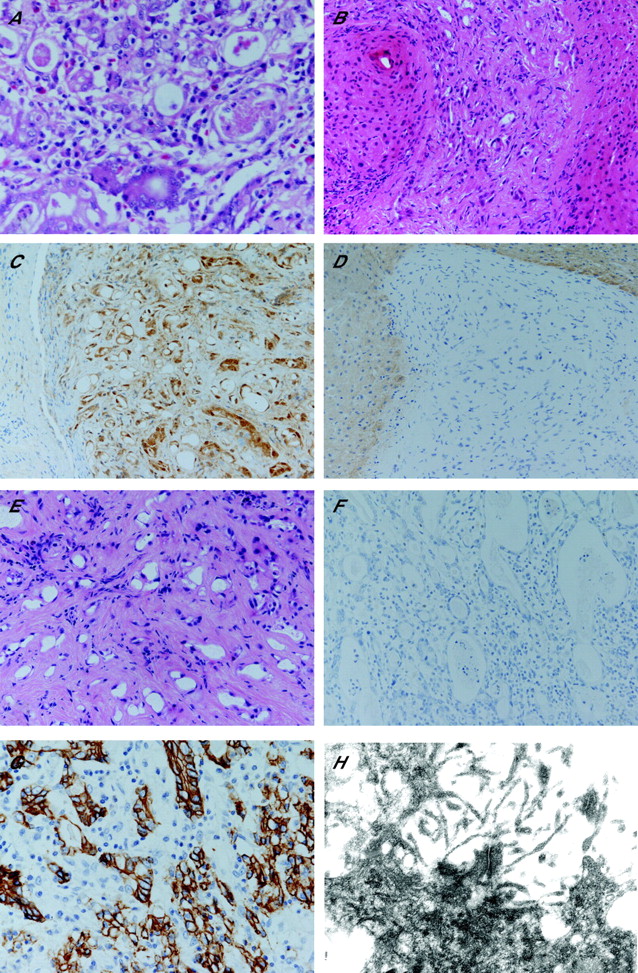
(A) H&E adenomatoid tumour peritoneum; (B) H&E adenomatoid tumour liver; (C) adenomatoid tumour from liver showing expression for calretinin; (D) adenomatoid tumour from liver showing no expression for BerEP4; (E) H&E gastric adenocarcinoma, note the similarity in the architecture to adenomatoid tumour; (F) gastric adenocarcinoma showing no expression for calretinin; (G) gastric adenocarcinoma showing expression for BerEP4; and (H) electron microscopy showing microvilli, and adenomatoid tumour from liver (×30 000).
The surgical team were now confident to proceed to gastrectomy, at which time a further lesion was detected, involving the surface of the posterior aspect of the left lobe of the liver. It appeared pale yellow and well circumscribed, similar to the peritoneal lesions, and measured 0.5 cm in diameter. On frozen section examination, it appeared unencapsulated, with a non-infiltrating and well-circumscribed margin, and with a morphology similar to that seen in the peritoneal lesions. Its circumscription, together with a lack of cytological atypia, led to an opinion that it was not malignant.
The architecture was angiomatoid, raising the possibility of it representing a vascular neoplasm, which was probably benign owing to a lack of cytological atypia (fig 1B). On frozen section examination, it was stated that the lesion appeared unusual, and hence paraffin wax sections were required to make a confident diagnosis.
The liver lesion had a histochemical and immunohistochemical profile identical to that of the peritoneal lesions, and the features were thought to be classic of an adenomatoid tumour (fig 1C,D).
Examination of the gastric tumour (fig 1E) showed a well-moderately differentiated adenocarcinoma, with focal penetration into the submucosa (stage pT1pN0pMx). Interestingly, the morphology of this tumour showed many similarities to the adenomatoid tumours from the peritoneum and liver. There was no evidence of lymphovascular space invasion, and none of the lymph nodes sampled showed any evidence of metastatic disease. A similar panel of immunohistochemical markers was requested (fig 1F,G). Table 1 summarises the histochemical and immunohistochemical findings on examination of paraffin-wax-embedded sections.
Histochemical and immunohistochemical findings of adenomatoid tumours from the peritoneum and liver, and of gastric adenocarcinoma
Ultrastructural examination performed on the liver lesion showed prominent desmosomes, long microvilli on the luminal surface of tumour acini, and a collagenous stroma, typical of an adenomatoid tumour7 (fig 1H).
Discussion
Following the first descriptions of adenomatoid tumours in the literature,1 there was much debate regarding the cell type of origin, and the candidate cell has been proposed by different authors to be of mesothelial, mesonephric, müllerian and endothelial origin.7 Adenomatoid tumours have subsequently been shown to consistently demonstrate mesothelial differentiation, following detailed histochemical, immunohistochemical and ultrastructural investigation, and therefore this benign tumour is now thought to be of mesothelial derivation.8 They have most often been described in the genital region, particularly in the epididymis, Fallopian tube and uterus,7,9,10 but are also described at other sites such as the omentum,2 pleura,3 small bowel mesentery8 and adrenal gland.6 The reason for an apparent predominance in the genital tract compared with other mesothelial locations has not been explained. These tumours are classified as benign, owing to their indolent behaviour and lack of metastasis. They are usually solitary, and are most often discovered as incidental findings during radiological examination, surgery or autopsy.3 Few case reports describe multiple adenomatoid tumours in individual patients.11–13 None of these reports involve adenomatoid tumours occurring exclusively outside the genital tract, but one report does include an adenomatoid tumour of the appendix.12
We consider the diagnosis of adenomatoid tumour to be potentially problematic, especially when the peritoneum or liver is involved. Adenomatoid tumours have a distinct but varying morphology. Acinar, angiomatoid, cystic and solid histological patterns have been described, and many tumours also show a mixture of patterns.6 All of the lesions which we describe showed acinar and angiomatoid architectural features, and they contained cells with a signet-ring morphology, resembling metastatic well-differentiated adenocarcinoma. Tumours were however well circumscribed, with no significant pleomorphism. Histochemical analysis of these tumours showed a lack of mucin, a feature aiding distinction from metastatic adenocarcinoma. The immunohistochemical profile was identical to that reported by other authors, who have described expression of adenomatoid tumours for broad-spectrum CKs, EMA, CK5/6, calretinin and HBME-1, and an absence of expression for BEREP4, CD31 and CD34.3,4,6,7 We detected a similar pattern of membranous staining for HBME-1, and membranous and cytoplasmic staining for EMA, as described by other authors.7 Ultrastructural examination of the liver lesion showed a collagenous stroma, prominent desmosomes and long microvilli on the luminal surface of tumour acini. These are the typical features of an adenomatoid tumour, as described by Dalahunt.7
The adenomatoid tumour of the liver that we report was seen to be embedded in the hepatic parenchyma. This should not be interpreted as a sinister feature, as adenomatoid tumours have been described to be similarly embedded in the heart,4 adrenal gland6 and uterus,12 even showing an infiltrating pattern at these sites.4,6
The angiomatoid architecture of the liver lesion on frozen section examination led to the suggestion that it represented a haemangioma. Immunohistochemistry was important in excluding this possibility, as only small capillaries showed expression of the vascular markers CD31 and CD34, and there was an absence of expression within lesional cells surrounding the tubular structures.
The occurrence of adenocarcinoma of the stomach, together with multiple adenomatoid tumours, is considered likely to be coincidental. The distinction from metastatic adenocarcinoma was important, and obviously a potential pitfall, as the patient was in the process of staging and treatment for a gastric adenocarcinoma. A mistaken diagnosis of metastatic adenocarcinoma would almost certainly have denied the patient surgery, which is potentially curative.
Considering the rarity of multiple adenomatoid tumours in the peritoneum and liver, in normal circumstances we would consider signet-ring-type cells in these locations in a patient with adenocarcinoma to be highly suggestive of metastasis. It is, however, important to be aware of other tumours such as lymphoma, malignant melanoma, myeloma and epithelioid haemangioma,14 in addition to adenomatoid tumour, all of which can have a signet-ring morphology.
A lack of cytological atypia does not necessarily help in distinguishing adenomatoid tumour from metastatic signet-ring-type adenocarcinoma, as the latter often appears non-infiltrative and cytologically bland. We consider the claw-like, angiomatoid architecture accompanying the signet ring cells of adenomatoid tumour to be a helpful pointer against metastasis. In such circumstances, it is not possible to make a definitive diagnosis at frozen section examination, or indeed on H&E-stained paraffin wax sections, as histochemical and immunohistochemical analyses of paraffin wax sections would be required to make an accurate diagnosis. Immunohistochemistry for calretinin, HBME-1 and BEREP4 easily distinguishes adenomatoid tumour from adenocarcinoma15 (table 1).
Immunohistochemical and ultrastructural examinations were helpful in establishing a mesothelial nature for these lesions. Once the mesothelial nature of the lesions was established, they had to be distinguished from mesothelial hyperplasia and malignant mesothelioma. We are uncertain whether or not the adenomatoid tumours that we describe are related in some way to each other. The frequent coexistence of chronic inflammation and fibrosis with adenomatoid tumours has suggested to some authors that, in at least some cases, they may represent a peculiar form of nodular mesothelial hyperplasia.14 Indeed, the multifocal distribution of our tumours could lend some support for this theory. Mesothelial hyperplasia has been reported to be associated with a form of insult to the peritoneum, such as a hernia, ectopic tubal pregnancy, cirrhosis and abdominal tuberculosis.5,16
We consider that these lesions are neoplastic rather than hyperplastic in nature, as they were nodular, with absence of any associated inflammation. They were also noted to have an angiomatoid and acinar architecture, characteristic of adenomatoid tumour, rather than a papillary architecture often associated with reactive mesothelial hyperplasia.2 The unlikeliness of these lesions being hyperplastic is supported by the absence of any known insult to the peritoneum and liver. In addition, the liver and peritoneum separate from the tumours appeared normal.
The differential diagnosis of distinct mesothelial tumours within the liver and peritoneum should also include well-differentiated papillary mesothelioma.2,4 Reported cases have been seen mostly in the peritoneum of young women, and they are thought to represent a rare variant of mesothelioma with a low malignant potential. Histologically, they have a characteristic appearance, containing papillary formations with thin fibrovascular cores, lined by mesothelial cells exhibiting a minimal degree of cytological atypia. A further differential diagnosis is localised malignant mesothelioma, which is extremely rare and histologically identical to diffuse mesotheliomas.18–20 We excluded these possibilities on morphological grounds, as our tumours did not have a papillary architecture and demonstrated a distinct lack of cytological atypia, with no evidence of necrosis.
Localised benign mesothelial tumours within the liver are extremely rare. On review of the literature, we found a single case of a benign cystic mesothelioma.21 These tumours have also been described at sites outside the liver, and they occur infrequently following castration or menopause, which suggests a degree of hormonal independence. They are thought to be related to adenomatoid tumours, but are, however, dissimilar with regard to their presentation and pathological features. We report tumours in an elderly woman, which are negative for oestrogen receptor and are therefore unlikely to be hormonally responsive. The liver lesion described by Fleming, despite its mesothelial nature, does not resemble adenomatoid tumour, as it was large and partially cystic, measuring 8 cm in diameter and histologically highly vascular, containing cords of tumour cells with a hobnail appearance, separated by medium and large-sized vessels. It showed expression of markers HBME-1, calretinin and CK 5/6, confirming its mesothelial origin.
We do not advocate the use of histochemical, immunohistochemical analyses and electron microscopy in the investigation of all biopsy specimens of peritoneal nodules having signet-ring-type cells. If a suspicion of adenomatoid tumour is raised on morphological examination, then a panel consisting of epithelial, mesothelial and vascular markers is probably sufficient for diagnosis. In addition, ultrastructural examination is not necessary to routinely diagnose adenomatoid tumours, It was performed for our interest, as multiple adenomatoid tumours within the peritoneum and liver have not been described previously, and we therefore wished to confirm that the ultrastructural features are identical to those reported by other investigators.
The infrequent reporting of adenomatoid tumours involving mesothelial surfaces, despite the common frequency of abdominal surgery, is surprising, and this is probably an indication of their rarity, but it is possible that they may go undetected. The immunohistochemical and ultrastructural features are consistent with a mesothelial derivation, and this provides further evidence supporting the mesothelial origin of adenomatoid tumours. We conclude that adenomatoid tumour should be added to the list of lesions that may be falsely diagnosed as malignancy within the liver, such as macroregenerative nodule in cirrhosis, focal nodular hyperplasia and liver cell adenoma.
References
Footnotes
-
Competing interests: None declared.