Article Text

Abstract
Histiocytoid breast carcinoma is an uncommon entity that is mostly regarded as a variant of lobular carcinoma. Its occurrence with apocrine lobular carcinoma in situ and consistent expression of gross cystic disease fluid protein 15 suggest apocrine differentiation. Its recognition is often challenging, particularly when histiocytoid tumour cells occur in a metastatic site before the primary diagnosis of breast carcinoma, or in limited core biopsy or cytology material. In the breast, its bland histological appearances can lead to a benign diagnosis. Clues to the correct conclusion include finding tumour cells with more cytological atypia, the presence of cytoplasmic vacuoles and secretions, coexistence with more traditional invasive lobular carcinoma patterns and/or lobular neoplasia, and the use of immuohistochemistry to confirm their epithelial nature. Close clinicoradiological correlation and awareness of histological mimics are needed to achieve an accurate diagnosis of this enigmatic condition that should be appropriately subsumed within the invasive lobular histological subtype.
- Apocrine
- benign mimics
- breast pathology
- cancer
- histiocytoid
- immunohistochemistry
- lobular
Statistics from Altmetric.com
Histiocytoid breast carcinoma was first described by Hood et al1 in 1973, in which 13 cases of tumour metastatic to the eyelid were documented, and eight of these metastases featured histiocytoid appearances that caused diagnostic challenges. It has since been variously ascribed to lobular or apocrine carcinoma,2–9 as well as being linked to lipid-rich carcinoma,1 10 with some authors advocating that it be considered a distinct entity.11
Despite the several small series and individual case reports that have been presented in the literature,2 6–9 11–15 there is still no universal agreement on how this enigmatic tumour ought to be specifically classified, or whether its recognition has prognostic or predictive significance.16 17 Its mimicry of benign and other conditions, however, is well recognised, and remains a pitfall for histopathologists, especially when assessing limited material on core biopsy or cytology.
In this review, we appraise the information on histiocytoid breast carcinoma since its initial description and discuss differential diagnoses that may pose interpretive issues.
Background and history
Although the first documentation of histiocytoid breast carcinoma by Hood et al1 was formally published in 1973, the authors alluded to having recognised this ‘peculiar variant’ of metastatic breast carcinoma for several years. Their 13 cases had histological confirmation of both primary breast tumours and metastases to the eyelid, with five cases presenting clinically with the eyelid lesion before discovery of the breast tumour, and in which initial erroneous histological diagnoses of the eyelid lesion included xanthelasma, chronic inflammation and benign xanthoma. In a report of 13 cases of lipid-rich carcinoma of the breast that followed soon after, Ramos and Taylor10 mentioned the tendency for lymph node metastases to mimic ‘some form of histiocytosis’, and implied that histiocytoid carcinomas in the paper by Hood et al1 were synonymous with lipid-rich cancers, although Hood et al1 had explicitly stated that fat stains were negative in their three cases that were tested, with mucin detected in tumour cell cytoplasm instead, contrasting with the presence of neutral lipid in four cases assessed in the series of Ramos and Taylor.10 Neutral fat was also demonstrated in a case report of lipid-rich carcinoma,18 indicating its difference from histiocytoid breast carcinoma.
Eusebi et al3 noted the association of histiocytoid invasive breast carcinoma with apocrine lobular carcinoma in situ, and concluded that histiocytoid tumour cells manifested apocrine differentiation based on their immunohistochemical reactivity with gross cystic disease fluid protein 15 (GCDFP-15). The authors later described a series of 10 aggressive pleomorphic invasive lobular cancers of which five included histiocytoid cells with granular foamy cytoplasm,5 and commented that apocrine differentiation in lobular carcinoma could assume typical apocrine, histiocytoid and pleomorphic appearances. In another paper documenting 13 histiocytoid breast cancers that were also termed ‘myoblastomatoid’,4 they demonstrated apocrine differentiation in all using immunohistochemistry and in-situ hybridisation, and cautioned about the potential misdiagnoses of these cases as benign conditions. Precise prognostic judgements could not be made for this group of histiocytoid breast carcinomas. Kasashima et al19 concurred with the controversial prognosis of lobular carcinoma with histiocytoid features, but found more frequent lymph node metastasis, shorter disease-free survival and higher mortality in their eight cases when compared with 14 age and tumour size-matched examples of classic invasive lobular cancer. They suggested that the expression of MUC2 and MUC5AC in histiocytoid tumours augured a worse outcome.
Gupta et al20 reported a series of 11 histiocytoid breast tumours, all of which had an invasive lobular pattern, with eight cases showing accompanying lobular carcinoma in situ. E-cadherin immunohistochemistry was negative in eight cases, but disclosed moderate to strong membrane reactivity in the remaining three tumours, although the figure depicted in their paper showed incomplete membrane staining in some cells suggesting aberrant E-cadherin expression. GCDFP-15 was present in all cases. Based on the presumed ductal differentiation with positive E-cadherin staining in three of their cases, the authors concluded that histiocytoid breast carcinoma did not belong to a specific phenotype and that both lobular and ductal origins were possible.
Pathological findings
Light microscopy
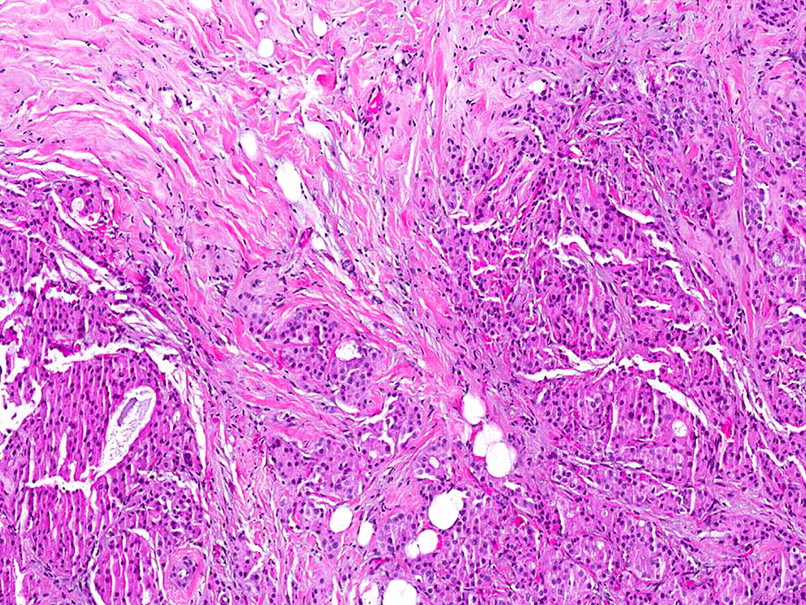
These tumours consist of sheets of pale histiocyte-like cells with ample, delicate, sometimes finely vacuolated to granular cytoplasm and inconspicuous cell membranes percolating the breast parenchyma (figure 1A). A ‘ground-glass’ appearance has been used to describe the cytoplasm.1 Nuclei are dark to vesicular and are centrally as well as eccentrically placed. Small nucleoli can be discernible. Mitoses are generally scarce. Nuclear pleomorphism is mild to moderate, with mostly grade 1 to 2 nuclei described,20 21 despite the incorporation of histiocytoid features in conjunction with pleomorphic lobular breast cancer.5 Rare cytoplasmic vacuoles can be discerned (figure 1B). The cells are disposed individually and in loose aggregates, without obvious glandular differentiation, and occasionally stream as linear arrays around resident benign lobules in a ‘targetoid’ arrangement1–3 6 9 11 15 22 (figure 1C). Accompanying lobular neoplasia (atypical lobular hyperplasia or lobular carcinoma in situ) may be identified (figure 1D), which may feature histiocytoid cells or those of conventional or apocrine lobular phenotype.
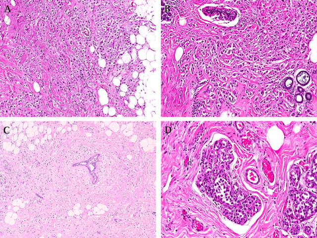
(A) Invasive histiocytoid carcinoma consists of sheets of pale tumour cells with ample cytoplasm with dark nuclei that are sometimes eccentrically placed. (B) Signet ring cells and cells with cytoplasmic vacuoles containing luminal secretions are present. (C) Pale histiocytoid cells streaming around a lobule in a targetoid fashion. (D) Atypical lobular hyperplasia is present.
Histochemistry
The majority of reports have demonstrated cytoplasmic mucin with Alcian blue and/or periodic acid Schiff stains1 3 5 6 15 22 (figure 2A). Eusebi et al3 referred to the positive cytoplasmic reactivity as ‘intracytoplasmic blue halos’, condensed blue crescent-like staining, or red granules, respectively.2 When performed, lipid stains have been mostly negative,1 21 22 although one report mentioned the presence of fine fat droplets in a few histiocytoid cells.15 Grimelius stains are negative.3

(A) Mucicarmine stain shows pink cytoplasmic secretions. (B) Immunohistochemistry for the epithelial marker AE1/3 shows diffuse positive reactivity in the histiocytoid tumour cells. (C) E-cadherin is negative in the tumour cells, while benign ductular epithelium and myoepithelial cells are positive. Lobular neoplastic cells of atypical lobular hyperplasia are also negative. (D) Diffuse positivity for GCDFP-15 immunohistochemistry.
Immunohistochemistry
Immunohistochemistry reveals positive reactivity of the histiocyte-like cells for epithelial markers such as AE1/3, MNF116, Cam 5.2, CK7, EMA confirming an epithelial origin2 4 22 (figure 2B). Strong positivity for CEA has also been reported.11 S100 is generally negative,21 with Eusebi et al4 mentioning weak staining in one of 13 histiocytoid carcinomas of the breast in their series.
E-cadherin is predominantly negative (figure 2C).6 20 Gupta et al20 described three positively stained cases, leading to their conclusion that histiocytoid breast carcinoma lacked a distinct immunophenotype, and could therefore belong to either lobular or ductal subtypes. Their E-cadherin-positive case illustration, however, showed a few cells with apparent incomplete membrane staining, which may reflect aberrant protein expression by a dysfunctional E-cadherin gene.23 Rakha et al24 also recently described E-cadherin expression in 16% of 239 cases of histologically confirmed invasive lobular carcinomas, of which the majority were associated with abnormal expression of one or more members of the E-cadherin–catenin complex, implicating a lack of functionality of the E-cadherin gene.
GCDFP-15 is consistently positive in studies in which it was applied3–5 20 22 (figure 2D), and this has been interpreted as evidence of apocrine differentiation.3 25–27 Oestrogen and progesterone receptor expression and cerbB2 status are variable.19 22
Kasashima et al19 extended their investigation into mucin profiles of eight histiocytoid and 14 classic invasive lobular carcinomas using immunohistochemistry. Histiocytoid lobular cancers showed positive reactivity for MUC2 and MUC5AC in 75% and 50% of cases, respectively, while almost all classic forms were negative. Both histiocytoid and classic lobular carcinomas were positive for MUC1 and negative for MUC4 and MUC6. It was postulated that the expression of non-mammary mucins (MUC2 and MUC5AC) could account for the worse prognosis of histiocytoid carcinomas in their study.
Electron microscopy
Ultrastructural studies show abundant cytoplasm with poorly developed organelles and small numbers of mitochondria, several lysosomes and Golgi apparatus. Mitochondria can be large with incomplete cristae.3 Membrane-bound electron-dense granules ranging from 300 to 800 nm22 and from 166 to 320 nm3 are present. Nuclei are round to oval with small nucleoli, thin nuclear membranes and fine chromatin. Intercellular junctions are rudimentary.15 22
Molecular studies
There are scant specific molecular data on histiocytoid breast carcinoma. Extrapolating from its presumed apocrine differentiation due to uniform GCDFP-15 expression, apocrine invasive lobular cancer has been shown to harbour molecular overlap with the pleomorphic variant,28 29 with both tumours demonstrating genetic similarity to classic invasive lobular tumours on expression profiling. In a recent detailed study on 31 in-situ pleomorphic lobular breast carcinomas, of which 13 were of apocrine cytomorphology, Chen et al30 discovered that pleomorphic lobular carcinoma in situ demonstrated 16q loss and 1q gain similar to the classic variety, with apocrine forms displaying significantly more alterations.
Differential diagnoses
The relatively bland cytomorphology of histiocytoid breast carcinoma can lead to potential misdiagnoses, differentials of which include the following entities.
Histiocytic inflammatory reaction
As its name implies, tumour cells of histiocytoid breast carcinoma resemble histiocytes with their abundant cytoplasm and relatively banal and sometimes eccentrically placed nuclei. An inflammatory histiocytic process needs to be considered in the differential diagnosis. Histiocytes can be seen in association with duct ectasia where histiocytes occupy the dilated duct lumen as well as spill out into the duct walls,31 sometimes effacing the lining epithelium that can be obscured by the inflammatory process, such that only a collection of histiocytes remain, leading to appearances reminiscent of histiocytoid breast carcinoma (figure 3A). Sometimes, foreign body type giant cells and cholesterol clefts may be seen, and the term xanthogranulomatous mastitis may be used to refer to this histological finding.32

(A) Histiocytes of duct ectasia, where in the absence of an intact duct, the histiocytic population can resemble the tumour cells of histiocytoid breast carcinoma. Inset shows CK7 decorating the residual epithelium of a duct that is partially effaced by histiocytes. (B) Histiocytes in post-chemotherapy changes, with a duct in the lower field containing pleomorphic cells of residual ductal carcinoma in situ.
Clues to a benign histiocytic inflammatory process are the presence of other accompanying inflammatory cells such as lymphocytes and plasma cells, and the generally limited localisation of inflammatory cells centred around a disrupted duct. Lobular neoplasia is not a usual association. Immunohistochemically, histiocytes are negative for epithelial markers and will react positively with CD68.
An exceedingly rare histiocytic process that has been described in the breast is Erdheim–Chester disease, which is a non-Langerhans cell histiocytosis of unknown aetiology that more commonly affects long bones, skin, orbit, pituitary and retroperitoneum.33 Histologically, a xanthomatous infiltrate is punctuated by Touton-type giant cells and patchy lymphocytes, which occasionally zone to a perivascular location. Immunohistochemically, CD68 decorates the histiocytes, with negative reactivity for S100, CD1a and cytokeratins.
Post-neoadjuvant chemotherapy, the accumulation of foamy histiocytes may mark the location of chemotherapy-induced tumour dissipation (figure 3B).
Fat necrosis
The microscopic appearances of fat necrosis include histiocytes occurring in relation to necrotic adipocytes (figure 4). A foreign body giant cell reaction and other inflammatory cells are frequently present. Fat necrosis in the breast is often encountered in association with reactive and reparative changes in the context of a previous needling/core or surgical biopsy procedure for which the history should be readily available. However, it can also present as a spontaneous breast lump in a woman, with or without a history of breast trauma. Fat necrosis can occur post-radiation treatment for breast carcinoma,34 and very rarely, lupus panniculitis has been reported in the breast of patients with systemic lupus erythematosus.35
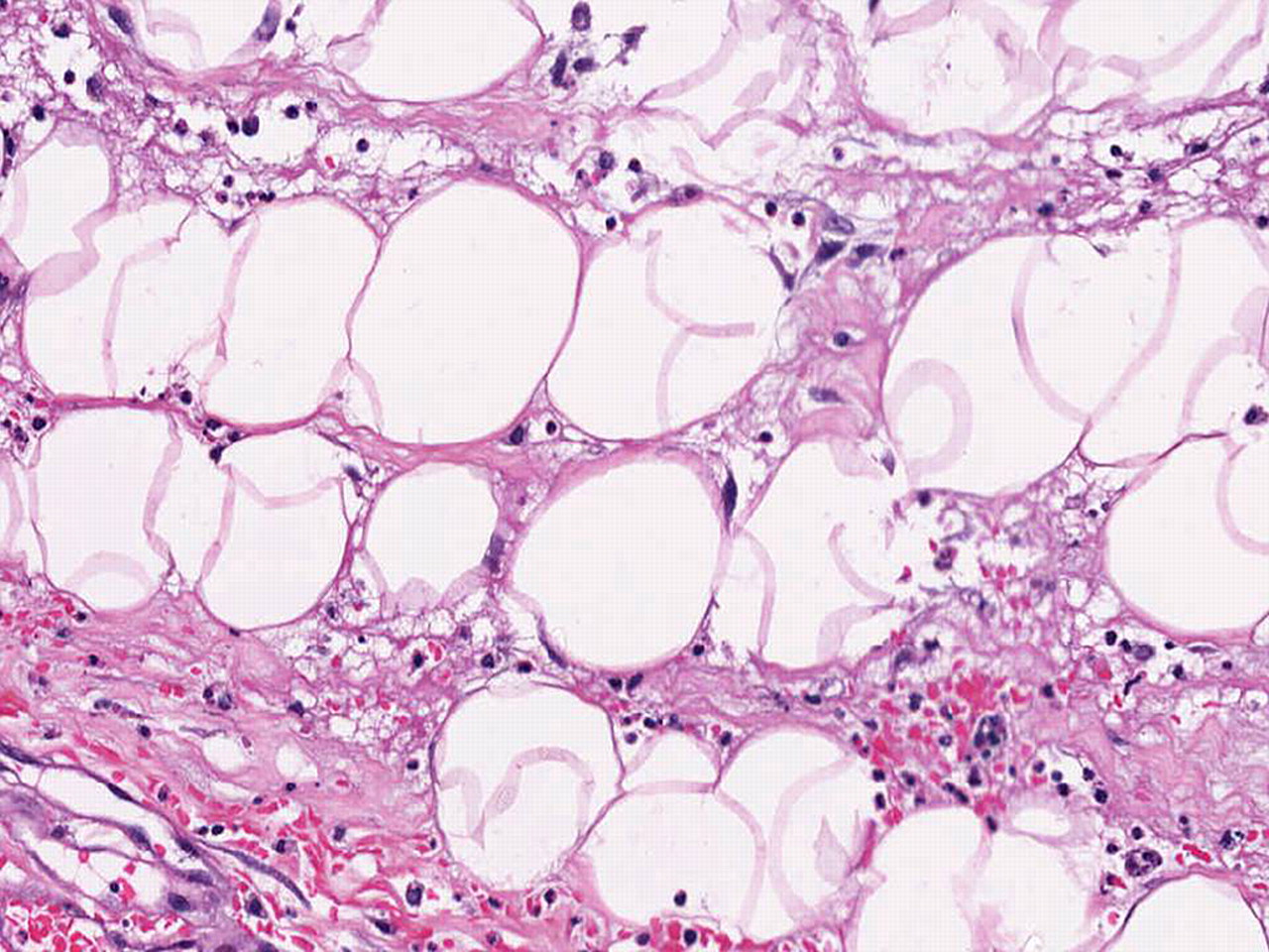
Fat necrosis shows necrotic adipocytes with histiocytes and other inflammatory cells.
The histological confusion of fat necrosis with a malignant process is particularly problematical during intraoperative frozen sections in which nuclear atypia of reactive histiocytes can appear accentuated. The correct diagnosis is aided by identifying accompanying inflammatory cells with reactive alterations and foreign body giant cells, as well as obtaining a proper clinical history.
Granular cell tumour
The granular cell tumour is a rare, usually benign tumour of Schwann cell derivation that can be discovered in the breast (figure 5).31 Clinically and radiologically mimicking breast malignancy, the granular cell tumour consists of sheets and nests of polygonal to occasionally spindle cells with ample cytoplasm containing eosinophilic granules that are periodic acid Schiff positive and diastase resistant. Some vacuolised and clear cells may be identified. Nuclei are vesicular with modest pleomorphism. Occasional distinct nucleoli are found. Nerve twigs may be observed in association with the granular cells.

Granular cell tumour shows cells with abundant pink granular cytoplasm and inconspicuous cell borders.
Immunohistochemically, the cells are positive for S100 and CEA, and are negative for oestrogen and progesterone receptors. They are usually negative for histiocyte-associated antigens including α1-antitrypsin and α1-antichymotrypsin, although some reactivity for CD68 has been described.
While generally benign and cured by complete excision, less than 1% of granular cell tumours are reported to be malignant. Histologically, the malignant forms display mitoses, pleomorphism, necrosis and there may be metastases,36 but instances of malignant behaviour have been observed even in the absence of these microscopic features.31
Rosai–Dorfman disease
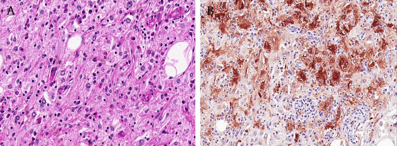
Rosai–Dorfman disease (RDD) or sinus histiocytosis with massive lymphadenopathy (SHML) is primarily a nodal-based, idiopathic, benign proliferative disorder of histiocytes, with 43% of these cases also involving extranodal sites. The breast is an unusual site of occurrence of RDD and can clinicoradiologically mimic cancer.37
Histology of RDD consists of sheets of characteristic large histiocytes displaying emperipolesis, a microscopic hallmark of this disease (figure 6A). The predominant histiocytic population is reminiscent of the appearances of histiocytoid breast carcinoma. Lymphoid aggregates with germinal centres and plasma cells are frequent accompaniments. Immunohistochemistry shows cytoplasmic staining of histiocytes for S-100, often with weak positivity for CD68, while electron microscopy confirms histiocytic engulfment of lymphocytes and plasma cells (figure 6B).

(A) Rosai–Dorfman disease shows voluminous histiocytes with lymphocytes and plasma cells residing within the cytoplasm, a phenomenon referred to as emperipolesis. (B) S100 immunohistochemistry shows positive cytoplasmic reactivity of the histiocytes, accentuating the presence of lymphocytes and plasma cells within the cytoplasm.
The aetiology and pathogenesis of RDD are obscure, with an infective or immune-mediated origin being proposed. Excision is often curative. Spontaneous resolution has also been described, although a more persistent and aggressive course is possible. RDD of the breast tends to be resected as malignancy is often clinically and radiologically suspected.
Invasive apocrine carcinoma
This histological variety is defined by the presence of apocrine cells in more than 90% of tumour cells (figure 7).21 Apocrine differentiation can be observed in any type and grade of breast carcinoma, and its recognition is of no current predictive value. Two cell types are described in apocrine cancer: type A cells, which are intensely eosinophilic and contain abundant granular cytoplasm (resembling conventional apocrine cells), and type B cells, which are foamy with ample finely vacuolated cytoplasm. The latter are histiocyte-like and would be histologically synonymous with histiocytoid breast carcinoma, affirming its apocrine immunophenotypic expression.3 4

Invasive apocrine carcinoma with irregular nests and islands of tumour cells with apocrine appearances.
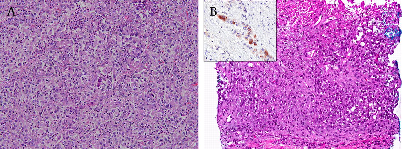
Metastasis to the breast
Unusually, the breast can be the site of metastatic disease.38 39 Examples of metastases to the breast that can histologically display plump polygonal cells resembling histiocytoid breast carcinoma are renal cell carcinoma, melanoma (figure 8A,B) and alveolar soft part sarcoma. Correlation with clinical history and radiological findings, together with adjunctive immunohistochemistry, can lead to the correct diagnosis.
(A) Metastatic renal cell carcinoma to the breast, showing polygonal cells with ample cytoplasm and vesicular nuclei containing distinct nucleoli. Polymorphs are also seen. (B) Metastatic melanoma to the breast with sheets of plump cells adjacent to necrosis. Inset shows melanA immunohistochemical positivity.
Conclusions
Histiocytoid invasive breast carcinoma is an unusual form of breast cancer, with the weight of evidence supporting its classification as a variant of invasive lobular carcinoma. Consistent GCDFP-15 expression suggests apocrine differentiation, although perhaps in a rudimentary form of cytomorphological expression. While histiocytoid breast carcinoma has been observed in relation to pleomorphic lobular carcinoma, whether its clinicobiological behaviour mirrors the aggressive characteristics of the latter variant is uncertain. Its resemblance to benign entities is a pitfall, and awareness of these mimics is needed in order to arrive at a correct diagnosis for appropriate patient management. Metastases to lymph nodes and skin can also be overlooked as benign sinus histiocytes and xanthomatous dermal lesions, respectively,1 with more disseminated spread to the uterus and parathyroid also being documented.40
Hints to the correct diagnosis include the presence of accompanying tumour cells that are more pleomorphic and mitotically active, cells with cytoplasmic vacuoles and targetoid secretions, architectural patterns resembling those of invasive lobular cancer with linear files and concentric encirclement of lobules, associated classic invasive lobular carcinoma or lobular neoplasia, and the use of adjunctive immunohistochemistry to verify the epithelial origin of lesional cells. Close clinicoradiological correlation is critical, as discordant findings on core biopsy or cytology should prompt histological pursuit of a conclusive diagnosis on open excision. In the presence of an earlier breast carcinoma, development of skin nodules comprising histiocyte-like cells should also be diligently assessed to rule out metastasis.
Take-home messages
Histiocytoid breast carcinoma is an unusual tumour that is most often regarded as a variant of invasive lobular cancer.
The bland histological appearances may mimic benign breast conditions, which pose diagnostic pitfalls especially on limited material such as core biopsies or fine needle aspiration cytology.
Clues to the correct diagnosis are the presence of cells with more overt cytological atypia and mitoses, cells with cytoplasmic vacuoles and secretions, accompanying components of classic invasive lobular carcinoma and/or lobular neoplasia, aided by immunohistochemistry to confirm the epithelial nature of the histiocytoid cells.
It is important for close clinicoradiological correlation, as any discordance should spur further diagnostic determination.
Interactive multiple choice questions
This JCP article has an accompanying set of multiple choice questions (MCQs). To access the questions, click on BMJ Learning: take this module on BMJ Learning from the content box at the top right and bottom left of the online article. For more information please go to: http://jcp.bmj.com/education. Please note: the MCQs are hosted on BMJ Learning the best available learning website for medical professionals from the BMJ Group. If prompted, subscribers must sign into JCP with their journal's username and password. All users must also complete a onetime registration on BMJ Learning and subsequently log in (with a BMJ Learning username and password) on every visit.
References
Footnotes
Competing interests None.
Provenance and peer review Not commissioned; externally peer reviewed.