Article Text

Abstract
Aims To correlate Ki67 expression with outcome in colorectal cancer (CRC).
Methods Ki67 labelling index (Ki67LI) was analysed by immunohistochemistry on a tissue microarray containing 1800 CRCs. The results were compared with clinicopathological and molecular parameters.
Results Ki67LI was considered low in 26.3%, moderate in 56.7% and high in 17.0% of 1653 interpretable CRCs. High Ki67 expression was associated with low tumour stage (p<0.0001) and nodal status (p=0.0315), but not with tumour grade (p=0.8639), histological tumour type (p=0.1542) or tumour localisation, and was an independent prognosticator of favourable survival (p=0.0121). High Ki67 expression was also significantly associated with high-level nuclear β-catenin and p53 expression (p<0.0001 and p=0.0095, respectively).
Conclusions In summary, our data show that high Ki67 expression in CRCs is associated with good clinical outcome. Ki67, p53 and β-catenin overexpression seem to be linked to CRC, and indicate a cellular state of high proliferative activity. Finally, our findings strongly argue for a clinical utility of Ki67 immunostaining as an independent prognostic biomarker in CRC.
- COLORECTAL CANCER
- MOLECULAR PATHOLOGY
- KI 67
- ANTIBODIES
- CANCER RESEARCH
This is an Open Access article distributed in accordance with the Creative Commons Attribution Non Commercial (CC BY-NC 4.0) license, which permits others to distribute, remix, adapt, build upon this work non-commercially, and license their derivative works on different terms, provided the original work is properly cited and the use is non-commercial. See: http://creativecommons.org/licenses/by-nc/4.0/
Statistics from Altmetric.com
Introduction
Colorectal cancer (CRC) is the fourth most common malignant disease with over one million novel cases and over 500 000 deaths each year worldwide.1 Although recent advances in the management of the disease have improved outcomes, CRC remains the second leading cause of cancer-related death in Western countries.1 In advanced metastatic CRC, surgery alone is not curative, and therefore, adjuvant chemotherapy is needed. Patients with nodal negative disease generally show a favourable clinical course. However, approximately one-third of node-negative CRCs recurs or shows progressive disease, suggesting failure to detect occult disease.2 Because aberrant genetic changes occur early in tumour progression, molecular profiling of specific tumour markers in the primary tumour might predict the tumour’s metastatic potential. Thus, patients with a potentially aggressive tumour, although not yet metastasised at the time of surgery, might also benefit from adjuvant therapy.
Ki67 is a nuclear antigen, which is expressed in proliferating cells from G1 to M-phase of the cell cycle.3 Many studies have shown a predictive role of Ki67 in a wide range of human malignancies, including gastrointestinal stromal tumours, gastrointestinal neuroendocrine tumours, and prostate and breast cancer.4–8 The quantification of Ki67 expression by immunohistochemistry, which is known as the Ki67 labelling index (Ki67LI: percentage of invasive cancer cell nuclei that are positive for Ki67 immunostaining over total invasive cancer cell nuclei present in a histological sample) has become a routine practice in clinical pathology to estimate the growth fraction of a tumour.9 For example, in hormone-positive breast cancer, Ki67 is currently used to predict the efficacy of pure hormone receptor blockade without chemotherapy in the neoadjuvant setting.10 ,11 In well-differentiated neuroendocrine tumours (NETs), Ki67 staining of core biopsies of the primary usually provides a reliable method of proliferation assessment for prognosis of metastatic NETs to the liver.12 However, only few studies exist on the prognostic role of Ki67 in CRC, and have partially shown contradictory results.13–16
To further elucidate the clinical relevance of Ki67 as a prognostic tumour marker, we performed an immunohistochemical study on a large cohort of patients with CRC using our pre-existing CRC microarray containing 1800 CRC specimens with attached clinical follow-up and extensive molecular data. The results of this study show that high Ki67 expression is an independent predictor of favourable outcome in CRC. Thus, patients with CRC that show low Ki67LI may benefit from adjuvant therapy.
Patients and tissue microarray construction
Two different tissue microarrays (TMAs) with a total of 1800 CRC samples were included in this study. The first TMA was manufactured from resection specimens of 1420 patients with CRC at the Institute of Pathology of the University Hospital of Basel. Patients were operated at the Department of Surgery of the University Hospital of Basel between 1988 and 1996. Raw survival data were obtained from the responsible physicians for all of the 1420 patients. The median follow-up time was 46 months (range 1–152 months). The second TMA included samples from 380 patients with CRC whose tumour resection specimens were examined at the Institute of Pathology of the University Medical Center, Hamburg-Eppendorf. Patients were operated at the Department of Surgery of the University Medical Center, Hamburg-Eppendorf from 1993 to 2006. Also for this TMA, raw survival data were available for all of the 380 patients with a median follow-up period of 36 months (range 1–179 months). None of the patients of both cohorts received neoadjuvant therapy. Postoperative therapy was executed according to the guidelines for the treatment of colorectal carcinoma.17 For both cohorts, no clinical data were available regarding the state of disease progression, especially, there was no separation of the patient population into groups based on RECIST guidelines of stable or responding disease and progressive disease.
TMA construction was as described by Kononen et al.18 In brief, H&E-stained sections were made from each block to define representative tumour regions. Tissue cylinders with a diameter of 0.6 mm were then punched from tumour areas of each ‘donor’ tissue block using a home-made semiautomated precision instrument and brought into empty recipient paraffin blocks. Four-micrometre sections of the resulting TMA blocks were transferred to an adhesive-coated slide system (Instrumedics, Hackensack, New Jersey). Patient information and clinical data such as age, sex, localisation and type of the tumour, pathological tumour-node-metastases stage and carcinoma grade were retrospectively retrieved from clinical and pathological databases (table 1). All tumours were reclassified by two pathologists (GS and AHM). Follow-up data were obtained from local cancer register boards or via attending physicians. For statistical analyses, tumour localisations were grouped as follows: right-sided cancer (caecum, ascending colon, transverse colon) and left-sided colon (splenic flexure, descending colon, sigmoid colon and rectum).19 The usage of tissues and clinical data was according to the Hamburger Krankenhaus Gesetz (§12 HmbKHG) and approved by our local ethical committee.
Clinical and pathological features of colorectal cancers
Ki67 immunohistochemistry
Standard indirect immunoperoxidase procedures were used for the detection of Ki67 (abcam, clone SPM171, dilution 1:150). Sections were heated in an autoclave at 121°C for 10 min in citrate puffer (pH 9.0). Diaminobenzidine was used as a chromogen, and sections were counterstained with Mayer's haematoxylin. Only unequivocal nuclear staining, regardless of its intensity, was counted.
For statistical analyses, the staining results were categorised into three groups (weak, moderate, high) according to the percentage of Ki67-positive tumour cells as follows: low Ki67, 0%–10%; moderate Ki67, more than 10% and up to 25%; high Ki67, 25% and more. This Ki67 labelling system is also currently being applied in grading breast cancer.20 The molecular database attached to this TMA contained results on β-catenin and p53 expression in 1800 cancers.
Statistics
Statistical calculations were performed with JMP V.10.0.2 software (2012 SAS Institute, North Carolina, USA). The one-way ANOVA (analysis of variance) statistical test with Ki67LI as a continuous variable was used to search for associations between molecular parameters and tumour phenotype. Survival curves were calculated according to Kaplan–Meier. The log-rank test was applied to detect significant survival differences between groups. Cox proportional hazards regression analysis was performed to test the statistical independence and significance between pathological and clinical variables.
Resuts
Technical issues
A total of 1653 (91.8%) tumour samples were interpretable in our TMA analysis (table 1). Reasons for non-informative cases (147 spots, 8.2%) included lack of tissue samples or absence of unequivocal cancer tissue in the TMA spot.
Ki67 expression in CRC
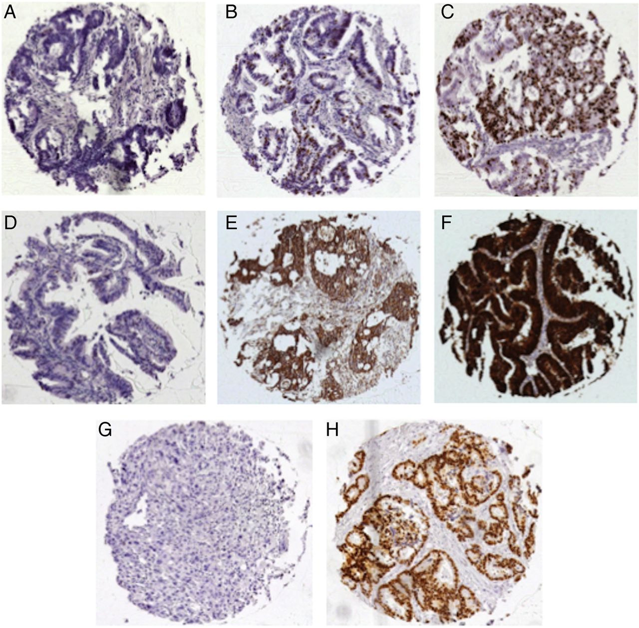
Ki67LI was considered low in 26.3%, moderate in 56.7% and high in 17.0% of 1653 interpretable CRCs. Representative images of Ki67 immunohistochemistry (IHC) are given in figure 1. Using Ki67LI as a continuous variable (ANOVA test), Ki67 expression was associated with low tumour stage (p<0.0001) and nodal status (p=0.0315), but not with tumour grade (p=0.8639), histological tumour type (p=0.1542) or tumour localisation (p=0.8571, table 1).

Representative images of Ki67 expression in colorectal cancer: (A) Ki67 low, (B) Ki67 moderate and (C) Ki67 high expression, magnification ×50 each.
Association with β-catenin and p53 expression
Strong immunohistochemical p53 and β-catenin positivity was observed in 41.5% and 75.8% of all cases, respectively, and was significantly associated with left-sided tumour localisation (p=0.0222 and p=0.0238, respectively, data not shown). Ki67 expression was significantly related to nuclear β-catenin and p53 expression (p<0.0001 and p=0.0095, respectively, table 1, figure 2).
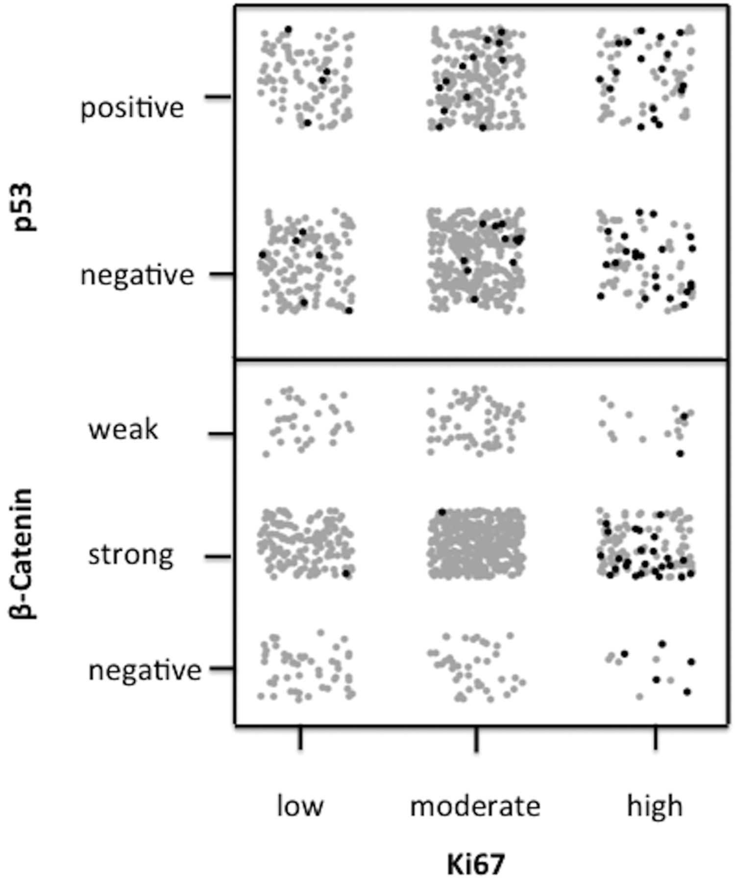
Scatterplot demonstrating correlation graphs of immunostaining of Ki67 with nuclear β-catenin and p53 expression.
Survival analysis
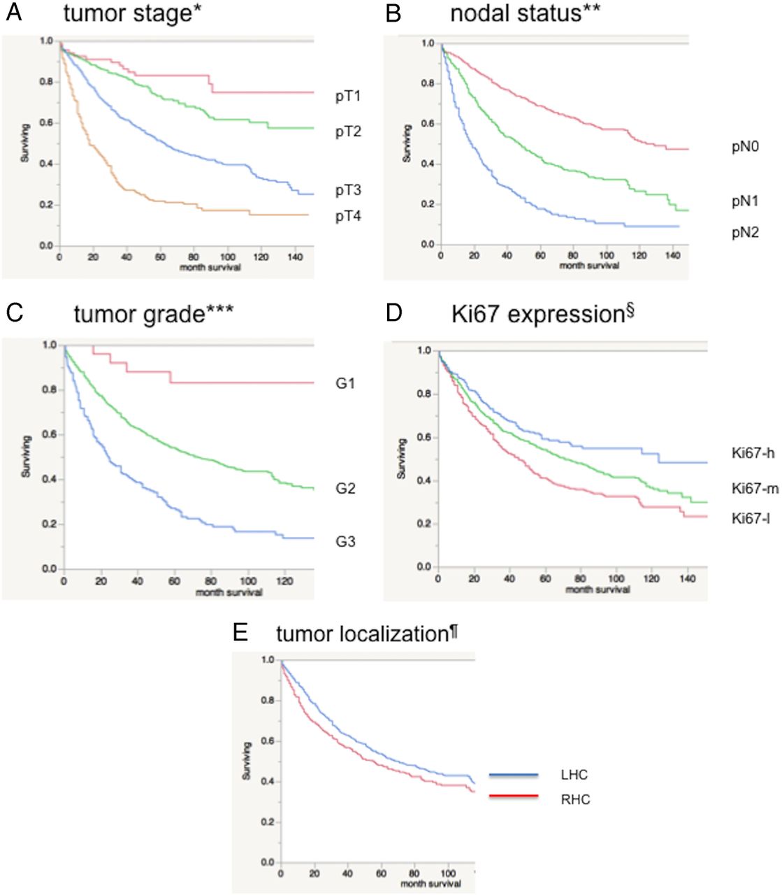
As expected, high tumour grade and stage as well as advanced nodal status were associated with poor patient survival (figure 3A–C, p<0.0001 each), while histological tumour type was unrelated to clinical outcome (p=0.4211). Left-sided CRCs (distal to the splenic flexure) were associated with a better prognosis (figure 3E, p=0.0161) as compared with tumours of the right-sided and transverse colon. High Ki67 expression of CRC was significantly related to improved patient survival (Ki67-low vs Ki67-moderate, p=0.0002; Ki67-moderate vs Ki67-high, p=0.0142; Ki67-low vs Ki67-high, p<0.0001; figure 3D). Immunohistochemical p53 and β-catenin status of the CRC did not show any impact on survival (p=0.4719 and p=0.2799, respectively). Separate Ki67 analysis of both cohorts alone resulted in similar findings regarding p values (cohort I: p<0.0001; cohort II: p=0.0035). This also held true for the other markers when they were separately tested for each cohort (data not shown).
Association between survival and (A) tumour stage, *p<0.0001; (B) nodal status, **p<0.0001; (C) tumour grade, ***p<0.0001; (D) Ki67 expression (Ki67-low, Ki67-moderate, Ki67-high), §p<0.0001 and (E) tumour localisation ¶p=0.0280.
Multivariable Cox proportional hazards regression model for survival
In a multivariate analysis (Cox proportional hazards model), including tumour stage, tumour grade, nodal status, tumour localisation, Ki67, p53 and β-catenin expression, only tumour stage, nodal status and Ki67LI retained significance (p<0.0001, p<0.0001 and 0.0121, respectively, table 2).
Multivariable Cox proportional hazards regression model for survival
Discussion
The results of this study show that in CRC, high Ki67 expression is an independent favourable prognostic marker. This finding is comparable with previous results. One earlier study by Salminen et al analysing 146 patients who had CRC with rectal and rectosigmoid cancer had also reported that high proliferative activity measured by Ki67 is associated with survival improvement compared with patients with low Ki67.14 In another study by Reimers et al on 285 patients with stage I–IV colon cancer, Ki67 and cleaved caspase-3 tumour expression were used to develop the combined apoptosis proliferation parameter and to correlate the results to patient outcome. Interestingly, patients with high levels of both apoptosis and proliferation showed the best outcome perspectives.15 Palmqvist et al16 showed that a low Ki67 index at the invasive tumour margin is associated with poor prognosis in Duke's stage B CRC. In contrast, in many other tumour types, including endocrine tumours, gastrointestinal stromal tumour, and head and neck, prostate and breast cancer, high Ki67 index has been linked to poor outcome.4 ,6–8 ,21–25 The reason for this discrepancy between the impact of Ki67 expression on prognosis in CRC and a wide range of other human malignancies is unclear. Duchrow et al compared the expression of Ki67 mRNA and protein in CRC, and showed that tumours with a high Ki67 protein level, but low mRNA expression, may proliferate more slowly than expected. They estimated that in a minimum of one-third of CRCs, a significant number of non-cycling tumour cells express Ki67, and in consequence, these tumours might grow more slowly than indicated by the Ki67LI. These Ki67-positive non-cycling tumour cells are probably more stable than tumour cells that cannot achieve cell cycle arrest, and thus, may be more resistant to adjuvant therapies or patient’s immune response.26 Interestingly, in oestrogen receptor-positive breast cancer, Ki67 expression was suggested to identify a subset of cancers, which may be sensitive to docetaxel treatment in the adjuvant setting.27 Comparably, Ki67 determination has also been suggested as a tool in selecting patients with rectal cancer for radiotherapy.13 It is well known that the proliferative activity is often heterogeneous within a tumour.12 Therefore, it has been recommended that the Ki67LI should be assessed in zones of high proliferation, which are often at the invasive margin of the tumour.11 ,16 However, this can only be accomplished by using whole section slides; on which, the invasive margin of the tumour is clearly identified. In routine clinical practice, core biopsies from the tumour or metastases are usually taken to assess the molecular characteristics of their malignancy. The question remains, whether the Ki67LI obtained from a core needle biopsy adequately represents the whole tumour or metastases. Recently, a study using TMAs of hepatic metastases from well-differentiated NETs demonstrated good correlation between Ki67LI in 1–3 random core biopsies and whole sections of G1 tumours in nearly 100% and in about 50% of G2 tumours. Thus, the authors concluded that a single needle core biopsy randomly taken from within a tumour or metastasis usually provides adequate proliferation assessment, despite the presence of intratumoral heterogeneity.12 Therefore, we assume that Ki67 expression analysis using TMAs constructed from one 0.6 mm core per patient yields representative results comparable with random core needle biopsies.
The cut-off levels of Ki67 expression used in our study (≤10% and ≥25%) are currently being applied in breast cancer diagnostics, and have been shown to be reproducible and significant. However, as suggested for breast cancer, a standardised method of Ki67 assessment is needed for other tumour types, including CRC.11 ,20 As expected, Ki67-h was strongly linked to high nuclear β-catenin and p53 expression in our cohort (table 1). This observation fits well with the known relationship of p53, β-catenin and Ki67 in cell proliferation.28–30 β-Catenin is a key factor of Wingless Int-1 (WNT) signalling, and nuclear translocation of β-catenin characterises cells with active WNT signalling.31 Active WNT signalling leads to enhanced cell proliferation and, thus, to elevated Ki67LI. Mutated p53 accumulates in tumour cells, and thus, serves as a marker of high proliferative activity too (reviewed in Refs 30 ,32 ,33).
Besides factors that influence cell proliferation, biomarker that plays a critical role in regulating epithelial–mesenchymal transition (EMT) in cancer cells has recently been evaluated for clinical significance. Toiyama et al34 have shown that increased expression of slug and vimentin, which play a critical role in regulating EMT via downregulation of epithelial markers and upregulation of mesenchymal markers, is significantly associated with poor prognosis. Another study by Satelli et al has suggested that detecting and measuring cell-surface vimentin on the surface of EMT-circulating tumour cells from blood of patients may predict progressive disease in CRC.35 However, the relationship between EMT-regulating factors and cell proliferation needs to be further elucidated. Studies on patients with CRC who are divided into groups based on Response Evaluation Criteria in Solid Tumors (RECIST) guidelines in stable or responding disease and progressive disease are needed to define a threshold of Ki67LI that is significantly predicting disease progression.
In summary, our data show that high Ki67 expression in CRCs is associated with good clinical outcome. Ki67, p53 and β-catenin overexpression seem to be linked to CRC, and indicate a cellular state of high proliferative activity.
Finally, our findings strongly argue for a clinical utility of Ki67 immunostaining as an independent prognostic biomarker in CRC that may contribute to the prognostic evaluation in patients with CRC.
Take home messages
High Ki67 expression in colorectal cancers (CRCs) is associated with good clinical outcome.
Ki67, p53 and β-catenin overexpression seem to be linked to CRC, and indicate a cellular state of high proliferative activity.
Ki67 immunostaining may serve as an independent prognostic biomarker in CRC.
References
Footnotes
NM and CMK contributed equally.
Handling editor Cheok Soon Lee
Competing interests None declared.
Ethics approval Local ethical committee.
Provenance and peer review Not commissioned; externally peer reviewed.