Article Text

Abstract
Aims Upper tract urothelial carcinoma (UTUC) is a rare malignancy with a poor prognosis which occurs sporadically or in few cases results from a genetic disorder called Lynch syndrome. Recently, examination of microsatellite instability (MSI) has gained importance as a biomarker: MSI tumours are associated with a better response to immunomodulative therapies. Limited data are known about the prevalence of MSI in UTUC. New detection methods using the fully automated Idylla MSI Assay facilitate analysis of increased patient numbers.
Methods We investigated the frequency of MSI in a multi-institutional cohort of 243 consecutively collected UTUC samples using standard methodology (Bethesda panel), along with immunohistochemistry of mismatch repair (MMR) proteins. The same tumour cohort was retested using the Idylla MSI Assay by Biocartis.
Results Using standard methodology, 230/243 tumours were detected as microsatellite stable (MSS), 4/243 tumours as MSI and 9/243 samples as invalid. In comparison, the Idylla MSI Assay identified four additional tumours as MSS, equalling 234/243 tumours; 4/243 were classified as MSI and only 5/243 cases as invalid. At the immunohistochemical level, MSI results were supported in all available cases with a loss in MMR proteins. The overall concordance between the standard and the Idylla MSI Assay was 98.35%. Time to result differed between 3 hours for Idylla MSI Assay and 2 days with the standard methodology.
Conclusion Our data indicate a low incidence rate of MSI tumours in patients with UTUC. Furthermore, our findings highlight that Idylla MSI Assay can be applied as an alternative method of MSI analysis for UTUC.
- urological neoplasms
- pathology
- molecular
- methods
Data availability statement
Data are available upon reasonable request.
This is an open access article distributed in accordance with the Creative Commons Attribution Non Commercial (CC BY-NC 4.0) license, which permits others to distribute, remix, adapt, build upon this work non-commercially, and license their derivative works on different terms, provided the original work is properly cited, appropriate credit is given, any changes made indicated, and the use is non-commercial. See: http://creativecommons.org/licenses/by-nc/4.0/.
Statistics from Altmetric.com
Introduction
Upper tract urothelial carcinoma (UTUC), including tumours in pyelocaliceal cavities and ureter, is a rare cancer with incidence rates close to 2/100 000 inhabitants per year in Western countries.1 Overall, UTUC accounts for only 5%–10% of all urothelial tumours, is more often found in people of advanced age, and three times more often in men than in women.2 3 In contrast to bladder cancer, UTUCs present as an invasive disease at diagnosis in 60% of cases and have a poor prognosis with a 5-year survival of less than 50%.2 UTUC can be sporadic and is significantly associated with exposure to tobacco and aromatics.2 4 On the other hand, an autosomal-dominant inherited tumour syndrome called the Lynch syndrome caused by germline mutations in genes of DNA mismatch repair (MMR), increases the risk for developing different tumour types, especially colorectal cancer and endometrial cancer. UTUC related to the Lynch syndrome is relatively rare, with an estimated risk of 6%–15%.5 6
The sporadic as well as the hereditary forms of UTUC are associated with microsatellite instability (MSI). A deficient DNA MMR system caused by germline or sporadic mutations of MMR genes lead to a nucleotide length variation of DNA repeat regions called microsatellites.7 Although previous reports on small patient cohorts indicate that the frequency of MSI in UTUCs is approximately 20%, a uniform description has not been defined to date.8 It is important to note that the MSI status represents an important prognostic and predictive tumour marker.9 In many tumour types, MSI is associated with a better outcome and improved response to adjuvant chemotherapy and immunotherapy regimes compared with tumours with stable microsatellite DNA regions.10–14
Currently, detection of MSI is mostly performed using the National Cancer Institute (NCI) consensus marker panel accompanied by immunohistochemistry analysis, which results in a time-to-diagnosis period of approximately two working days.15 With rising diagnostic numbers due to therapeutic options, procedures in terms of testing duration and the specific detection method need to be improved. The Biocartis Idylla MSI Assay is a fully automated, real-time PCR-based molecular test which implements a new set of seven markers for detection of MSI. The new marker set for MSI analysis by Biocartis was developed and reviewed in the work of Zhao et al.16 In previous reports, the new marker set has previously been approved by in vitro diagnostic regulation including Conformité Européenne (CE) marking as an alternative testing method for MSI analysis in colorectal carcinoma.17
The aim of this study was to assess the frequency of MSI among a large retrospective cohort of 243 consecutively collected, multi-institutional UTUC samples using standard NCI consensus methods along with immunohistochemistry compared with the Idylla MSI Assay.
Materials and methods
Analysed UTUC cohort
A consecutively collected, multi-institutional cohort of 249 primary tumours collected from 1995 to 2017 from the renal pelvis or ureter were retrieved from the archives from three collaborating Institutes of Pathology: Málaga (Spain), Marburg and Erlangen (Germany). We excluded six cases due to insufficient preserved tumour tissue. The UTUC cohort was histologically re-evaluated and classified according to the most recent tumour, node, metastasis (TNM) classification (2017) and the18 classification of genitourinary tumours by two uropathologists (VW and AH).18 Pathological as well as clinical characteristics are shown in table 1.
Clinicopathological characteristics of the analysed cohort
Microdissection and DNA isolation
DNA extractions of matched tumour and normal tissue from formalin-fixed paraffin-embedded (FFPE) tissues were isolated according to a standard protocol, described previously.19 Briefly, regions of UTUC tumour or normal tissue for microdissection were initially identified. Then 5–10 m histological tissue sections of tumour and normal tissues were fractionated via manual microdissection following deparaffinisation and staining with 5% methylene blue. DNA isolation was performed using the Promega Maxwell 16 LEV Blood DNA kit (Promega, Mannheim, Germany) according to the operator’s instructions.
Microsatellite analysis
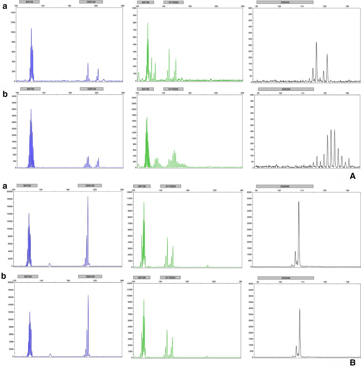
Microsatellite analysis was performed using tumour cells as well as corresponding normal tissue DNA. The Bethesda panel of five markers, consisting of two mononucleotide repeats (MNRs) (BAT25 and BAT26) and three dinucleotide repeats (D2S123, D17S250 and D5S346), was used as previously recommended by the NCI.15 Approximately 100 ng of DNA was used for PCR amplifications; primer sequences and PCR conditions were used as previously described.20 For samples with low amounts of normal tissue DNA, a panel of five MNRs (BAT25, BAT26, NR21, NR24 and NR27) was used for tumour DNA only and called the MNR panel. Reproducibility of this MSI method has been validated in a previous work.21 Both MSI analyses are implemented in routine diagnostic workflow of the Institute of Pathology, University Hospital Erlangen, with accreditation by the German Accreditation Office (DAKKs) according to DIN EN ISO/IEC 17020. Detailed information on primer sequences and conditions were previously described. Briefly, the amplification products were analysed by capillary electrophoresis using the ABI Prism 3500 genetic analyser, and fragment analysis was performed using GeneMapper software V.4.1 (Applied Biosystems, Foster City, California, USA). As illustrated in figure 1A, at least two of five markers are required to be different with novel peaks in order to classify a cancer as MSI.8 If this is not the case and length changes are seen with only one marker, as shown in figure 1B, they are defined as microsatellite stable (MSS). For cases with invalid as well as MSI results, a second microsatellite analysis was performed to check for reproducibility.

(A) Representative capillary electrophoresis results of MSI tumour of (a) normal tissue and (b) tumour tissue. X-axis is the number of bases; y-axis is the number of amplicons reflected in fluorescence intensity, scaled to the variable amount of analysed DNA. Blue, green and black peaks are products of the amplifications of the different MSI markers: BAT25, D2S123, BAT26, D17S250 and D5S346. Comparing (a) and (b), we saw a shift and extension of base amplification product in each marker of tumour tissue compared with normal tissue. (B) Representative capillary electrophoresis results of MSS tumour of (a) normal tissue and (b) tumour tissue. X-axis is the number of bases; y-axis is the number of amplicons reflected in fluorescence intensity, scaled to the variable amount of analysed DNA. Blue, green and black peaks are products of the amplifications of the different MSI markers: BAT25, D2S123, BAT26, D17S250 and D5S346. No shift and no expansion of the base amplification product are seen in the markers of tumour tissue (b) compared with normal tissue (a). MSI, microsatellite instability; MSS, microsatellite stable.
Immunohistochemistry
To validate microsatellite testing and to detect possible deficiencies in MMR proteins (mutL homologue 1 (MLH1), mutS homologue2 (MSH2), mutS homologue 6 (MSH6), PMS1 homologue 2 (PMS2)), an immunohistochemical staining was performed. A tumour tissue microarray (TMA) of each paraffin block from cases derived from Germany (n=197) was produced to gain a high degree of standardisation. Digitally scanned H&E slides (Panoramic P250; 3DHistech, Hungary) were marked for tumour centres as well as invasion borders using the computer software CaseViewer V.2.0 (3DHistech). Then two cores (diameter 1 mm) of each region were punched by using the TMA-Grandmaster (3DHistech). Immunohistochemistry staining with anti-MLH1, anti-MSH2, anti-MSH6 and anti-PMS2 was performed. Detailed information of the antibodies are displayed in online supplemental table 1. The expressions of MMR proteins MLH1, MSH2, MSH6 and PMS2 were assessed as described previously.22 Nuclear staining of surrounding stromal and immune cells was applied as internal positive control. A loss of expression of the four MMR genes can lead to a deficient MMR system.23
Supplemental material
Idylla MSI assay
The Idylla MSI Assay (Biocartis NV, Mechelen, Belgium) is a new diagnostic tool which connects the steps of MSI analysis into a single automated process. PCR amplification followed by a high-resolution melting curve analysis allows the detection of DNA mutations in seven new MSI biomarkers (ACVR2A, BTBD7, DIDO1, MRE11, RYR3, SEC31A and SULF2).24 The Idylla MSI Assay was prepared according to the operation manual. Idylla MSI Assay cartridges were loaded with FFPE tumour tissue sections (5–10 m, neoplastic cells 20%). After a 150 min automated workflow, the Idylla MSI Assay system automatically evaluates test results with an interpretation including MSI status and separate results of MSI biomarkers. Possible sample results are ‘microsatellite stable (MSS)’ or ‘microsatellite instability–High (MSI-H)’. A sample is defined as MSI-H if a DNA mutation is detected in at least two biomarkers. A sample is defined as MSS if a mutation is detected in less than two biomarkers In cases with invalid as well as MSI results, a second microsatellite analysis was performed to check for reproducibility.
Statistical analysis
Descriptive statistical analysis and differences between altered (MSI) and non-altered (MSS) were compared using Fisher’s exact test. Statistical analysis was performed using JMP SAS V.13.2. P values were two-sided, and a p value of <0.05 was considered statistically significant. To calculate the overall concordance, the quotient of number of concordant results and the number of all results was formed and multiplied by 100.
Results
MSI frequency in UTUC using standard methodology and associations with clinicopathological characteristics
Due to invalid results with the Bethesda panel, 15 cases were additionally retested with the MNR panel using only tumour DNA. In the first run, in 13 out of 243 tumours, no valid result could be assigned using both panels. MSI tumours were detected in four samples (two by Bethesda panel and two by MNR panel). In contrast, 226 cases were evaluated as MSS tumours. These preliminary invalid results were cross-checked in a second run. Thereby, four of the previously invalid results were detected as MSS (for a detailed description, see further). The final results of MSI testing with standard methodology showed 230 (94.7%) MSS tumours, 4 (1.6%) MSI tumours and 9 (3.7%) invalid cases. One MSI tumour was included in the Erlangen cohort, two cases from Marburg and one case from Málaga. The patients’ age ranged from 52 years to 83 years. Three cases were male, and one case was female. One pT1, one pT2 and two pT3 tumours with high-grade morphology were included. Localisation of the primary tumour in two cases was found in the renal pelvis and the other two in the ureter. No differences of MSI tumours compared with MSS samples in terms of clinicopathological characteristics were observed (data not shown).
Immunohistochemical analysis of MMR protein expression and comparison with MSI status
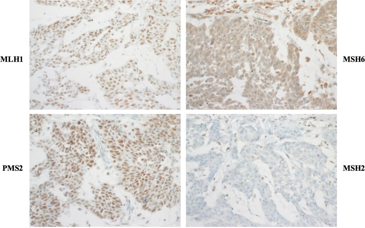
For immunohistochemical analysis, a cohort of 197 patients was available. Due to the lack of an internal positive control or tumour cells of some TMA cores, no reliable results were available in 41 cases. In 151 (96.8%) out of 156 tumours expression of all MMR proteins could be detected. In five tumours, loss of expression was observed: two cases showed an isolated loss of MSH6 expression; in two tumours, a loss in MSH2 and MSH6 expressions was detected, and one tumour showed a loss of PMS2 expression. Figure 2 illustrates the staining results of MMR-deficient tumours. Compared with MSI analysis, we identified 148 out of 150 MMR cases as MSS, the other two cases were invalid using DNA-derived MSI analysis. The two cases with MSH2 and MSH6 loss as well as one case with PMS2 loss were all identified as MSI tumours. One MSI tumour had an invalid immunohistochemistry analysis. The other two cases with MSH6 loss were MSS.

Whole slides of immunohistochemical staining of MMR-proteins MLH1, MSH2, MSH6 and PMS2. Note the strong staining of inflammatory and stromal cells and additionally the strong nuclear expression of tumour cells for MLH1 and PMS2. In contrast, immune and stromal cells are strongly stained (internal positive control), but the tumour cells are showing a nuclear loss of expression for MSH2 and MSH6. MLH1, mutL homologue 1; MMR, mismatch repair; MSH2, mutS homologue2; MSH6, mutS homologue 6; PMS2; PMS1 homologue 2.
MSI frequency using the Biocartis Idylla MSI assay
All 243 samples were analysed with the Idylla MSI Assay. In the first run, 5 out of the 243 tumours were identified as MSI-H. Furthermore, 235 tumour samples were analysed as MSS and 3 cases were invalid. To validate the results and check for reproducibility, a second run was performed (for details, see further). We obtained a final result of 234 (96.3%) MSS tumours, 4 (1.6%) MSI tumours and 5 (2.1%) invalid cases.
Reproducibility testing and comparison of the Bethesda panel and Idylla MSI assay
To prove reproducibility of Idylla MSI Assay testing, all seven cases with discordant results regarding MSS/MSI status as well as six concordant cases were selected. All seven mismatched cases, which presented with results from the NCI panel as ‘invalid’, and Idylla MSI Assay: ‘MSS or MSI’, were repeated with both methods. Table 2A summarises the repetition testing for both methods. In one case, no difference between the first and second runs was found. Three samples were tested as ‘MSS’ with the standard method (first run: NCI panel invalid) and MSS with Idylla MSI Assay. One case which was invalid using the NCI panel and MSS with Idylla MSI Assay in the first run was confirmed as invalid in the second run for both methods. One tumour that was initially identified as ‘MSI’ with Idylla was identified as MSS with both NCI panel and Idylla confirmed again in the second run and in the third Idylla run. In addition, the sample also showed preserved protein expression of MSH2, MSH6, PMS2 and MLH1. The last differing case was initially tested as invalid using the NCI panel and MSS with Idylla. In the repeating run, both the NCI panel and Idylla showed an MSI tumour. In a third repeat using Idylla, an invalid result was detected. Thus, no final result could be assigned to this tumour and it is therefore considered invalid. Table 2B summarises the concordant cases with the result MSI (three samples) or invalid (three samples), which were all repeated only with Idylla. All these results were confirmed in the second run.
Rerun of cases for reproducibility testing
Taken together, both the standard methodology and Idylla MSI Assay could identify four tumours as MSI. 230 tumours were classified as MSS by the standard method and 234 tumours by Idylla, respectively. Nine tumours and five tumours could not be classified by either standard method or Idylla, respectively, so considered invalid (table 3). The concordance rate between the standard methodology and Idylla for the detection of microsatellite instable tumours is 100%. The overall concordance rate was 98.35%. At the immunohistochemical level, MSI results were supported in three available cases with a loss in MMR proteins.
Summary of MSI results
Figure 3 depicts the different workflows and detailed working steps, including hands-on-time and time-to-result of Idylla versus standard method. Time to results differed between 3 hours using Idylla and 2 days for standard methodology.

Comparison of workflow of (A) standard method and (B) Idylla MSI assay. FFPE, formalin-fixed paraffin-embedded; MSI, microsatellite instability.
Discussion
In recent years, with the implementation of innovative therapeutic options, evaluation of MSI as a biomarker for immunomodulative therapy has gained importance in many different tumours. Due to personalised treatment options and with rising numbers of diagnostically evaluated tumours, analysis and processing times as well as the methodologies have to be reviewed with new options available.25 26 In this study, we evaluated the frequency of MSI in UTUC among a large cohort of 243 patients and identified a low frequency of MSI cases. Additionally, the results using standard methods were compared with the Idylla Assay in order to validate a new approach for MSI analysis.
UTUC is a rare entity, which mostly arise sporadically or due to the inherited Lynch syndrome.2 In the literature, variable frequencies of MSI among this malignancy have been reported. On the one hand, several studies have indicated a frequency of MSI between 13% and 28% and MMR deficiency of 30%–83% in UTUC.7 8 10 27–29 Conversely, our findings present lower tested frequencies of MSI (1.6%) and MMR deficiency (3.2%) in UTUC among a consecutively collected, multi-institutional cohort. Importantly, our results are consistent with the recent work of Necchi et al and Ericson et al, who estimated an MSI status in 3.4% and 4.6% of analysed cases, respectively.30 31 One possible interpretation for the discrepancies could be differences in tissue sample size. In studies presenting with high MSI frequencies and/or high numbers of MMR deficiency, analysis was performed with sample sizes of ≤128 cases.7 8 27–29 For example, Hartmann et al analysed 62 UTUC samples and identified a frequency of 21% MSI.8 Catto et al tested 71 UTUC samples and determined a 27% MSI frequency.27 In contrast, studies with larger cohorts, including more than 200 patients, observed a significantly lower number of MSI tested samples.30 31 Importantly, patient median age appears not to be a factor explaining the MSI differences since among the aforementioned studies patients had a median age from 65 to 71 years. Additionally, tumour stage and grade characteristics between the different cohorts were also comparable.8 10 27 30 Furthermore, it cannot be ruled out that differences in results could arise due to diverse methodologies for MSI analysis using another marker panel. Except Necchi et al, who performed MSI analysis in 114 homopolymer repeats via comprehensive genomic profiling, all other studies used the Bethesda panel for MSI analysis.8 10 27 30 In summary, our results show that MSI has a low incidence rate in UTUC. More assessments of biomarker identification for immunotherapeutic therapies are needed.
In this investigation, with the Idylla MSI Assay, a new, innovative easy to use and time-saving approach for MSI diagnostics was tested for UTUCs. To date, the newly designed marker set, consisting of seven mononucleotides, is clinically validated and applied for diagnostic use for colorectal cancer and is ongoing for verification of endometrial carcinoma.32 In our study of 243 UTUCs, we observed a high concordance of 98.35% of MSI testing between the two methods. These concordance rates are comparable with those of colorectal cancer. Compared with standard diagnostics, we propose that the Idylla MSI Assay is a more robust methodology with a lower invalid rate. Nine vs five invalid results were detected using standard method compared with the Idylla MSI Assay, respectively. Additional reasons for implementing the Idylla MSI Assay into routine laboratory testing are: (1) the turnaround time was shortened significantly to approximately 3 hours compared with a workload of 2 days using the NCI method and (2) implementing a fully automated analysis system as well as a reduction of preanalytical steps also significantly reduces human resources. Therefore, our study could determine that the same marker panel, applied for colorectal cancer samples, is transferable to UTUC with a high concordance to current established testing methods.
One case with preserved MMR protein expression was initially detected as MSI-H by Idylla, while no valid result could be obtained with the standard method. Thus, both assays were repeated twice and revealed an MSS tumour concordantly matching with preserved protein expression. The initial discordant result could be caused by lo-quality DNA or a possible heterogeneity in the tumour mass of the same tumour block, as it has been observed in colorectal cancer.33 Lastly, the isolated loss of the MSH6 protein with MSS status in two tumours could indicate a different way of activation among the MMR pathways. In the study of Gayhart et al investigating MMR proteins in UTUC, an isolated loss of MSH6 was described in 3 of 74 analysed cases. Unfortunately, there is no investigation about the relation with MSI status.28 Nonetheless, there are a few studies describing a discordance between MSI analysis and immunohistochemistry in colorectal cancer, where in some cases an isolated MSH6 loss is associated with an MSS status. This is attributed to a partial activity of the MMR pathway in some tumour cells.34
Limitations of our study include the use of a retrospective cohort. That is particularly reflected in the quality of the older aged tumour material, which leads to a possible difficulty and inaccuracy in evaluation, especially with immunohistochemical results. In addition, it should be noted that to implement the Idylla MSI Assay into routine diagnosis, the purchasing of the instrument is needed. Moreover, we were unable to obtain patient characteristics, like family history, information about Lynch syndrome and smoking history.
Conclusion
Taken together, our data support that MSI status has less influence and prevalence in UTUC compared with other tumour types, which are also part of the spectrum of Lynch syndrome-associated malignancies like colorectal or endometrial cancer. Our study confirmed Idylla MSI Assay as a valid MSI analysis tool with an extremely low failure rate. With regard to UTUC, other biomarkers, such as tumour mutational burden or PD-L1 expression, have to be analysed in terms of therapy selection.
Take home messages
We investigated the frequency of microsatellite instability (MSI) in upper urinary tract urothelial carcinoma with a large retrospective cohort and performed a comparison between the standard MSI methodology and the new diagnostic tool Idylla MSI Assay from Biocartis.
We validated Idylla MSI Assay as an alternative diagnostic approach for MSI analysis in UTUC, opening the possibility to simplify and fasten the diagnostic process in the future.
The rate of microsatellite instable tumours is low with four MSI cases out of 243 tested samples.
Data availability statement
Data are available upon reasonable request.
Ethics statements
Patient consent for publication
Ethics approval
This study was conducted in accordance with the Declaration of Helsinki, and the protocol was approved by the ethics committee of the Friedrich-Alexander University Erlangen-Nürnberg (number 329_16B) and the ethics committee of Biomedical Investigation of Andalucía (number 03619003 CCEIBA).
Acknowledgments
The present work was performed in fulfilment of the requirements of Friedrich‐Alexander University Erlangen‐Nürnberg for Friederike Kullmann for obtaining the degree 'Dr Med'. We are grateful to Verena Popp, Natascha Leicht, Christa Winkelmann and Claudia Schmied for their excellent technical support. EM-R is supported by the ’Ramón y Cajal’ Award (RYC2019-027950-I) from MICINN, Spain.
References
Supplementary materials
Supplementary Data
This web only file has been produced by the BMJ Publishing Group from an electronic file supplied by the author(s) and has not been edited for content.
Footnotes
Handling editor Runjan Chetty.
Contributors AH, RS and VW conceived the study. ME, SB, BW, DS, SW, HT, PO, HH, MFL, MLM, EM-R, MJL, DP, IH, BH-I, AH, JLB, TvD and VW collected samples and clinical data. FK, MFL, EMP, VW and RoS performed the experimental work. FK and VW completed statistical analysis. VW, RoS, BH-I, MFL, EM-R and AH supervised the experimental work. PLS, ReS, ME, FK and VW wrote the manuscript. All the authors reviewed and edited the article and approved the final version of the manuscript.
Funding This work was supported by the Interdisciplinary Centre for Clinical Research at the University Hospital of the Friedrich-Alexander University Erlangen-Nürnberg and by a grant of the ERA-NET TRANSCAN for 'Translational research on rare cancers'. Idylla MSI Assay cartridges were given free of charge by Biocartis NV (Biocartis NV, Mechelen, Belgium).
Competing interests None declared.
Provenance and peer review Not commissioned; externally peer reviewed.
Supplemental material This content has been supplied by the author(s). It has not been vetted by BMJ Publishing Group Limited (BMJ) and may not have been peer-reviewed. Any opinions or recommendations discussed are solely those of the author(s) and are not endorsed by BMJ. BMJ disclaims all liability and responsibility arising from any reliance placed on the content. Where the content includes any translated material, BMJ does not warrant the accuracy and reliability of the translations (including but not limited to local regulations, clinical guidelines, terminology, drug names and drug dosages), and is not responsible for any error and/or omissions arising from translation and adaptation or otherwise.