Article Text

Abstract
Aims—To ascertain the clinical importance of a strain of slide coagulase positive but tube coagulase negative staphylococcus species isolated from the blood culture of a 43 year old patient with refractory anaemia with excessive blasts in transformation who had neutropenic fever.
Methods—The isolate was investigated phenotypically by standard biochemical methods using conventional biochemical tests and two commercially available systems, the Vitek (GPI) and API (Staph) systems. Genotypically, the 16S ribosomal RNA (rRNA) gene of the bacteria was amplified by the polymerase chain reaction (PCR) and sequenced. The sequence of the PCR product was compared with known 16S rRNA gene sequences in the GenBank by multiple sequence alignment.
Results—Conventional biochemical tests did not reveal a pattern resembling a known staphylococcus species. The Vitek system (GPI) showed that it was 94% S simulans and 3% S haemolyticus, whereas the API system (Staph) showed that it was 86.8% S aureus and 5.1% S warneri. 16S rRNA gene sequencing showed that there was a 0 base difference between the isolate and S aureus, 28 base difference between the isolate and S lugdunensis, 39 base difference between the isolate and S schleiferi, 21 base difference between the isolate and S haemolyticus, 41 base difference between the isolate and S simulans, and 23 base difference between the isolate and S warneri, indicating that the isolate was a strain of S aureus. Vancomycin was subsequently prescribed and blood cultures taken four days after the start of treatment were negative.
Conclusions—16S rRNA gene sequencing was useful in ascertaining the clinical importance of the strain of slide coagulase positive but tube coagulase negative staphylococcus species isolated from blood culture and allowing appropriate management.
- 16S
- ribosomal
- RNA
- sequencing
Statistics from Altmetric.com
Traditionally, the identification of bacteria in the clinical microbiology laboratory is performed by isolating the organism and studying it phenotypically to elucidate its Gram staining, culture, and biochemical characteristics, which have been the gold standard of bacterial identification. However, these methods have two major drawbacks. First, they cannot be used for non-cultivable organisms. Second, we are occasionally faced with organisms with biochemical characteristics that do not fit into the patterns of a known genus and species.
Since the discovery of the polymerase chain reaction (PCR) and DNA sequencing, comparisons of the gene sequences of bacterial species have shown that the 16S ribosomal RNA (rRNA) gene is highly conserved within a species and among species of the same genus, and hence can be used as the new gold standard for the speciation of bacteria. Using this new standard, phylogenetic trees, based on base differences between species, are constructed and bacteria are classified and re-classified into new genera.1, 2 Furthermore, non-cultivable organisms and organisms with ambiguous biochemical profiles can be classified and identified.3, 4 Recently, we have reported the application of 16S rRNA gene sequencing in the identification of two clinical isolates with ambiguous biochemical profiles,5, 6 and a bacterium that was non-cultivable.7 In our present study, we report the application of such a technique to the identification of a strain of Staphylococcus aureus, which is slide coagulase positive but tube coagulase negative, isolated from a patient with myelodysplastic syndrome. The importance of identifying the organism is also discussed.
Methods
PATIENT AND MICROBIOLOGICAL METHODS
All clinical data were collected prospectively as described in our previous publication.8 Clinical specimens were collected and handled according to standard protocols, and all suspect colonies were identified by standard conventional biochemical methods.9 In addition, the Vitek System (BioMerieux Vitek, Hazelwood, Missouri, USA) and the API system (BioMerieux Vitek) were used for the identification of the bacterial isolate.
EXTRACTION OF BACTERIAL DNA FOR 16S rRNA GENE SEQUENCING
Bacterial DNA extraction was modified from our previously published protocol.10 Briefly, 80 μl of NaOH (0.05M) was added to 20 μl of bacterial cells suspended in distilled water and the mixture was incubated at 60°C for 45 minutes, followed by the addition of 6 μl of Tris/HCl (pH 7.0), achieving a final pH of 8.0. The resultant mixture was diluted 100× and 5 μl of the diluted extract was used for PCR.
PCR, GEL ELECTROPHORESIS, AND 16S rRNA GENE SEQUENCING
PCR amplification and DNA sequencing of the 16S rRNA gene were performed according to our previous publications.5–7 Briefly, DNase I treated distilled water and PCR master mix (which contains dNTPs, PCR buffer, and Taq polymerase) were used in all PCR reactions by adding 1 U of DNase I (Pharmacia, Uppsala, Sweden) to 40 μl of distilled water or PCR master mix, incubating the mixture at 25°C for 15 minutes, and subsequently at 95°C for 10 minutes to inactivate the DNase I. The bacterial DNA extract and control were amplified with 0.5μM primers (LPW57, 5′-AGTTTG ATCCTGGCTCAG-3′ (nucleotides 0–18); and LPW58, 5′-AGGCCCGGGAACGTAT TCAC-3′ (nucleotides 1365–1383)) (Gibco BRL, Rockville, Maryland, USA). The PCR mixture (50 μl) contained bacterial DNA, PCR buffer (10mM Tris/HCl, pH 8.3, 50mM KCl, 2mM MgCl2, and 0.01% gelatin), 200μM of each dNTP and 1.0 U Taq polymerase (Boehringer Mannheim, Mannheim, Germany). The mixtures were amplified for 40 cycles at 94°C for one minute, 55°C for one minute, and 72°C for two minutes, followed by a final extension at 72°C for 10 minutes in an automated thermal cycler (Perkin-Elmer Cetus, Gouda, the Netherlands). DNase I treated distilled water was used as the negative control. A 10 μl aliquot of each amplified product was electrophoresed in 1.0% (wt/vol) agarose gel, with a molecular size marker (φX174 HaeIII digest, Boehringer Mannheim) run in parallel. Electrophoresis in Tris/borate/EDTA buffer was performed at 100 V for 1.5 hours. The gel was stained with ethidium bromide (0.5 μg/ml) for 15 minutes, rinsed, and photographed under UV light illumination.
The PCR product was gel purified using the QIAquick PCR purification kit (QIAgen, Hilden, Germany). Both strands of the PCR product were sequenced twice with an ABI 310 automated sequencer according to the manufacturer's instructions (Perkin-Elmer, Foster City, California, USA), using the PCR primers (LPW57 and LPW58) and additional primers designed from the sequencing data of the first round of the sequencing reaction (LPW252, 5′-AGCCACGGCTAACTACGT-3′ (nucleotides 494–511); and LPW253, 5′-AGTT TCAACCTTGCGG-3′ (nucleotides 928–943)). The sequence of the PCR product was compared with known 16S rRNA gene sequences in the GenBank by multiple sequence alignment using the CLUSTAL W program.11
Results
PATIENT AND IDENTIFICATION OF THE BACTERIAL STRAIN BY TRADITIONAL METHODS
During consolidation chemotherapy with idarubicin, cytarabine, and etoposide, a 43 year old woman with refractory anaemia with excessive blasts in transformation developed fever. Physical examination showed an oral temperature of 39°C. There were no signs of exit site infection of the central catheter. The following test results were found: haemoglobin, 108 g/litre; total white blood cell count, 0.2 × 10⁹/litre; neutrophils, 0; lymphocytes, 0.2 × 10⁹/litre; monocytes, 0.0 × 10⁹/litre; and platelets, 8 × 10⁹/litre. Blood cultures were performed through the two lumens of the central catheter and peripheral puncture and were incubated using the BACTEC 9240 blood culture system (Becton Dickinson, Sparks, Maryland, USA). Intravenous imipenem (500 mg every six hours) was given as empirical treatment for neutropenic fever.
On day 1 post-incubation, one pair of blood cultures turned positive with a Gram positive, facultative anaerobic coccus in clusters. The bacterium grew on blood agar, chocolate agar, and MacConkey agar to sizes of 1–2 mm in diameter after 24 hours of incubation at 37°C in ambient air. It was yellow, non-haemolytic, catalase positive, slide coagulase positive, but tube coagulase negative. The Vitek system (GPI) showed that it was 94% S simulans and 3% S haemolyticus, whereas the API system (Staph) showed that it was 86.8% S aureus and 5.1% S warneri (table 1). Therefore, sequencing of the 16S rRNA gene was performed to speciate the isolate. The strain was resistant to penicillin G, methicillin, gentamicin, erythromycin, clindamycin, and amoxicillin/clavulanate, but sensitive to vancomycin, cotrimoxazole, fusidic acid, rifampicin, and minocycline. Vancomycin was subsequently prescribed and blood cultures taken four days after the start of treatment were negative.
Biochemical profiles and identification of the blood culture isolate by conventional biochemical tests, the Vitek GPI system, and the API Staph system
16S rRNA GENE SEQUENCING
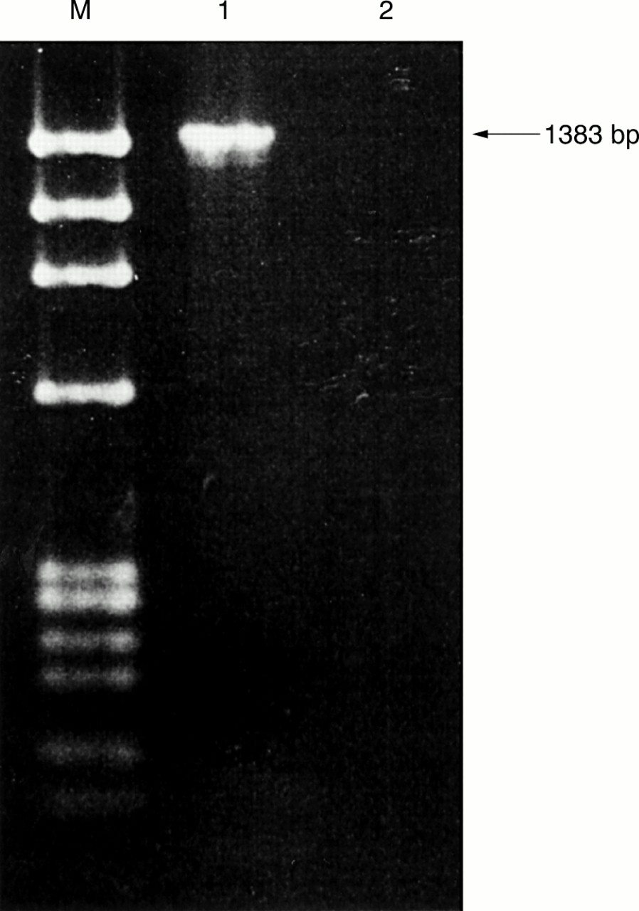
PCR of the 16S rRNA gene of the bacterium showed a band at 1383 bp (fig 1). The base sequences of the purified band and the corresponding region in S aureus (GenBank accession no. L37597), S lugdunensis (GenBank accession no. AB009941), S schleiferi (GenBank accession no. AB009945), S haemolyticus (GenBank Accession no. D83367), S simulans (GenBank accession no. D83373), and S warneri (GenBank accession no. AJ276810) were compared. There was a 0 base difference between the isolate and S aureus, 28 base difference between the isolate and S lugdunensis, 39 base difference between the isolate and S schleiferi, 21 base difference between the isolate and S haemolyticus, 41 base difference between the isolate and S simulans, and 23 base difference between the isolate and S warneri, indicating that the isolate was a strain of S aureus.

DNA products from PCR of 16S rRNA gene. Lane M, molecular marker (φX174 HaeIII digest); lane 1, blood culture isolate from patient; lane 2, negative control containing DNase I treated distilled water.
Discussion
Small rRNA gene sequencing, particularly 16S rRNA sequencing in bacteria, has led to advances on multiple fronts in microbiology. First, the construction of a universal phylogenetic tree classifies organisms into three domains of life: bacteria, Archaea, and Eucarya.1, 2, 11 Second, it revolutionises the classification of microorganisms, and makes the classification of non-cultivable microorganisms possible.3, 4 Third, it helps to elucidate the relation of unknown bacterial species to known ones. New species of bacteria such as Gemella sanguinis, Mycobacterium heidelbergense, and Massilis timonae have been discovered using 16S rRNA sequencing.12–14 Bacteria such as Pseudomonas veronii, Mycobacterium celatum, and Methylobacterium zatmanii, which were not known to cause infections in humans, have been identified in clinical specimens using this technique.7, 15, 16 Furthermore, bacteria difficult to identify were speciated successfully using this technique, and we have recently used it in the identification of a strain of Mycobacterium neoaurum with ambiguous biochemical and whole cell fatty acid profiles isolated from a patient with acute lymphoblastic leukaemia, and a strain of Escherichia coli with an ambiguous biochemical profile isolated from a bone marrow transplant recipient with acute myeloid leukaemia.5, 6
Tube coagulase negative S aureus have been reported in the literature since 1988.17 In some reports, the authors used other phenotypic tests, such as strong DNase activity, production of phosphatase and slide coagulase, nitrate reduction, and utilisation of certain sugars, to identify the isolates as S aureus.17–19 However, phenotypic tests cannot identify isolates with ambiguous biochemical profiles. In fact, for the first tube coagulase negative S aureus reported,17 the isolate was mannitol and ribose negative, but maltose and Voges Proskauer reaction positive. This made the identification of the isolate as a strain of S aureus questionable. Recently, it was reported that amplification of the coagulase gene can be used to identify tube coagulase negative S aureus,20 and it was found that coagulase deficiencies in these isolates can involve both transcriptional and post-transcriptional defects.21 However, the tube coagulase gene can be present in other staphylococci, such as S intermedius and S hyicus. Therefore, it is not absolutely specific for S aureus.
We describe the first identification of a strain of tube coagulase negative S aureus by 16S rRNA gene sequencing. We chose 16S rRNA sequencing as the means of identification because it is extremely specific—the identity can be clearly defined by the number of base differences between the isolate and the existing species. The organism in our study was isolated from the blood culture of a patient with myelodysplastic syndrome. Conventional biochemical tests failed to identify the bacterium to species level. Tube coagulase negative but slide coagulase positive staphylococci include S lugdunensis and S schleiferi. Reactions pointing against S lugdunensis include arginine hydrolysis (1%), utilisation of mannitol (0%), and alkaline phosphatase (16%); whereas reactions pointing against S schleiferi include utilisation of maltose (0%), mannitol (0%), sucrose (0%), and lactose (1%), and urease production (0%). Because there is no base difference between the 16S rRNA gene sequence of the isolate and that of S aureus, the conclusion that the isolate was a strain of tube coagulase negative S aureus can be made unequivocally.
Identification of the organism is important because the management of the patient can differ radically. If the organism was a strain of S aureus (as in this case), vancomycin should be prescribed. In contrast, if it was a coagulase negative staphylococcus, blood cultures should be repeated for the differentiation between genuine coagulase negative staphylococcal bacteraemia and blood culture contamination, because prudent use of vancomycin is crucial for the battle against the emergence of vancomycin resistant Gram positive organisms.22
16S rRNA gene sequencing will continue to be the gold standard for the identification of bacteria, and the automation of the technique could enable it to be used routinely in clinical microbiology laboratories, as a replacement of the traditional phenotypic tests. Modern technologies have made it possible to construct a high density of oligonucleotide arrays on a chip with oligonucleotides representing the 16S rRNA gene sequence of various bacteria. Such a design will facilitate automation of the annealing and detection of the PCR products of 16S rRNA gene amplification, and hence routine identification of most clinical isolates will be possible. The use of 16S rRNA gene sequencing has several advantages. First, the turnaround time is short. Because amplification of the 16S rRNA gene takes only four to six hours, and the annealing and detection of PCR products takes only another few hours, theoretically the identification can be completed within one day. Second, it can be used for slow growing bacteria, unlike most commercially available kits that are based on phenotypic tests that require the detection of growth of the organism in the presence of certain specific substrates, and hence the slow growing bacteria are usually “unidentified” when the growth control shows a negative result. Third, the problem of “unidentifiable strains” will be overcome and there would be minimal misidentification—the identification of a clinical strain is clearly defined by the number of base differences between it and the existing species. Fourth, oligonucleotides representing all bacterial species, including those rarely encountered clinically, can be included in the array, making it easy to identify the rare species. Lastly, such a technique will be applicable not only to pyogenic bacteria, but also to other organisms such as mycobacteria, which are not identified in most clinical microbiology laboratories because special expertise and equipment, such as gas liquid chromatography, are required. Therefore, eventually it should reduce not only manpower, but also capital costs and costs of consumables. Although the cost effectiveness of using 16S rRNA gene sequencing in routine clinical microbiology laboratories remains to be evaluated, our study reports the first case of tube coagulase negative S aureus bacteraemia identified by 16S rRNA gene sequencing.
Acknowledgments
This work was partly supported by the committee of research and conference grants, the University of Hong Kong.