Article Text

Abstract
Aim—To develop a simple method of recording digital images of surgical specimens on to a personal computer (PC) for use in presentations for teaching and reporting of their pathology.
Methods—A perspex box was constructed to international A4 size 100 mm deep. This box had a base of 3 mm clear perspex with sides and top of 5 mm white perspex. This box was partially filled with distilled water and a specimen immersed in it. It was then placed on top of a standard A4 scanner. The specimen was then scanned into a PC using image capture software.
Results—The images produced showed noticeable improvement over normal photographs, especially with specimens prone to wet highlights.
Conclusions—The method has proved to be a rapid and efficient means of producing macroscopic images of surgical specimens.
- scanning
- personal computer
- macroscopic images
- surgical specimens
Statistics from Altmetric.com
Our laboratory has for some years been using a Polaroid land camera for recording—for reference or teaching—macroscopic images of interesting surgical specimens. A disadvantage of this method is the inability to obtain further pictures or produce slide transparencies for meetings or teaching.
When reasonably priced digital cameras became available one was purchased as an alternative to the Polaroid camera. The results from this camera, although an improvement, were disappointing for many reasons. Having no aperture control limited the available depth of field and the colour response to tungsten lamps used on our copying stand was not good, probably because of high sensitivity of the CCD (charge coupled devices) in the camera to the infra red emission of the bulbs.
Because we had already been using a personal computer (PC) with scanning attachment for producing reports, some experiments were conducted using clear plastic bags to keep specimens immersed and to protect the scanner from contamination. From this the following system was developed.
Method
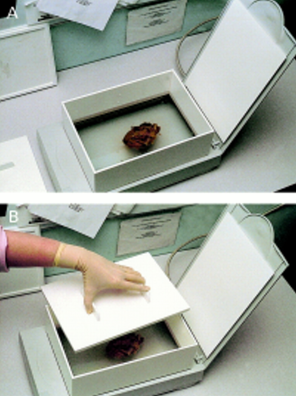
A perspex box of international A4 size was constructed (fig 1). The box was 100 mm deep, had a sliding heavy insert to keep specimens immobile, and had a close fitting lid. The base was 3 mm clear perspex, the remainder of the box being constructed from 5 mm white perspex. The cost of materials for this was approximately £50.

(A) The perspex box mounted on the scanner. (B) Application of the heavy sliding insert.
The box was filled to a depth of 25 mm (for an average specimen) with distilled water or alcohol (for colour enhancement) and the specimen immersed and orientated, being careful to avoid trapped bubbles. The insert is then added to prevent the specimen moving and finally the lid secured. The box is then placed on the platten of an inexpensive A4 flatbed SCSI (small computer systems interface) scanner and the specimen scanned into the PC and manipulated using suitable image capture and manipulation software. In our department we use Adobe Photoshop but any similar software would provide comparable results.
For most specimen images the usual digital manipulation techniques used were minor brightness and contrast adjustments and an unsharp mask was used to sharpen the image (all scanned images require a certain amount of sharpening). Sometimes the background was “cleaned” up for aesthetic purposes. Details of specific manipulation techniques are to be found within the respective manual of the software used.
To avoid possible contamination or spillage in the vicinity of the computer the scanner was sited in the cut up room and connected with an extension cable to the PC in an adjacent room. We were able to use an existing PC but suitable systems are available for less than £1000.
Results
The images produced by scanning needed minimal manipulation as described previously. The images produced showed noticeable improvement over those taken with a standard point and shoot digital camera, this improvement being most noticeable with specimens prone to wet highlights, such as open bowels (fig 2A and B). It is possible to remove the highlights from camera images such as fig 2B with image manipulation techniques; however, this is extremely time consuming and degrades the accuracy of the image. This was unnecessary with the scanned image, thus preserving its integrity. The depth of field available on the scanner appears to be about 30 mm, which is adequate for most average sized surgical specimens. Slices of whole organs were also scanned with good results (fig 3).
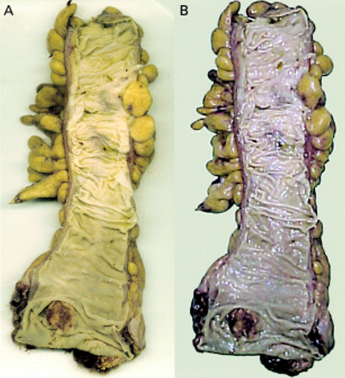
(A) Scanned opened bowel with small carcinoma near distal margin. (B) Same specimen photographed using digital camera showing severe highlights and poor colour reproduction. Both digital images were subjected to the same image manipulation.

Kidney slice with large carcinoma in upper pole.
Discussion
It has long been considered desirable to keep images of interesting or complex specimens to record the appearances and origin of wax blocks taken during cutup. Previously, photocopying of organ slices has been advocated.1 Digital images are far more adaptable than any other media because images saved in the computer can be used for presentations using standard software, such as Microsoft Powerpoint, or added to typed histopathology reports to enhance the information presented. The images can also be easily archived on disk using a compact disk writer installed in the PC.
The method is not intended to replace the high quality photographic methods in current use by medical photographers or the results obtainable by using professional standard digital cameras such as the Nikon D1 SLR with modern lighting systems. However, the cost of the former (in excess of £5000) is beyond the resources of most histopathology laboratories.
For the district general hospital this method is a rapid and inexpensive means of producing good quality photographic reproductions of surgical specimens.