Article Text

Abstract
Aims: To evaluate two detailed step sectioning protocols for sentinel lymph nodes (SLNs).
Methods: After vital dye or combined dye and radiocolloid guided biopsy, SLNs were fixed in formalin and embedded in paraffin wax. In protocol A, SLNs from 123 patients were sectioned in steps of 50–100 μm, whereas in protocol B, SLNs from 123 patients were sectioned at steps of 250 μm. Epithelial marker immunohistochemistry (IHC) was performed on multiple levels in cases with negative haematoxylin and eosin findings.
Results: In groups A and B, 74 and 47 patients were found to have tumour cells in their axillary SLNs, and 19 (28%) and 18 (19%) patients, respectively, were upstaged as compared with the standard histological assessment. Nodal involvement detected by deeper sections was often micrometastatic or in isolated tumour cells
Conclusions: Serial sectioning and IHC are recommended for the evaluation of SLNs. The optimal extent of the histopathological work up should be studied further.
- breast cancer
- immunohistochemistry
- micrometastasis
- sentinel lymph nodes
- step sectioning
- HE, haematoxylin and eosin
- IHC, immunohistochemistry
- SLN, sentinel lymph node
- SLNB, sentinel lymph node biopsy
- VP, virtual protocol
Statistics from Altmetric.com
- HE, haematoxylin and eosin
- IHC, immunohistochemistry
- SLN, sentinel lymph node
- SLNB, sentinel lymph node biopsy
- VP, virtual protocol
The sentinel lymph node (SLN) theory in breast cancer suggests that the metastatic spread of mammary carcinomas through the lymphatic vessels follows an orderly pathway, and that only one or, at most, a few lymph nodes, the SLNs, are reached first by the process of metastasis. These are therefore the most likely sites of lymphogenic metastases. Identification, biopsy, and histological assessment of these nodes seems an ideal procedure for the qualitative staging of breast cancer, to clarify whether the tumour has spread to the axilla or not.1–4 It is generally believed that patients who have a positive SLN require axillary dissection to ensure a more quantitative staging, by indicating the number of lymph nodes involved. Axillary dissection can also provide a form of regional disease control, but if this option is not taken then treatment should include radiotherapy to the axilla. Alternatively, those patients who have negative SLNs are not likely to benefit from dissection or further treatment of the axilla.
“The sentinel lymph node theory in breast cancer suggests that the metastatic spread of mammary carcinomas through the lymphatic vessels follows an orderly pathway”
Because the SLNs are the most likely sites of regional metastasis, their histopathological assessment provides an opportunity for a more accurate staging by way of a more intensive investigation.5,6 However, there are many controversial issues regarding the extent of the histopathological work up, which lacks uniformity.7 Our study reports on the yields of two extensive protocols for the histopathological evaluation of axillary SLNs.
MATERIALS AND METHODS
The method of SLN biopsy (SLNB) first involved the peritumoral injection of patent blue dye, but a combined dye and radiocolloid guided technique preceded by lymphoscintigraphy is now preferred. Both tracers were administered peritumorally, although a few patients with negative lymphoscintigrams also received a smaller dose of radioactive tracer subareolarly. Technical details of the two biopsy methods, and their comparison, have been reported elsewhere.8,9 Briefly, blue lymphatics were always looked for first, and this was followed by a search for the remaining radioactive nodes. SLNs were defined as blue nodes, nodes at the end of a blue lymphatic, or radioactive nodes with an ex vivo radioactivity level ≥ 10 times the background value. SLNs were sent for pathological assessment separately.
At the beginning of our study, which comprised our institutional validation phase, all patients (n = 191) but five underwent level I and II or complete axillary dissection after the SLNB. Four of these five patients had in situ carcinomas diagnosed preoperatively as malignant, but not as in situ carcinomas, and one had a cribriform carcinoma measuring 1 mm. Later, axillary dissection was planned only if the SLNs were positive (n = 55).
All SLNs were fixed in 7% buffered formalin. Smaller nodes were embedded in paraffin wax in toto, whereas larger nodes were bisected and both halves were processed.
In group A (protocol A), all of the SLNs were sectioned serially until the wax blocks were completely used up. Not all 3 to 5 μm thick sections were taken for histology, but every 10th to 20th section was examined (depending on nodal size) and, for a given node, the depths between the examined sections were approximately the same. This method of sectioning resulted in the examination of sections relatively equidistant (50–100 μm) from each other, depending on the overall nodal size, the distances being greater for larger SLNs. After every sixth section taken for haematoxylin and eosin (HE) staining, one was taken for cytokeratin immunohistochemistry (IHC) (NCL-PAN-CK; Novocastra, Newcastle, UK; 1/100 dilution; or MNF116 (M0821); Dako, Glostrup, Denmark; 1/100 dilution). At the beginning of our study, epithelial membrane antigen (EMA; MU-182-UC; Biogenex, San Ramon, California, USA; 1/50 dilution) immunostaining was also performed in each node assessed, alternating with sections investigated by means of anticytokeratin antibodies. Immunostains were investigated only in the event of negative HE findings.
In group B (protocol B), the SLNs were sectioned at steps of 250 μm. These sections were stained with HE. Sections for IHC were taken after every third section stained, and were further processed in cases of negative HE findings.
A few obviously metastatic nodes were examined on only one or a few sections in both groups.
Tumours and metastases were classified on the basis of the TNM system.10–12
In the comparison of the two equally sized groups, the χ2 test was used for categorical data, and the Student’s t test for continuous variables. Significance was set at p < 0.05.
RESULTS
Although the aim of our study was the description and possible comparison of two detailed histopathological analyses of SLNs, data on the results of SLNB itself are given here as background data required for a better understanding.
As reported previously, during the validation process of our study, the accuracy of the use of the SLNs to predict the overall axillary nodal status was 95.7% (176 patients with SLN status matching the axillary nodal status/184 patients with successful SLNB), with a false negative rate of 7.5% (eight patients with negative SLNs/106 patients with a positive nodal status). As part of this process, performance of the combined SLNB method on 72 patients during the validation phase yielded accuracy and false negative rates of 98.6% and 3.3%, respectively (one false negative SLNB/30 node positive patients).9
In a previous report on the first 58 patients investigated by protocol A, a mean of 49 levels/SLN (range, 25–102) was reported.13 For protocol B, the mean number of levels/SLN was 21 (range, 7–46).
Table 1 compares the tumours of the patients in groups A and B. The tumours were significantly larger in the former group (χ2 = 14.25; p < 0.05), their mean (SD) sizes being 2.30 (0.98) cm and 1.91 (0.90) cm, respectively (t = 3.15; p < 0.01).
Comparison of tumour sizes categorised on the basis of the TNM system10
Table 2 compares the metastases identified by the two protocols. Even when all the categories of table 2 were considered, there was a significant difference between the numbers of metastases identified in the two groups (χ2 = 15.91; p < 0.025). It was mainly of interest to compare the metastatic yields of the two protocols, and accordingly the tumours were also grouped into metastatic and non-metastatic to the SLNs; protocol A identified significantly more metastases. When isolated tumour cell positivity was included as nodal involvement, 60.2% of the tumours in group A and 38.2% of the tumours in group B had nodal involvement (χ2 = 11.86; p < 0.001) (table 3). When isolated tumour cell positivity was taken into account as N0, as suggested by TNM experts,12 52.8% of the tumours in group A and 34.1% of the tumours in group B had nodal involvement (χ2 = 8.75; p < 0.01). Table 3 shows the figures when any nodal involvement was taken as a positive result; the numbers of patients with positive SLNs in a given tumour size category and their percentage of the total number of tumours in the same category are shown.
The absolute and relative numbers of involved sentinel lymph nodes in the two groups stratified by tumour size
Figures 1 and 2 show the stepwise yields of sectioning protocols A and B, respectively.
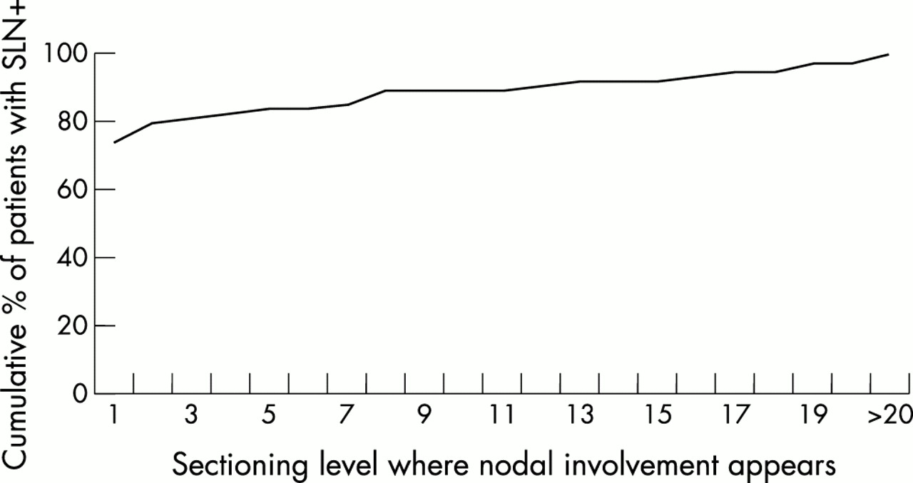
Stepwise cumulative yield of step sectioning at 50–100 μm (protocol A). The rate of identification of sentinel lymph nodes (SLNs) with any involvement expressed as a percentage of the total number of SLN positive patients.

Stepwise cumulative yield of step sectioning at 250 μm (protocol B). The rate of identification of sentinel lymph nodes (SLNs) with any involvement expressed as a percentage of the total number of SLN positive patients.
Table 4 shows the proportion of isolated tumour cells, micrometastases, and macrometastases identified by different levels of the step sectioning protocols. It is evident from this table that metastases detected by deeper levels are more likely to be micrometastases or isolated tumour cells.
Proportion of isolated tumour cells and/or micrometastases and macrometastases to the total number of metastases detected by each protocol stratified by levels
After analysing the real protocols, virtual protocols were also created on the basis of protocol A. In these virtual protocols (VPs) only every third (VP3), fourth (VP4), fifth (VP5), or sixth (VP6) level of the SLNs from group A were hypothetically examined. Table 5 shows the results of their analysis.
Analysis of virtual protocols derived from protocol A
DISCUSSION
The identification of SLNs makes these nodes suitable for a more intensive histological work up. Serial sectioning and IHC would be too expensive for all axillary nodes, but these special methods may be cost effective for the detection of occult metastases in a limited number of specific nodes, the SLNs.5 However, because most of our knowledge of the prognostic relevance of nodal metastases is based on standard pathological assessments, metastases detected by special methods are of uncertain biological relevance, and some authors recommend only standard assessment of the SLNs in the routine setting.14,15 Despite the fact that the biological meaning of minimal nodal involvement (micrometastatic disease and isolated tumour cell involvement) is unclear, there is growing evidence that their presence may indicate a somewhat worse outcome, at least in some subgroups of patients.16–18 Currently, most studies reporting on SLNB use special techniques to detect minimal involvement.6 However, the extent of the histopathological work up varies from study to study.6
“Many laboratories still use a single level assessment for the determination of the nodal status of malignant tumours, despite the fact that the inadequacy of such methodology was pointed out several decades ago”
In our present study, the yields of two extensive histological protocols for SLNs were assessed without considering the issue of the biological importance of the metastatic deposits identified. Because of the stricter inclusion criteria, the tumours studied later in the course of our SLNB series were smaller, and this could impact on the rate of their metastatic involvement; therefore, the results of the two methods used must be compared with caution. The rates of isolated tumour cells and micrometastases in groups A and B were 23% and 32%, respectively. This suggests that the difference in tumour size contributed considerably to the difference in the rate of nodal involvement identified, which is also reflected by the virtual protocols, where the upstaging rate of VP6 (21%) was above that of protocol B (19%), despite the larger steps in sectioning. When the nodal involvement detected in each tumour size category was considered (table 3), it became clear that the higher yield of protocol A was also present in most categories, particularly in the pT1c and pT2 tumours, which were the most common ones found in the series. Because of the different tumour sizes in the two groups, the yields of the two histopathological protocols studied are best viewed separately.
Many laboratories still use a single level assessment for the determination of the nodal status of malignant tumours, despite the fact that the inadequacy of such methodology was pointed out several decades ago.19,20 Some of the recent recommendations consider a single section from each lymph node to be sufficient,21 although there are statements on the macrosectioning of larger nodes for better fixation and examination.14,22 The National Health Service breast screening programme in the UK recommends taking up to four separate blocks from each node, depending on its size,23 and it is believed that this assessment would be suitable for SLNs too.14 The recommendation of the College of American Pathologists’ surgical pathology committee for handling SLNs states that a minimum protocol for SLNs should include three HE stained levels.24 A standard assessment of SLNs was used in several studies on these nodes, but many have used a more detailed approach, with either serial or step sectioning or IHC, or both.6 Molecular analysis of SLNs with the reverse transcription polymerase chain reaction has also been applied as a method of ultrastaging in some studies,6 but a critical assessment of these results is beyond the scope of this article.
The specific role that SLNs may play in breast cancer staging and in the omission of axillary dissection (formerly required for adequate staging) has stimulated particular interest in the identification of small metastases in SLNs and the study of their effect on non-SLN metastases. The size of the SLN metastases was found to correlate with the probability of occurrence of non-SLN metastases.25–29 However, it has also been established in a few studies that, even if the chances of associated non-SLN metastases are lower, small SLN metastases may be associated with non-SLN involvement.27–33 The rate of non-SLN metastases may reach 11% when SLN involvement is demonstrated by cytokeratin IHC,30 and if the SLN micrometastases are > 1 mm, the incidence of non-SLN involvement may be as high as 36%.29 Therefore, if the decision on axillary dissection is to be based on the result of SLNB, micrometastases also warrant axillary dissection (or irradiation), at least until the results of the American College of Surgeons’ oncology group Z-11 trial (randomising patients with SLN metastasis to axillary dissection versus no further surgery) are available.34
Both of our protocols identified a substantial proportion of the metastases in the first or the first few sections (figs 1, 2), and this is a general phenomenon seen in multiple studies assessing the role of a more intensive histopathological work up summarised in table 6. A single level HE stain approach would have identified somewhat more than 60–70% of the metastatic nodes. Although metastases in the SLNs show a predilection for the site where the tumour draining lymphatic vessel reaches the lymph node,51 the identification of this site requires vital dye labelling of the SLN, in addition to a piece of the blue stained lymphatic vessel. There has also been a suggestion by Turner et al that the site most likely to contain metastatic deposits is often located opposite the hilum of the node.38 Often, neither the inflow point of the tumour draining lymphatic vessel, nor the plane opposite to the hilum are easy to find, especially in SLNs identified by radioactivity alone.52,53 Deeper levels are also likely to demonstrate metastatic tumour cells, and this has been shown in all our real and virtual protocols, and in many other studies (table 6).
Review of studies assessing the role of serial or step sectioning and immunohistochemistry in the assessment of sentinel lymph nodes
It is clear from table 6 that the use of epithelial marker (cytokeratin) IHC as a more sensitive method for detecting metastases results in the upstaging of between 2.6% and 24.4% of the patients with SLNs found to be free of tumour cells by standard HE assessment. The upper limit of this conversion rate can be criticised: two of the three cases cited in the article by Mann et al had nodal involvement identified by IHC only, but this could have been seen on HE staining also.45 It is thought that a more accurate estimate of upstaging by IHC alone is between 2.6% and 19%.38,39 The addition of serial sectioning, as a method of more intensive sampling of the SLN, resulted in the upstaging of from 4.8% to 30.7% of the patients initially found to be node negative by standard HE assessment of the SLNs. The standard assessment usually identifies macrometastases (> 2 mm), whereas subsequent levels are more likely to identify micrometastases or isolated tumour cells. As far as we are aware, no study has given details on the proportion of these extremely small metastatic foci with regard to the levels at which they are detected. Thus, it can be concluded from these series that the chances of identifying macrometastases diminish with the number of levels assessed, and even in a population suitable for SLNB based selective axillary treatment or omission of axillary dissection (group B in our study), the chances of detecting macrometastases may be as high as 17% after examining five levels at 250 μm.
“The costs and affordability of step sectioning and immunohistochemistry must be balanced against each other to attain a reasonable assessment protocol for the work up of sentinel lymph nodes”
Three studies are of particular relevance, because they reported on a sufficient number of patients and evaluated SLNs cut until the blocks were completely used up.40,46,47 Because metastases are not uncommonly distributed away from the central cross section 13, these studies are those that document the highest rate of conversion from standard HE negative SLNs to involved SLNs. With the use of extensive step sectioning, the role of IHC in identifying metastatic cells or groups of metastatic cells decreases; IHC identified only 5.4% and 12.8% of the involved nodes in our protocols A and B, respectively, whereas it helped to strengthen the suspicion of metastatic cells in only three cases in the Milan series.40 The smaller the sectioning steps, the higher the rate of metastatic cells or micrometastases detected and the lower the value of IHC.
The combination of step sectioning and IHC gives the best rates of identification of occult metastatic deposits. The costs and affordability of step sectioning and IHC must be balanced against each other to attain a reasonable assessment protocol for the work up of SLNs. It must be accepted that histopathology is based on tissue sampling and has a rate of unidentified lesions (including occult metastases). If it is assumed that the identification of an isolated tumour cell and a group of 10 tumour cells randomly placed in a 1 cm SLN would require 312 and 139 sections, respectively, each of the SLN histology protocols studied to date must have missed a few cases of minimal nodal involvement of unknown importance.52 Complete examination of any routinely assessed specimen is not feasible and is unwise. Rather, a compromise between workload, costs, and sensitivity should be found. Our study is one step towards finding such a compromise. At least, it can be concluded from our results that the combination of step sectioning and IHC may be recommended for the work up of SLNs. Whether the detection of small metastatic foci (isolated tumour cells or micrometastases) detected in deeper sections is clinically relevant or not is not known. As discussed previously, the size of SLN metastases might be important in either predicting further nodal involvement (and therefore indicating some type of regional treatment) or in influencing survival and prognosis (and therefore influencing the indication of adjuvant systemic treatment). SLNB has a recognised false negative rate, which seems acceptable to many. A certain risk of further nodal involvement may also be acceptable, in addition to a minor prognostic disadvantage. Therefore, the extent of step sectioning and the impact of extremely small metastatic foci in SLNs on non-SLN involvement and on the fate of the patients remain to be elucidated in prospective clinical trials and probably cost benefit analyses.
Take home messages
-
Serial sectioning and immunohistochemistry (IHC) increase the rates of identification of metastases in sentinel lymph nodes (SLNs) and are therefore recommended for the evaluation of SLNs
-
Complete examination of any routinely assessed specimen is not feasible, but a compromise between workload, costs, and sensitivity should be found
-
The optimal extent of the histopathological work up should be investigated further
-
It is still unclear whether the detection of small metastatic foci (isolated tumour cells or micrometastases) in deeper sections is clinically relevant
Acknowledgments
This work was supported by grant ETT-176/2001 from the Hungarian Ministry of Health.