Article Text

Statistics from Altmetric.com
Neonatal thromboembolic events, both arterial and venous, are rare but increasingly recognised problems in tertiary care neonatology. The pathophysiology of these events in the context of the neonatal haemostatic system and the importance of both inherited and acquired prothrombotic disorders remain poorly defined. Similarly, optimal diagnostic and therapeutic approaches in this setting are not currently based on sound clinical evidence, and there is a clear need to investigate these issues, which will inevitably require large multicentre studies.J Clin Pathol 2000;53:419–423
Epidemiology
The peak incidence of thromboembolic events in the paediatric age group occurs in neonates and infants less than 1 year of age.1 The published literature on neonatal thrombosis consists largely of case reports and small, single centre case series, and is therefore open to bias. Recently, however, two neonatal registries from Canada and Germany have collected prospective data on unselected cases from multiple centres. In the Canadian registry, the incidence of clinically apparent thrombosis (excluding stroke) was 2.4/1000 admissions to neonatal intensive care units, whereas in the German study, symptomatic thrombosis was recorded in 5.1/100 000 births.2,3
Clinical features
Table 1 shows the distribution of events in these two studies. Although broadly similar, there are some important differences, with the German registry recording renal vein thrombosis as the most common venous event, whereas in the Canadian study other venous events predominated. In addition, in the Canadian registry, despite the exclusion of stroke, there was a relatively high incidence of arterial events compared with the German group. The reasons for these differences are not clear, but might simply represent the relatively small numbers involved. Cerebral events were included in the German registry, where there were nine in a total of 79 arterial events with, perhaps surprisingly, no cases of sinovenous thrombosis recorded. In both studies, renal vein thrombosis tended to present early, particularly in term infants, with other venous and arterial events presenting later.
Distribution of thombotic events
The single most important risk factor for the development of thrombosis in both studies was the presence of an indwelling central line and, consequently, both arterial and venous events tended to involve those large vessels used most commonly for catheterisation. Other risk factors identified include asphyxia, septicaemia, dehydration, and maternal diabetes.
The importance of indwelling catheters in the aetiology of neonatal thrombosis is a recurring theme in all studies in this age group. Owing to its size and easy accessibility, the umbilical artery is a commonly used vessel for catheterisation, and the incidence of clinically overt thrombosis at this site has been estimated at 1%.4 It is clear, however, that the incidence of clinically silent thrombosis associated with catheters is much higher. In a recent study of umbilical venous catheters, thrombus formation was documented by venography in 14 of 48 cases, none of which was clinically suspected.5 The short term consequences of catheter related events include catheter occlusion and acute problems related to large vessel obstruction, including organ and limb dysfunction as well as embolic events (fig 1). The longer term consequences of both symptomatic and asymptomatic events are less well defined and are clearly an area that requires further study.

An acute ischaemic limb in a noenate secondary to a catheter related thrombosis.
Spontaneous, non-catheter related events are relatively uncommon in the neonatal period and it is these events that most commonly involved the renal vein. Renal vein thrombosis usually presents soon after birth, and it is likely that many of these events initially develop antenatally. Haematuria, proteinuria, and a non-functioning kidney are the most common presenting features. Approximately 25% of cases are bilateral, with a smaller proportion extending into the inferior vena cava.
Neonatal cerebral thromboembolic events comprise both acute ischaemic stroke and sinovenous thrombosis. In an analysis from the Canadian stroke registry, de Veber presented data on 98 neonates with radiologically confirmed events—acute ischaemic stroke (61 of 98; 62%) and sinovenous thrombosis (37 of 98; 38%)—and noted that sinovenous thrombosis appears to be more common in this age group than in older children.6 This study emphasised the often subtle presentation of stroke in this age group, with only a small proportion of cases presenting with focal motor signs, which might result in underdiagnosis unless there is a high index of clinical suspicion. Risk factors for both acute ischaemic stroke and sinovenous thrombosis included asphyxia, sepsis, dehydration, cardiac disease, and coagulation disorders, but in a large proportion of patients there were no obvious precipitating factors.
Diagnosis
In both the Canadian and German registry studies, Doppler ultrasound methods were by far the most common imaging technique used for diagnosis, accounting for 86% and 87% of cases, respectively.2,3 Contrast angiography and magnetic resonance angiography were the other methods used, particularly for cerebral events. The advantages of Doppler techniques, in terms of being non-invasive and convenient to perform, are clear. Using Doppler, it is also easy to perform sequential scans to monitor the effects of treatment. Recently, however, concern has been expressed regarding the sensitivity and specificity of Doppler techniques as compared with contrast angiography.
In Roy and colleagues' study of neonatal umbilical venous catheter related thrombosis, only three of 11 venographically diagnosed thrombi in the right atrium and inferior vena cava were also diagnosed by Doppler flow echocardiography and, in addition, there were three false positive diagnoses using Doppler.6 Further comparative studies should help to resolve this issue, but in the meantime complete reliance on Doppler for the accurate diagnosis of neonatal thrombosis at all sites might not be optimal, and caution is required where clinical suspicion is high but initial Doppler studies are negative.
Management
The treatment of thromboembolic events occurring during the neonatal period remains controversial. Here again, the published literature consists largely of single case reports and it is difficult to draw valid conclusions from the analysis of such data. From the registries, treatment modalities fell into three main categories—supportive care, anticoagulation, and thrombolysis, with a small number of neonates in the Canadian study also undergoing surgery (table 2).2,3 Given the heterogeneous nature of the events treated in these studies, it is difficult to conclude whether one treatment modality is more effective than another in a given situation. Therefore, at the present time, management decisions in this area continue to be highly individualised, requiring careful consideration of both the potential benefits of treatment and the risks, particularly of bleeding, with more aggressive treatment.
Neonatal thrombosis registries: treatment
Supportive care alone might well be appropriate for small, asymptomatic catheter related events, particularly where the catheter has been removed. It is important, however, that the size of the thrombus is monitored objectively to detect early signs of extension, which might be an indication for more aggressive treatment.
In both registries, anticoagulation was the most frequently used treatment modality. Heparin remains the anticoagulant of choice, although there is now increasing experience with low molecular weight heparin, which might have considerable advantages compared with standard unfractionated heparin.7 Oral anticoagulants are not usually recommended for use in this age group because treatment is difficult to control.
The unique features of the neonatal haemostatic system result in important interactions with therapeutic agents, including heparin. This might cause both a relative heparin resistance, secondary to reduced concentrations of antithrombin, and difficulties with in vitro monitoring.8 Current treatment regimens use higher doses of unfractionated heparin than those required for older children and, similarly, pharmacokinetic studies of low molecular weight heparin indicate increased requirements in neonates.7,9 Where heparinisation appears difficult to achieve, concomitant administration of antithrombin concentrate might be appropriate. The optimal duration of anticoagulation is undefined but, in general, short course treatment with regular objective monitoring of the thrombus is recommended.
Thrombolytic treatment is most frequently used in the presence of extensive thrombosis, where either organ function or limb viability is threatened. Streptokinase, urokinase, and tissue plasminogen activator (t-PA) have all been used in neonates, but overall experience is relatively limited.10 Thrombolytic agents act by converting plasminogen to plasmin and are therefore likely to be affected by the reduced concentrations of plasminogen in newborn plasma. There is, however, some in vitro evidence to suggest that this might be less pronounced with t-PA and urokinase than with streptokinase. There is no consensus on optimal treatment regimens, and the duration of treatment is usually determined largely by the response to treatment.10 The risk of major bleeding, including intracranial haemorrhage, does not appear to be excessive in term infants; however, in premature neonates the risk remains unclear at this time.11,12
Investigation of thrombophilia in neonatal thrombosis
The haemostatic system is profoundly affected by age, a feature that is particularly pronounced during fetal and neonatal life. Coagulation proteins do not cross the placenta but are synthesised in the fetus from an early stage. In the term neonate, concentrations of several procoagulant proteins, particularly the vitamin K dependent and contact factors, are reduced when compared with adult values.13 The naturally occurring inhibitors of coagulation, antithrombin, heparin cofactor II, protein C, and protein S, are also reduced at birth, although α2 macroglobulin is significantly increased.13 The fibrinolytic system also appears different, with plasminogen concentrations around 50% of adult values.14 These features all tend to be gestationally dependent and are therefore more pronounced in the preterm infant.15 The system appears very dynamic during the 1st few weeks and months of life as the concentrations of many proteins adjust towards normal adult values.
Despite these differences, the healthy term infant does not appear to be particularly susceptible to spontaneous thrombotic problems, which remain uncommon events. The same is not the case in the presence of coexisting illness, particularly in the preterm infant, and it is primarily in this context that neonates appear to be at higher risk of thrombosis than older children. Although the exact mechanisms involved in the pathogenesis of neonatal thrombosis are unresolved, it has been suggested that antithrombin and the protein C system might be less important in the control of thrombin generation in newborn plasma. Despite this, abnormalities of antithrombin and the protein C system have been documented in association with neonatal thrombotic problems, and assessment of these proteins is currently an area of considerable interest.
Inherited thrombophilia
At present, the impact of inherited prothrombotic defects on thrombotic events occurring in the neonatal period remains inadequately defined. Evidence in the literature supporting an association is derived from case reports, registry data, and a limited number of small case series. Antithrombin deficiency, protein C/S deficiency, and activated protein C resistance have all been reported in neonates with thrombosis, including spontaneous events.16,17 In the German registry, inherited defects were recorded in seven of 35 cases investigated, and this analysis did not include testing for the relatively common factor V Leiden mutation.3
Published case series often include older infants and children as well as neonates, which makes the data difficult to analyse. This is illustrated in a study by Aschka et al, who examined the prevalence of the factor V Leiden mutation in 125 infants and children with a variety of thromboembolic events, and noted an increased prevalence of this mutation in the age groups 0–0.5 years and 10–18 years.18 Similarly, Nowak-Gottl et al recorded a high incidence of inherited thrombophilia in catheter related thrombotic events in infants and children ranging in age from neonates to 18 years.19 This contrasts with data presented recently by Manco-Johnson et al in a study exclusively in neonates, where no such association with catheter related events could be found.20
Data in the literature on inherited thrombophilia in neonatal stroke are also conflicting. Hagstron et al documented the presence of the factor V Leiden mutation in six of 22 neonates with arterial central nervous system events, whereas Zenz et al found a significant increase in the presence of this mutation only in stroke occurring beyond the neonatal period.21,22 Clearly, these are areas that require further examination in well designed prospective studies.
Acquired thrombophilia
Acquired deficiencies of protein C and protein S appear to be relatively common in sick, preterm infants, and there are some limited data to suggest an increased risk of thrombosis in these patients.23 Neonatal thrombotic events have also been reported occasionally in association with maternal systemic lupus erythematosus, as a result of the transplacental passage of antiphospholipid antibodies.24
Thrombophilia screening in neonatal thrombosis
Recommended investigations for neonates with a clinical history of thrombosis are likely to evolve as information in this area accumulates, but are currently similar to those used in adult practice (table 3). It is important that inhibitor values are interpreted in conjunction with age adjusted normal ranges, and it should be remembered that acquired deficiencies are relatively common at this time and will require follow up testing. Concomitant screening of parents might be helpful in the interpretation of results, and maternal screening for the presence of antiphospholipid antibodies might also be relevant.
Thrombophilia screening
Screening asymptomatic infants for thrombophilia
The incidence of spontaneous thrombosis in healthy term neonates is extremely low and routine screening for underlying prothrombotic defects is not indicated. Similarly, at this time, the benefit of identifying such abnormalities in the sick preterm infant to facilitate thromboprophylaxis is unclear. One potential exception is where there is a family history of antithrombin deficiency, where prophylactic administration of antithrombin concentrate has been advocated by some authors. At present, however, although the administration of antithrombin concentrate might be useful in the management of thrombosis in antithrombin deficient neonates, its use as prophylaxis is of unknown benefit, and cannot therefore be recommended as routine practice.
Homozygous inherited thrombophilia
HOMOZYGOUS ANTITHROMBIN DEFICIENCY
Homozygous antithrombin deficiency has been recorded only very rarely. Type I defects are thought to be incompatible with life, and it is presumed that most affected fetuses die of thrombosis related problems in utero. Homozygous type II heparin binding site defects have occasionally been documented, and there is a single report in the literature of a neonatal presentation with severe venous thrombosis.25
HOMOZYGOUS PROTEIN C/PROTEIN S DEFICIENCY
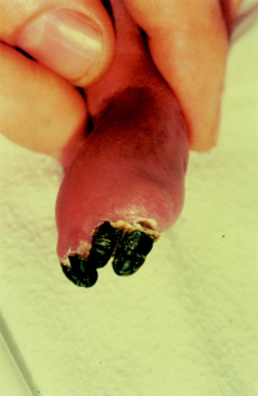
The incidence of homozygous (or compound heterozygous) protein C deficiency is estimated at 1/160 000 to 1/360 000.26 Homozygous protein S deficiency is probably even less common and there is only one well documented case in the literature at present.27 Parental consanguinity is a common feature in affected patients. The presentation is usually early and catastrophic, with purpura fulminans and disseminated intravascular coagulation developing within the 1st few days of life (fig 2).28 Cerebral, ocular, and renal thrombosis are also common, and in some cases these appear to have occurred as intrauterine events.
Digital gangrene in a neonate with protein C deficiency.
The definitive diagnosis is based on low or undetectable concentrations of protein C or protein S. The diagnosis is complicated initially by the presence of physiologically reduced concentrations of these inhibitors in neonates which, in the acute untreated phase, are further reduced secondary to disseminated intravascular coagulation. A high degree of clinical suspicion and assessment of parental values might help to confirm the diagnosis at this stage. The molecular defects responsible for these conditions have now been documented in a number of patients and can be used for future prenatal diagnosis.29
Management in the acute phase involves replacement of the deficient inhibitor protein.30 Protein C is now available as a plasma concentrate, whereas protein S replacement requires the administration of fresh frozen plasma. In the longer term, continued replacement treatment or the use of anticoagulant agents, primarily warfarin, are required to prevent recurrent episodes of purpura fulminans and other thrombotic events.
HOMOZYGOUS FACTOR V LEIDEN/PROTHROMBIN 20210A DEFECTS
The factor V Leiden and prothrombin 20210A mutations are relatively common in the general population, occurring with an estimated frequency of 3% and 1%, respectively, in the UK. Homozygosity for these defects will therefore be observed relatively frequently, but typically these defects do not seem to present clinically until adult life.