Article Text

Abstract
The effectiveness of targeted therapies with tyrosine kinase inhibitors in non-small-cell lung cancer (NSCLC) depends on the accurate determination of the genomic status of the tumour. For this reason, molecular analyses to detect genetic rearrangements in some genes (ie, ALK, ROS1, RET and NTRK) have become standard in patients with advanced disease. Since immunohistochemistry is easier to implement and interpret, it is normally used as the screening procedure, while fluorescence in situ hybridisation (FISH) is used to confirm the rearrangement and decide on ambiguous immunostainings. Although FISH is considered the most sensitive method for the detection of ALK and ROS1 rearrangements, the interpretation of results requires detailed guidelines. In this review, we discuss the various technologies available to evaluate ALK and ROS1 genomic rearrangements using these techniques. Other techniques such as real-time PCR and next-generation sequencing have been developed recently to evaluate ALK and ROS1 gene rearrangements, but some limitations prevent their full implementation in the clinical setting. Similarly, liquid biopsies have the potential to change the treatment of patients with advanced lung cancer, but further research is required before this technology can be applied in routine clinical practice. We discuss the technical requirements of laboratories in the light of quality assurance programmes. Finally, we review the recent updates made to the guidelines for the determination of molecular biomarkers in patients with NSCLC.
- immunohistochemistry
- diagnostic techniques and procedures
- oncogenes
This is an open access article distributed in accordance with the Creative Commons Attribution Non Commercial (CC BY-NC 4.0) license, which permits others to distribute, remix, adapt, build upon this work non-commercially, and license their derivative works on different terms, provided the original work is properly cited, appropriate credit is given, any changes made indicated, and the use is non-commercial. See: http://creativecommons.org/licenses/by-nc/4.0/.
Statistics from Altmetric.com
Introduction
ALK and ROS1 gene rearrangements occur in approximately 4% and 2% of lung adenocarcinomas, respectively.1 Although the frequency of these genomic alterations is low, their diagnosis offers patients with lung cancer the opportunity to receive highly effective targeted therapies.2 3 The story of ALK and ROS1 fusions reflects the current exciting state in lung cancer research.1 This review summarises the opinion of experts on ALK and ROS1 testing in the molecular diagnosis of non-small-cell lung cancer (NSCLC) and its relevance in routine clinical practice.
Pre-Analytical considerations
The quality of a histological diagnosis, as well as the additional molecular determinations, is strongly influenced by the pre-analytical management of the sample from the time it is obtained. For that reason, standardisation of sample handling and processing is necessary to reduce technical variability.4
Biopsies
Biopsy samples should be fixed by immersion in 10% neutral buffered formalin rapidly after they are obtained. A fixation time of 6–24 hours for small biopsies (including the duration of the fixation steps in the protocol of the processing machines), or 24–72 hours for surgical pieces, is recommended.5 6 In lung resection specimens, it is advised to insufflate the sample and make a cut along the tumour to achieve a homogeneous fixation throughout its length. The use of acid fixatives such as Bouin solution or heavy metals (ie, mercury) should be avoided.7
Cytology
Cytological smears fixed in alcohol or an alcoholic solution are suitable in protocols aimed at biomarker determination.8–10 National and international guidelines recommend, when possible, creating cell blocks for subsequent paraffin processing. This facilitates the application of similar protocols as those of tissue biopsies.1 11 There is no standardised procedure commonly used among laboratories.1 12
Sample optimisation
During tissue sample processing, it is necessary to adapt procedures and workflows to optimise the resources available in each laboratory. The role of pathologists and laboratory technicians is essential in the coordination and management after samples are obtained. Most molecular testing is performed on small samples (biopsies and/or cytological), and a minority on surgical resection specimens, which may represent both the primary tumour and the metastatic lesions. In case of multiple primary lesions, a sample of each lesion should be obtained.13 14 Regardless of the technique or procedure used, the objective of tumour tissue sampling is to obtain enough tumour cells with adequate quality for diagnosis and biomarker determination.
The minimum number of tumour cells has not been specified, although generally the presence of at least 100 tumour cells, or the minimum percentage of tumour cells established for the methodology used (such as in EGFR determinations), is recommended.15 16 For a proper assessment of fluorescence in situ hybridisation (FISH), a minimum of 50 cell nuclei with an adequate signal pattern are required. For molecular studies, it is also important to avoid tumour necrosis areas, or those with extensive fibrosis, and select a sample with the highest percentage of tumour cells with respect to accompanying non-tumour tissue.
In cytological smears, those areas with sufficient tumour cells in monolayer should be selected. Marking the appropriate area with diamond glass on the foil facilitates saving the probe in FISH techniques by limiting the area that should be covered.8
The application of sample management protocols, from the initial steps of the process and adapted to each laboratory, optimises profitability and avoids unnecessary losses of tissue by successive repetition of the paraffin block cutting process.1 15 16 In general, two important aspects of proper management include simultaneous planning of the required techniques and tissue preparation when possible, and avoiding the use of tumour samples with unnecessary broad-spectrum immunohistochemical panels. A single slide with one to two sections for the initial evaluation that allows both confirming the presence of a tumour and the morphological typing must be performed by trained personnel using the minimum amount of possible tissue. Some types of NSCLC without morphological features of squamous or glandular differentiation may require additional immunohistochemistry (IHC) techniques for tumour subtyping and for the exclusion of other diagnoses (non-epithelial or metastatic from another origin).
The current recommendation is to limit the use to two IHC markers if a clear morphological differentiation is not observed: TTF-1 as a marker of glandular differentiation and p40/p63 for squamous differentiation.15–17 Performing additional IHC is not recommended. It is important to minimise the number of techniques, supporting the diagnosis in the multidisciplinary evaluation with clinical-radiological data and prioritising the tissue for the biomarker’s determination. Finally, estimate the percentage of tumour cells and select the optimal area for macrodissection or microdissection, depending on the technical means available at each laboratory.
Performing biomarker tests on pre-cut tissue that has been stored for months is not recommended due to the apparent loss of antigenicity.18 19 Cutting the material for the determination of biomarkers at the beginning of the study and preserving the tissue in the paraffin block is preferred.
Therapeutic targets and diagnosis techniques
ALK IHC
ALK lung carcinomas express the protein resulting from the rearrangement of ALK with different genes. However, the protein amount produced is small compared with other tumours in which the expression is the result of other alterations. Thus, IHC detection has to be refined with primary antibodies of higher affinity and at an increased concentration, and with a more rigorous step of antigenic recovery and signal amplification procedures by polymers or synthetic molecules such as tyramine.1 However, the use of signal amplifiers alters staining evaluation because it invalidates the classification according to the intensity, and the cases are only scored as positive or negative. Further, excessive amplification could cause artefacts that could be misinterpreted as false positives.
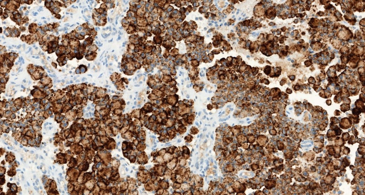
Comparisons between the different antibodies show that D5F3 (Cell Signaling Technology, Danvers, Massachusetts, USA) (figure 1), with the D5F3-based immunoassay (Ventana ALK [D5F3] CDx Assay, Tucson, Arizona), and 5A4 (Novocastra, Newcastle, UK) with the ADVANCE system (Agilent/Dako, Carpinteria, California, USA) present similar sensitivity and specificity.20 21 The Ventana ALK D5F3 CDx Assay is the only IHC test approved by the US Food and Drug Administration (FDA) as a companion diagnostic assay for four ALK inhibitors.22 The clone ALK1 (Dako) is less sensitive and it should not be used for this purpose. A new 1A4 antibody (Origene, Rockville, Maryland, USA) seems comparable to D5F3 and 5A4, although it has less specificity,23 is used without signal amplification,24 and positive results should be confirmed with a second technique.

Example of a lung adenocarcinoma immunohistochemistry (IHC)-positive for ALK using the Ventana ALK D5F3 antibody (Cdx assay) (×200).
Positive controls on the same slide where the sample is placed are mandatory, although there are parameters, like all those related to the pre-analytical phase, that do not ensure the quality of staining.25 26 The College of American Pathologists (CAP) recommends the use of positive controls on the same slide (IHC critical assay performance controls or iCAPCs).27 The last important technical point to emphasise is the analytical validation process. The CAP recommends a minimum of 10 positive samples for the technical validation of an IHC technique, although issues with sample acquisition in uncommon pathologies must be considered.28
There are assessment issues that can occur in signet ring cells when mucin vacuoles displace the ALK signal, resulting in false negatives. Also, neuroendocrine cells intermingled in some neoplasms can generate false positives, as does the non-specific background staining of mucin. In contrast, the non-specific apical membranous staining in pneumocytes or in neoplastic cells should not be considered positive.29
Finally, it should be noted that there can be discordant cases of positive IHC expression of ALK that are negative when using other techniques, especially FISH, due to several causes.30 First, there can be difficulty in the diagnosis with the interpretation of FISH in cases close to the threshold estimated as positive; second, due to amplification of the ALK gene, which is very common and usually provides a low staining intensity; third, due to a IHC false positive, usually because of interpretive error; fourth, because of the existence of complex rearrangements that difficult the interpretation of FISH, that is, false negatives of FISH; and fifth, due to alterations of ALK promoters that could overexpress the protein without altering the gene itself.29
ROS1 IHC
For assessment of ROS1 rearrangements, the international and national guidelines recommend the use of IHC as a screening method and confirmation of positive cases by cytogenetic techniques (mainly FISH) or molecular techniques such as real-time PCR (RT-PCR) or next-generation sequencing (NGS).11 16 31 32
Currently, there is no IHC assay FDA-approved for clinical practice, but there are two commercial antibodies available: the D4D6 clone (Cell Signaling Technology, Danvers, Massachusetts, USA), which is the most frequently used in published studies, and the SP384 antibody (Ventana Medical Systems, Tucson, Arizona, USA), which is the first and the only in vitro diagnostic (IVD) ROS1 IHC assay (figure 2).

Example of a lung adenocarcinoma positive for ROS1 using the D4D6 antibody (A) and SP384 antibody (B) (×200).
Using both clones, ROS1 IHC shows high sensitivity in most comparative studies with FISH or RT-PCR.1 11 16 33 However, the specificity ranges from 70% to 100%, depending on the positivity criteria used,1 11 16 33 34 which certainly can be improved by higher cut-offs.33 Different interpretation criteria were suggested (such us considering the intensity of staining (0–3+) or quantifying with an H-score), as well as different cut-off points (eg, positivity defined with moderate/strong intensity (2+/3+) or with H-score >100 or>150).16 33 35–37 Currently, there is no standard assessment accepted. Thus, it is recommended that each laboratory validates its own interpretative range.11 16 34 Although it is too soon to draw definitive conclusions about the differences between both clones, the few published comparative studies showed lower sensitivity for the D4D6 clone.33 38
It is important to consider that, unlike ALK IHC (which shows high specificity), ROS1 expression can be found in up to one-third of tumours without underlying ROS1 rearrangements,37 39 40 but with other genomic alterations (eg, mutations of EGFR, KRAS, BRAF or HER2, and ALK rearrangements).40 41 However, staining observed in these cases is usually focal.1 11 34 On the contrary, non-specific immunostaining has also been observed in the histological subtype of infiltrating mucinous adenocarcinoma37 and in non-tumour tissue (hyperplastic type II pneumocytes, alveolar macrophages and osteoclast-type giant cells).1 33 34
To ensure both the analytical phase and the interpretation of ROS1 IHC, a positive control in each case must be included.34 Unlike ALK IHC, when appendix ganglion cells are an adequate external positive control, there is no normal tissue for ROS1 IHC that can be used as an adequate positive control. In this case, the use of a known ROS1-positive tumour or a cell block of the HCC78 line (carrier of the SLC34A2-ROS1 fusion) is recommended.34 The frequent staining of non-neoplastic type II pneumocytes, especially with the SP384 clone, can be used also as an in situ control.33
Positive staining of ROS1 is characteristically cytoplasmic. However, variations in the immunostaining pattern have been described depending on the specific type of fusion (granular with focal or diffuse globular aggregates in tumours with the variant CD74-ROS1; weak cytoplasmic with membranous reinforcement in the fusion EZR-ROS1; solid cytoplasmic in cases with rearrangements SLC34A2-ROS1 and SDC4-ROS1; and vesicular in the variant GOPC-ROS1).1 33 34 36 37
In summary, ROS1 IHC is a screening technique of high quality due to its high sensitivity, rapid response time and lower cost compared with other techniques such as FISH or NGS. Given the variability in specificity, confirmation of positive or doubtful immunostaining by FISH and/or other molecular techniques (RT-PCR or NGS) before considering a tumour as ROS1-positive is recommended.1 11 16 31 32 34
ALK and ROS1 FISH
FISH has been considered the most sensitive method for the detection of ALK and ROS1 rearrangements, with a good correlation with IHC assays.42 However, it is necessary to use guidelines for interpretation that include technical details.
After histological diagnosis by H&E staining, the tissue sample needs to be examined to ensure that there are sufficient tumour cells for FISH testing. Representative tumour areas can be marked on the H&E-stained serial slide. The thickness of the paraffin-embedded sections can also affect FISH results (4±1 µm recommended), and the use of positively charged slides is highly recommended, as the adherence of the tissue during the procedure is improved. Pretreatment and digestion of the tissue need to be optimal to obtain an excellent morphology and probe signal intensity with low background noise.
FISH with ‘break-apart’ (BA) probes is a reliable method for the detection of ALK and ROS1 rearrangements in NSCLC. The proximity of the ALK (2p23) and EML4 (2p21) genes and the several fusion partners described for ROS1 make the BA design the best strategy for the detection of rearrangements. These probes are designed by labelling the 3′ (telomeric) of the fusion breakpoint with one fluorochrome and the 5′ (centromeric) with the other. Typically, 3′ ALK is labelled in orange and 5′ ALK in green. In contrast, the common design for ROS1 is the opposite: 3′ ROS1 is labelled in green and 5′ ROS1 in orange. Currently, the only approved FISH assay for the detection of ALK positivity, as a companion diagnostic tool for crizotinib-based treatment eligibility, is the Vysis ALK Break Apart FISH Probe Kit (Abbott Molecular, Des Plaines, Illinois, USA). Multiplex FISH panels that analyse ALK and ROS1 genes simultaneously are now available and could be useful to minimise issues of tissue scarcity in some cases.
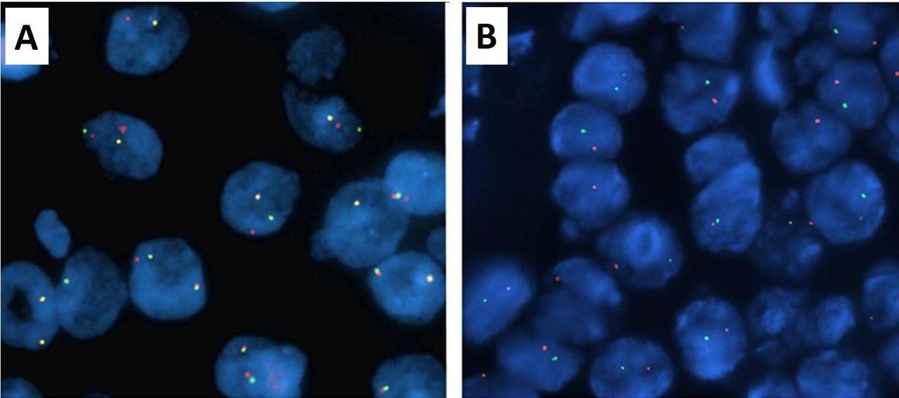
A nucleus is interpreted as rearranged when 3′ and 5′ signals are separated (figure 3). Due to structural variants of the rearrangement, deletion of one of the probes can occur resulting in an isolated 3′ or 5′ FISH signals. Isolated 3′ nuclei are categorised as positive, while nuclei with isolated 5′ should be classified as negative. Due to diversity of FISH patterns, fluorescence background, poor hybridisation or nuclei/FISH signals overlap, it is necessary to establish robust thresholds for the interpretation of the results. A minimum of 50 tumour cells are required for scoring and when the positive cells are ≥50%, it is considered positive for both ALK and ROS1 genes.34 43 In uncertain cases (range 10%–15%), a correlation with another diagnostic test is recommended (IHC or NGS).44 The use of higher cut-offs and/or imaging systems are strategies that ensure specificity.1 11 33 34 45
Example of a lung adenocarcinoma positive for ALK (fluorescence in situ hybridisation (FISH) pattern: 1F1O1G) (A) and a lung adenocarcinoma positive for ROS1 with a deletion of the non-rearranged allele (FISH pattern: 1O1G) (B) using break-apart probes (×100).
Recently, false-positive FISH results have been detected by NGS technology: an isolated 3′ pattern could represent a deletion that produces a non-functional ALK or ROS1 fusion.46 47 False-negative FISH findings are also well documented and could be explained by the presence of atypical FISH patterns (red-doublet pattern for ALK gene) generated by complex gene rearrangements/cryptic insertions not clearly detected by FISH.
ALK and ROS1 analysis in cytological samples
Tissue material is often preferred for biomarker analysis in clinical trials because paraffin blocks are routinely processed in pathology laboratories, and these blocks provide multiple sections for various analyses. However, as many as 40%–70% of all advanced NSCLC are diagnosed by cytological evaluation alone, with no concurrent histological examination of tissue material, emphasising the necessity to expand ALK and ROS1 analysis to cytological specimens.9 10 48–51
Processing such specimens for formalin-fixed paraffin-embedded (FFPE) cell blocks has become the preferred method in many laboratories, as cell blocks can be handled in a similar way as histological specimens, and the same protocols for biomarker analysis can be applied. However, some cell blocks contain too few or no cancer cells for molecular analysis, and differentiating tumour cells from adjacent reactive cells is more challenging than in conventional cytology, especially during FISH analysis.52 Consequently, it is necessary optimising cytological samples for their study.53 54
FISH analysis is applicable to almost all types of cytological specimens including conventional smears. Also, FISH technology was used to evaluate cell lines or disaggregated intact nuclei from histological tumour specimens before it became applicable to tissue sections.1 55 The protocol for cell blocks is usually the same as the one used for histological samples. The use of non-FFPE cytology specimens (ie, direct smears, cytospins and liquid-based cytology) can give excellent results following previously described recommendations and protocols, supported by stringent validation studies. Destaining Giemsa-stained smears is recommended before starting the FISH assay.1 9 34 In fact, cytological specimens have several advantages:1 53–56 first, intact nuclei, which allows for the detection of the true number of FISH signals in a nucleus; second, the use of previously stained slides (Papanicolaou & Diff-Quick are valid), without the need for additional unstained slides,53 so a real percentage of tumour cells among normal/inflammatory cells can be determined; and third, better DNA quality in both air-dry and alcohol-fixed smears than in FFPE samples, which can lead to cross-linking and chemical modification of nucleotides.
Currently, smears are being reported as one of the preferred cytological samples.1 8 51 53 54 57 The use of adhesive-coated or positively charged slides is recommended, as the adherence is improved and the cells are prevented from floating off during FISH/IHC procedures.1 55 57 FISH also applies well to specimens stained immunocytochemically if 3-amino-9-ethylcarbazol is used as a chromogen. The use of 3,3′-diaminobenzidine can interfere with the FISH signal because of autofluorescence.55 58
The threshold for a positive ALK and ROS1 FISH result and the patterns of positivity have been established on the basis of analysis of histological samples, and similar scoring is used with cytological samples.8 58 59 Concerns regarding the loss of the slides dedicated to FISH are addressed by capturing representative images or by scanning the whole slide before FISH analysis. It is also possible to stain slides again after FISH analysis.53 58
IHC to detect overexpression of the ALK and ROS1 proteins has emerged as a valuable method to screen NSCLC for subsequent FISH analysis and for further evaluation of uncertain FISH findings.59 ALK IHC works well in cell blocks and in other types of cytological samples such as conventional and liquid-based cytological preparations. There are reports showing a 100% concordance of IHC in cell blocks using the ALK (D5F3) CDx Assay on the Benchmark XT automated immunostainer (Ventana) with FISH.60 The accuracy of ALK IHC on Papanicolaou-stained slides is equally high.54 61
ROS1 IHC is highly accurate for prescreening cytological samples for FISH in both cell blocks and smears. However, as mentioned before, there is a need to expand ALK and ROS1 analysis to cytological specimens and further published studies on ROS1 IHC in cytology are required.
IHC performed on cytological smears and/or cytospin slides may be significantly influenced by various factors during the pre-analytical phase because of high variability in preparation, fixation and staining methods of cytological specimens.1 62 When dealing with fine-needle aspiration cytology, ROSE (Rapid On-Site Evaluation) helps in keeping quality and quantity of the cytological samples in order to guarantee the performance of ancillary analysis for biomarker information.
Other techniques for molecular diagnosis in lung cancer
Breakpoints of ALK are located at exons 19 and 20, and the most frequent ALK fusion partners include EML4, NPM1, CLTC, TPM3, RANBP2, STRN, ATIC, KIF5B, TPM4, TFG and HIP1, among others. Breakpoints of ROS1 are sited at exons 32, 34, 35 and 36, and the most frequent partners include SLC34A2, CD74, TPM3, SDC4, EZR, LRIG3, FIG or GOPC, MSN, KDELR2 and CCDC6.63 64
In addition to in situ tissue determinations such as FISH and IHC, several non-in situ assays based on RT-PCR or NGS for the detection of ALK and ROS1 gene rearrangements have been developed. RT-PCR to explore ALK and ROS1 gene rearrangements is a simple and relatively inexpensive method, but extracting RNA from FFPE samples may be challenging. In this procedure, RNA is converted into complementary DNA (cDNA) by reverse transcriptase, which is then PCR-amplified with fusion-specific primers to detect the presence of a certain fusion variant. Therefore, multiple assays are necessary to discriminate among the previously described variants.65 In addition, a very sensitive PCR-based method was reported to detect overexpression of ALK fusion transcripts expressed in lung cancer,66 with protocols including multiplex RT-PCR and analyses of the relative expression of the 5′ and 3′ fragments of the ALK gene transcript. However, this method cannot be easily applied to ROS1, since the gene is also expressed in normal and hyperplastic lung tissue.66 67
A recent meta-analysis on the use of ALK determination by RT-PCR, including 21 studies and involving 2800 patients with NSCLC, confirmed a sensitivity of 92.4% and a specificity of 97.8% for detecting rearrangements in FFPE samples.68 RT-PCR for ROS1 has been successfully applied to identify positive cases with a sensitivity of 100% and a specificity of 85%–100%, using FISH as the reference standard method.67 69 However, in addition to the mentioned limitations, the list of ALK and ROS1 fusion partners is still growing, and RT-PCR is likely to miss rare variants. These reasons have limited the use of the technique in clinical practice.
The NanoString technology, capable of detecting known fusion gene transcripts from FFPE samples employing a dual capture and reporter probe system, provides an alternative assay that has shown good concordance with FISH and IHC results for ALK and ROS1.70 Agreement in detecting ROS1 was 87.2% and 86% with IHC and FISH, respectively.
Some limitations prevent the full implementation of these technologies in the clinical setting. Standard methods, as described, easily highlight already known fusions, but may misdiagnose new variants and fusion partners due to the low precision of the 3′−5′ imbalance value. A series of innovative approaches to detect ALK and ROS1 gene fusions using NGS have been developed. These comprehensive assays require just nanograms of RNA, show relatively low failure rates in paraffin-embedded tissues and allow for the detection of the specific alteration irrespective of fusion partner.71 72 Amplicon-based NGS approaches have been assessed for the detection of gene fusions in patients with NSCLC, including the Oncomine (Thermo Fisher) and the Archer FusionPlex panels.73–76 Reported studies using NGS for detecting fusions demonstrated a sensitivity of 100% and a specificity of 97.7% compared with the gold standard. These studies make it possible to confirm the presence or not of a rearrangement in FISH samples with 3′ isolated ALK or ROS1 signal FISH pattern finding discrepancies in some cases.77 Interestingly, novel translocations partners have been identified, highlighting the ability of NGS to detect novel translocation partners not inherently captured by FISH.78 79 The global concordance of NGS for fusions was 67% when compared with both FISH and IHC (F. Rojo, unpublished data). Additionally, NGS has also been shown to detect novel and complex ALK fusions. The clinical relevance is due to the benefit of using of crizotinib in the IHC-negative or FISH-negative gene rearrangement setting.80 Finally, the use of comprehensive genomic profiling based on a hybrid capture-based NGS in a large cohort of patients has revealed a prevalence of alterations in ALK of 4.1% and of ROS1 in 1.5%.81 Implementation after clinical and analytical validations should be done following recommendations.82–84
The development of liquid biopsies has changed substantially the treatment of patients with solid cancers, particularly in those with advanced lung cancer. Addition of liquid biopsy in the advanced lung cancer setting can significantly impact the initial detection of actionable oncogenic drivers, the identification of resistance mutations in patients relapsing on targeted therapies and the assessment of the response to treatment.85 Several recent studies showed that gene rearrangements could be detected in circulating tumour cells (CTCs) by immunocytochemistry and/or FISH.86–89 Free plasma DNA is routinely used to analyse mutations in EGFR in patients with advanced NSCLC. In contrast, plasma RNA is very rarely used for detection of rearrangements. Nonetheless, the detection of these genomic alterations can be performed in plasma using RT-PCR. While RT-PCR can detect rearrangements in the blood, it still has some limitations for its application in clinical practice, including RNA preservation in plasma, as blood RNA degrades rapidly after sampling, or the low detection limit.90 Several studies have shown that platelets can sequester tumour RNA by taking up circulating microvesicles.91 These tumour-educated platelets can be isolated and constitute an enriched source of tumour RNA for the detection of ALK rearrangements.90
However, CTC detection techniques are not available in all laboratories and thus not easily accessible to patients. The detection of rearrangements in plasma or platelets can only be performed if the pre-analytical phases are perfectly controlled. In addition to methodology, it should be noted that the number of CTCs, the plasma RNA or the RNA associated with platelets, may vary depending on patients, tumour biology and tumour burden. In this context, NGS approaches have been developed to identify genomic alterations in purified free nucleic acids in plasma.92 93 Based on the hybrid-capture approach, these techniques do not only search for rearrangements but also for point mutations, insertions, deletions and copy number variations. However, to date, these approaches probably still lack sufficient sensitivity and collaborative studies will be necessary before using them in routine clinical practice.
The pathology report
The goal of the pathology report is to provide the oncologists with the information needed to readily select the best therapy and to explain the results to their patients. Pathologists should be able to learn sufficient details from the reports to help determine if repeated testing is appropriate, or to help resolve discrepancies between clinical and laboratory findings, or between results from different laboratories.94
Both for ALK and ROS1, the pathology report should contain at least the following information16: patient and physician identification; anatomopathological diagnosis; type and date of sample; identification of external code in the case of reference centres; medium in which the sample is received (eg, fresh, frozen, paraffin); sample anatomical origin; date of request; sample reception and results; technique used (for ALK and ROS1, specification on whether gene rearrangements or protein overexpression were examined); in the case of use of commercial reagents, the label ‘in vitro diagnostic products’ (IVD) should be indicated, and trade name and batch number; quality of sample (including the percentage of tumour cells and whether the sample has been enriched by macro or microdissection, as well as DNA concentration and purity); specification if sample was adequate or inadequate; and the analysis result (defining the type of molecular alteration detected or the absence of molecular alterations). It should also include the number of cells analysed, and the number and percentage of cells with each finding, professional responsible, laboratory manager, additional information important for the physician, and information of the laboratory about accreditation or participation in Quality Control (QC)/Quality Assurance programmes (QAP).
Technical and quality requirements of laboratories
Although NGS procedures are becoming a reliable alternative to standard methods of diagnosing translocations involving ALK or ROS1, we will refer mainly to the most practical methods in place in pathology laboratories: IHC and FISH. Since IHC is easier to implement and interpret, it is normally used as the screening procedure, while FISH is used to confirm the rearrangement and decide on ambiguous immunostainings.
IHC is currently performed in most laboratories following validated procedures that use antigen retrieval methods, antibodies and signal enhancing reagents that have been thoroughly tested. Alternatively, laboratories should have controls obtained from their cases or through participation in a QAP.16
FISH is becoming available in most pathology laboratories.15 Several suppliers provide fluorescent probes (either orange-red or green) flanking the breakpoint at either ALK or ROS1 (BA design). While FISH procedures have become quite simple and even automated in some laboratories, the selection of the area to be studied is relevant. FISH results are often interpreted by biologists who have not been involved in the overall diagnostic procedure. Thus, it is of great importance that biologists and pathologists work together effectively so the correct cells are being evaluated. The correct interpretation of FISH signals must follow current guidelines,1 taking into account the type of abnormalities that are considered positive (eg, both split signals and ‘single red’ signals in standard ALK FISH with Vysis probes).
Laboratories involved in this type of diagnosis must regularly participate in a recognised QAP. The Spanish Society of Pathology (SEAP) has a very practical programme for ALK. The European Molecular Genetics Quality Network also provides accredited external quality assessment to laboratories performing molecular diagnosis. A list of the available providers can be found in a previous work.16 Some of the results of ring trials or QAP have been published.95 96
Updated guidelines for selection of biomarkers
In 2013, the CAP, together with the International Association for the Study of Lung Cancer and the Association for Molecular Pathology, published the first guidelines for the determination of molecular biomarkers in patients with NSCLC.94 Continuous therapeutic and technological progress, together with a better knowledge of tumour molecular biology, made it necessary to update these guidelines.97 98 This update was published at the beginning of 2018, receiving approval by the American Society for Clinical Oncology (ASCO) a few months later.11 31 More recently, the European Society for Medical Oncology (ESMO) and the SEAP, together with the Spanish Society of Medical Oncology (SEOM), have updated their respective guidelines.16 32
Box 1 shows the main modifications or novelties of these guidelines.
Summary of the main topics reported in the updated guidelines
Which biomarkers should be studied and in which patients?
EGFR and ALK
The study of EGFR mutations and ALK rearrangements is strongly recommended in:
Patients with lung adenocarcinoma, regardless of their clinical characteristics, or with other histology when clinical data indicate high probability of molecular alterations
The study of the EGFR T790M resistance mutation is strongly recommended in:
Patients with lung adenocarcinoma with EGFR mutations that confer sensitivity to anti-EGFR tyrosine kinase inhibitors and have progressed after treatment
ROS1
The ROS1 rearrangement study is recommended in:
Patients with lung adenocarcinoma, regardless of their clinical characteristics
BRAF*, RET, HER2, MET and KRAS
The molecular study of BRAF, RET, HER2, MET and KRAS in an integrated way within large molecular panels is recommended both initially and when the results of EGFR/ALK/ROS1 are negative
Individual study is not recommended outside the context of clinical trials
When should biomarkers be studied?
At the time of diagnosis
In patients with advanced stage disease (IIIB and IV) or at the time of progression in patients who initially had a lower stage
Which samples are optimal for biomarkers study?
Tissue samples
It is recommended to use samples fixed in formalin and included in paraffin, frozen fresh samples, or alcohol fixed samples
It is recommended to use tissue optimisation procedures that allow not only the pathological diagnosis but also the analysis of biomarkers
Cytological samples
Both cell blocks and cytological extensions can be used
Liquid biopsy
The use of molecular methods based on cfDNA for the diagnosis of lung adenocarcinoma is currently not recommended
In some clinical contexts in which the tissue is limited and/or insufficient, cfDNA can be used for EGFR determination
In patients with EGFR-mutated adenocarcinomas treated with anti-EGFR tyrosine kinase inhibitors who have progressed or developed acquired resistance, cfDNA can be used for the determination of the EGFR T790M mutation; if the result is negative, it is recommended to perform the molecular study in tissue
How long should it take for the results to be available?
It is recommended that molecular study results be available in 2 weeks (10 working days) from the moment the sample is received in the molecular pathology laboratory
What techniques should be used?
EGFR mutations
Techniques capable of detecting molecular alterations in samples with at least 20% of tumour cells should be used
EGFR T790M resistance mutation
Techniques capable of detecting EGFR T790M mutations in as little as 5% of viable cells should be used
ALK rearrangements
Both FISH and IHC can be used
ROS1 rearrangements
IHC can be used as a screening method, confirmation by cytogenetic or molecular techniques being necessary in ROS1 IHC positive cases
BRAF*, RET, HER2, MET and KRAS
To identify other treatment options beyond EGFR/ALK/ROS1, comprehensive sequencing panels are preferred over individual tests
How should the results be reported?
It is recommended that discordant, equivocal or unexpected results be confirmed or resolved with an alternative method or another sample
It is strongly recommended that laboratories follow similar quality controls, quality assurance policies and procedures as for other clinical laboratory trials
It is strongly recommended that laboratories use clinically validated methods with appropriate performance characteristics, following standardised best practice standards for each technology
With regard to the selection of molecular biomarkers, together with the already recommended EGFR mutations and ALK rearrangement testing, ROS1 rearrangements and BRAF mutations assessment should be included.16 31 32 Other biomarker assessments such as MET, RET, HER2 and KRAS are not recommended as independent tests but are recommended within large molecular panels both initially and when the results of EGFR/ALK/BRAF/ROS1 are negative.
When clinical data indicate high probability of molecular alterations in non-adenocarcinoma cases, the previous recommendations have remained valid for the patients, similar to other international guidelines.11 16 31 32
In relation to the optimal sample and its handling, in addition to the paraffined material (tissue samples or cell blocks from cytological samples), cytological extensions can be used.9 The use of protocols for tissue utilisation that allow both the pathological diagnosis and biomarker assays were recommended.6 15 16
On analytical aspects, for ALK testing, IHC was included as a method equivalent to FISH. For ROS1 testing, the use of IHC was recommended as a screening method with confirmation of positive cases by cytogenetic or molecular techniques.
Finally, molecular determinations in blood and the use of comprehensive sequencing techniques were incorporated (box 1).
Take home messages
Both immunohistochemistry (IHC) and fluorescent in situ hybridisation (FISH) can be used to search for ALK fusions.
IHC can be used as a screening method to search for ROS1 fusions, but confirmation by cytogenetic or molecular techniques is necessary in positive samples.
In case of contradictory, ambiguous, or unexpected results, verification with alternative methods or another sample is recommended.
Ethics statements
Patient consent for publication
Acknowledgments
Manuscript formatting support was provided by Cristina Sacristán and Francisco López de Saro (Trialance SCCL), funded by Pfizer.
References
Footnotes
Handling editor Runjan Chetty.
Contributors All authors contributed equally to the development of this review.
Funding All the authors received an honorarium from Pfizer in connection with the development of this manuscript.
Competing interests None declared.
Provenance and peer review Not commissioned; externally peer reviewed.